Fibrosis suppression by inhibiting integrin alpha-8 beta-1 function
a technology of fibrosis suppression and integrin alpha-8 beta-1, which is applied in the field of antifibrosis agents, can solve problems such as hepatic cirrhosis, and achieve the effect of inhibiting fibrosis
- Summary
- Abstract
- Description
- Claims
- Application Information
AI Technical Summary
Benefits of technology
Problems solved by technology
Method used
Image
Examples
example 1
Generation of Anti-integrin α8β1 Antibody
(1) Immunization
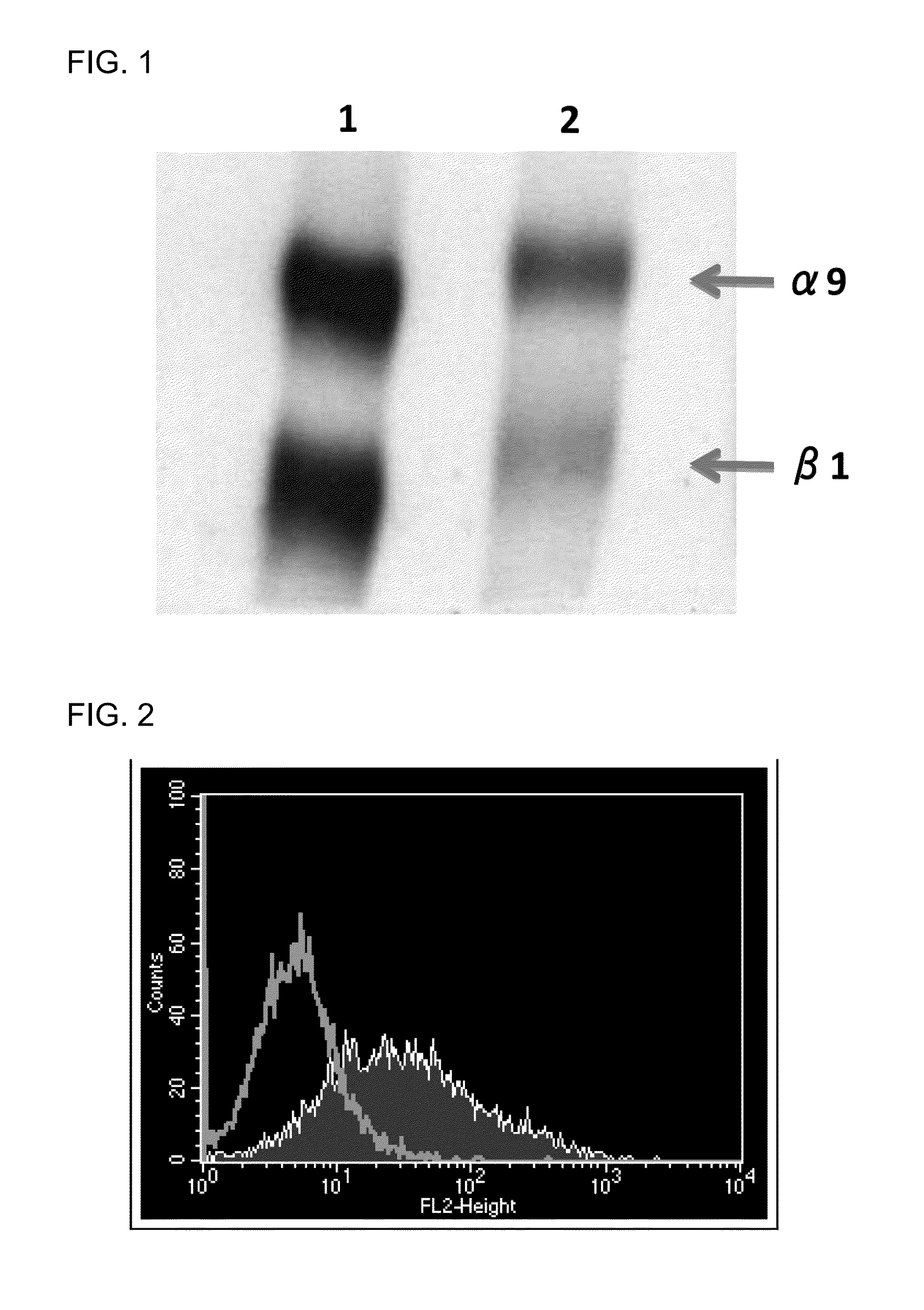
[0120]A cDNA encoding the mouse integrin α8 chain was cloned into a mammalian expression vector. Next, the expression vector was transfected into a chicken lymphoblastoid cell line by electroporation. Then, an antibiotic was added, and vector-expressing cells were selected. A chicken was hyperimmunized with the resulting mouse integrin α8-expressing cells. An antibody titer in the chicken serum was determined by flow cytometry (FACS) analysis. The FACS analysis was performed in accordance with a typical protocol of FACSCalibur (BD, USA).
[0121]Note that it was verified that integrin α8 and β1 chains form a heterodimer in any of birds and mammals (Bossy B et al., EMBO J, 10(9): 2375-2385, 1991; J Cell Sci, 108: 537-544, 1995). In this regard, however, specific experimental data on whether or not a mammalian a chain and a bird β chain can form a heterodimer have not been published. In view of this situation, the following procedu...
example 2
Epitope Evaluation
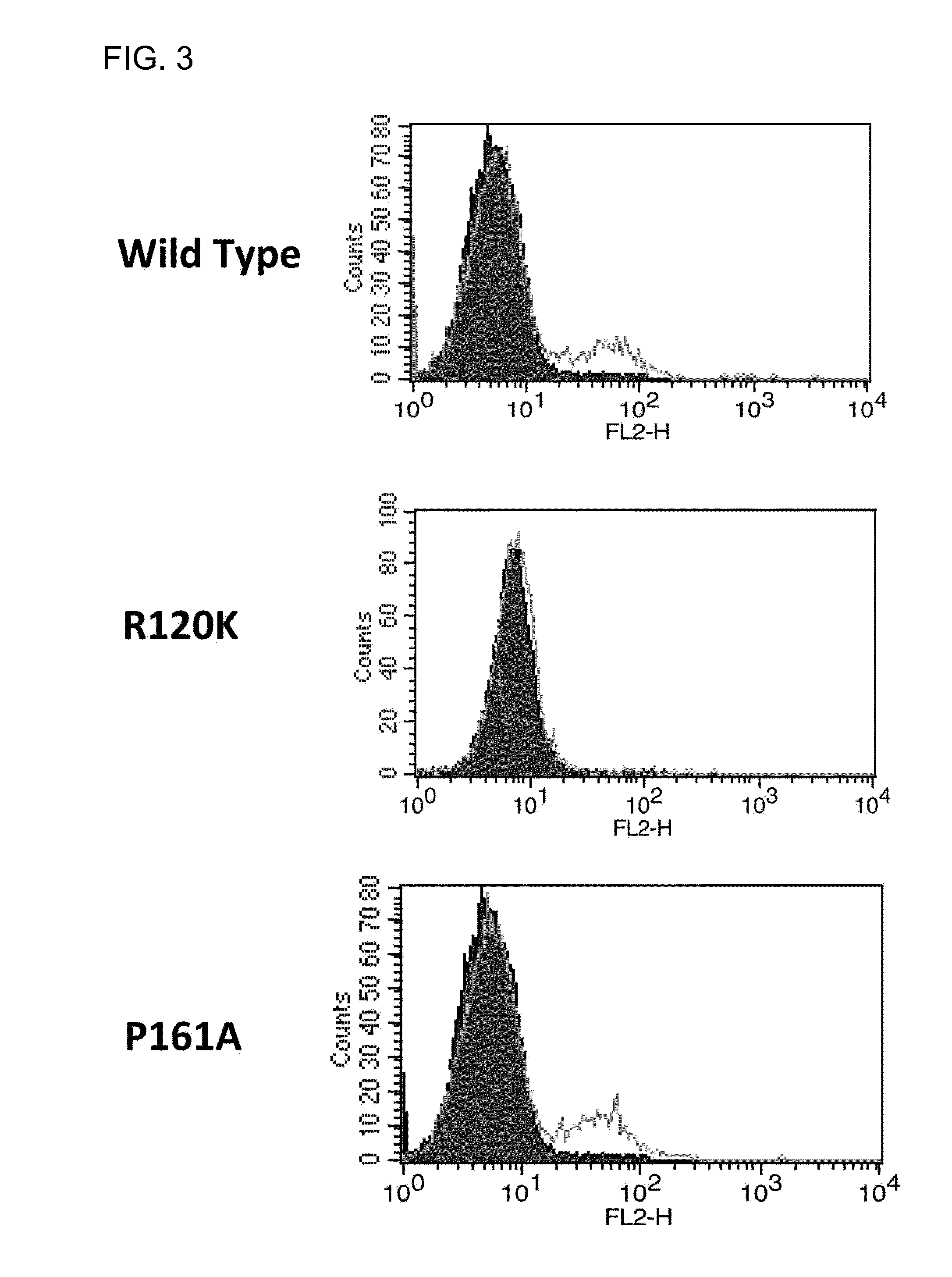
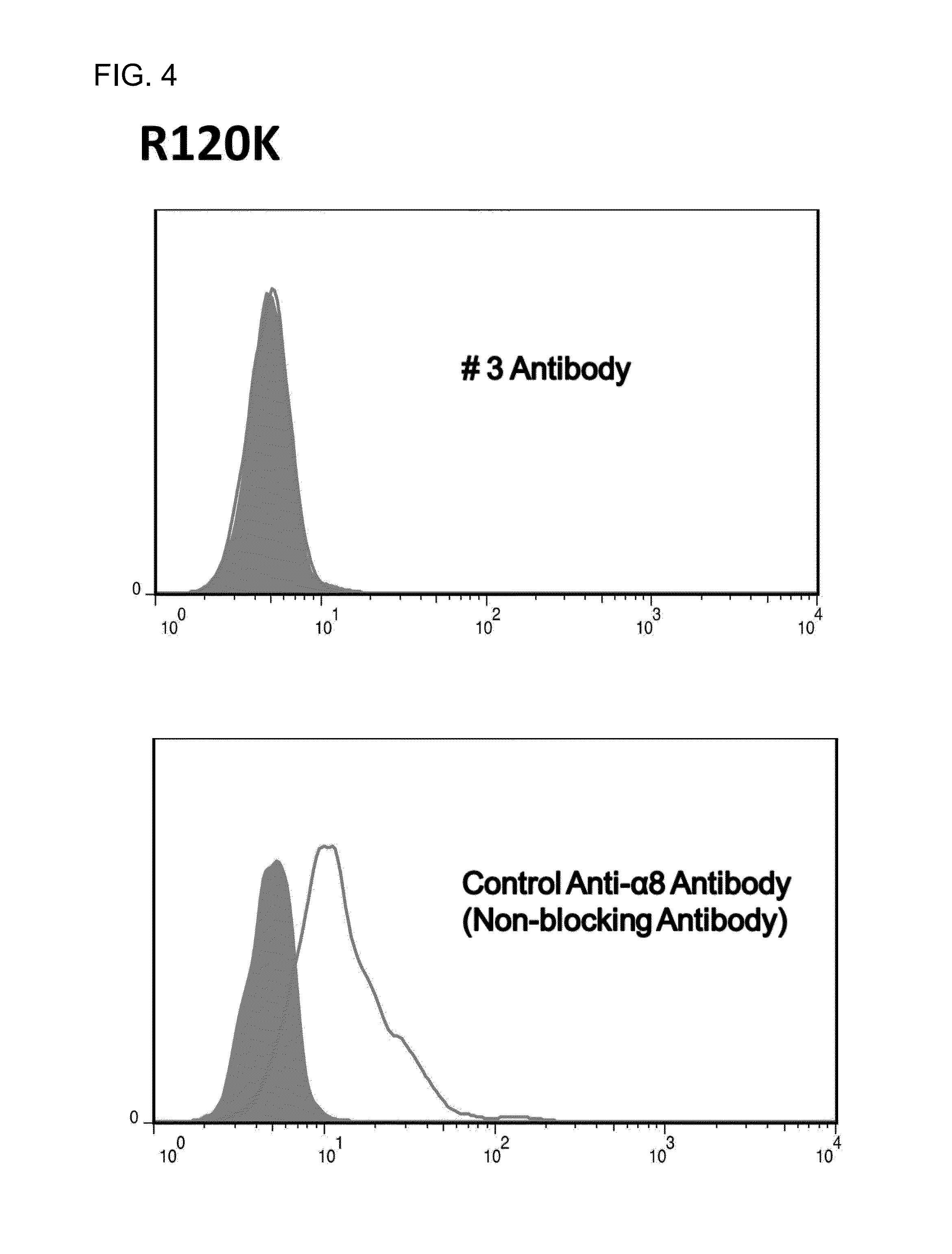
[0130]First, cDNAs encoding 8 different human α8 chains (R120, K125, 5132, P161, Y199, A238, A260, and 5261) were constructed. Next, these cDNAs were each transiently expressed on CHO cells and the CHO cells were reacted with the #3 antibody. The #3 antibody was reacted with the wild-type and 7 mutants (K125, 5132, P161, Y199, A238, A260, and S261), but was not reacted with the R120 mutant. FIG. 3 shows the results of FACS analysis in which the #3 antibody was reacted with the wild-type, R120 mutant, or P161 mutant. Then, in order to exclude the possibility that the R120K mutant was insufficiently expressed due to its transient expression, a stable cell line was established and the antibody was likewise reacted with the mutant. The reactivity was still not observed. In addition, the R120K mutant was well reacted with a control anti-α8 antibody. This result verified that the R120K mutant was sufficiently expressed (FIG. 4). The above results indicate that an epitope...
example 3
Evaluation of Blocking Activity toward Integrin α8β1
[0131]First, mouse osteopontin (2.5 mg / ml) was immobilized on a 96-well plate. Next, α8-expressing K562 cells were added at 1×10 E5 cells / well to the plate, and the #3 antibody was also added at concentrations designated in FIG. 5. In FIG. 5, absorbance of adherent cells was detected at 570 (A570) nm A lower value at A570 nm indicates less binding between the α8-expressing K562 cells and osteopontin.
[0132]These results demonstrated that the #3 antibody blocked the binding between the α8-expressing K562 cells and osteopontin (FIG. 5). That is, the #3 antibody functioned as an antagonist for integrin α8131. In addition, when the value in the case of no antibody addition was set to 100, 60% or higher blocking activity was observed at an antibody concentration of 0.05 μg / ml. Further, 70% or higher blocking activity was observed at an antibody concentration of 0.10 μg / ml and 95% or higher blocking activity was observed at an antibody co...
PUM
| Property | Measurement | Unit |
|---|---|---|
| Weight | aaaaa | aaaaa |
Abstract
Description
Claims
Application Information
 - R&D
- Intellectual Property
- Life Sciences
- Materials
- Tech Scout
- Unparalleled Data Quality
- Higher Quality Content
- 60% Fewer Hallucinations
Browse by: Latest US Patents, China's latest patents, Technical Efficacy Thesaurus, Application Domain, Technology Topic, Popular Technical Reports.
© 2025 PatSnap. All rights reserved.Legal|Privacy policy|Modern Slavery Act Transparency Statement|Sitemap|About US| Contact US: help@patsnap.com


