
Pet/spect agents for applications in biomedical imaging

a biomedical imaging and agent technology, applied in the field of ligands and radioisotopic tracers, can solve the problems of not clinically available sup>18/sup>f-based agents, further urgency, and less than ideal cardiac perfusion studies, so as to enhance the detection of myocardial viability, enhance the extraction of first pass into heart tissue, and reduce liver retention

Active Publication Date: 2014-11-20
WASHINGTON UNIV IN SAINT LOUIS
View PDF1 Cites 0 Cited by
  • Summary
  • Abstract
  • Description
  • Claims
  • Application Information

AI Technical Summary

Benefits of technology

The present invention aims to provide MPI probes with enhanced first pass extraction, linearity with true blood flow, enhanced detection of myocardial viability, reduced liver retention, and more efficient clearance from non cardiac and adjoining tissues compared to currently available probes. Additionally, the invention seeks to develop agents that can be used for heart imaging by incorporating a radionuclide and with high myocardial tissue uptake, high heart / blood ratios, and rapid clearance from the livers. The synthesis, purification, and formulation of the agents can be accomplished in less than 60 minutes.

Problems solved by technology

However, no 18F-based agent is yet clinically available.
In addition, recent threats to the stable production and supply chain of 99mTc have added further urgency to the search for a viable PET perfusion agent.
Among the several agents that are commercially available for perfusion imaging, all of these suffer from one or more shortcomings that render them less than ideal for cardiac perfusion studies.
While the former factor can decrease sensitivity, the latter component can increase background noise from adjacent tissues thereby atficting signal-to-noise ratios.
The resulting image quality can be less than optimal for interpretation by clinicians in nuclear medicine or physicians such as radiologists.

Method used


Image

  • Pet/spect agents for applications in biomedical imaging
  • Pet/spect agents for applications in biomedical imaging
  • Pet/spect agents for applications in biomedical imaging

Examples


example 1

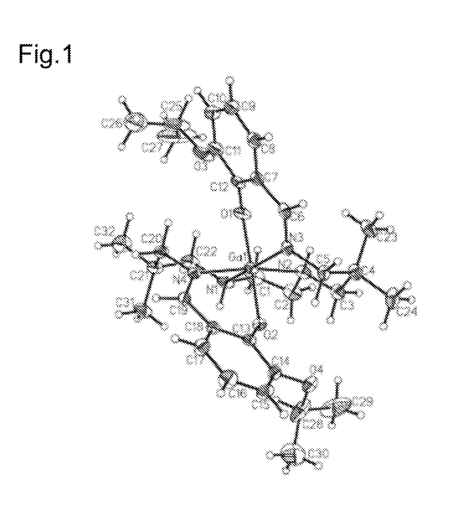
[0188]This Example illustrates the structure of a complex of the present teachings. The crystal structure of [ENBDMP-3-isopropoxy-PI-Ga]+ displayed in FIG. 1 shows a symmetrical engagement of the four nitrogen atoms in the equatorial plane and two axial phenolate atoms. FIG. 1 presents a projection view of cationic gallium (II) complex [ENBDMP-3-isopropoxy-PI-Ga]+ (1), but without iodide (I−) as the counter anion. FIG. 1 includes the crystallographic numbering scheme. Atoms are represented by thermal ellipsoids corresponding to 50% probability. 1H NMR, proton-decoupled 13C NMR, and HRMS analysis can also be used to validate the structure.

example 2

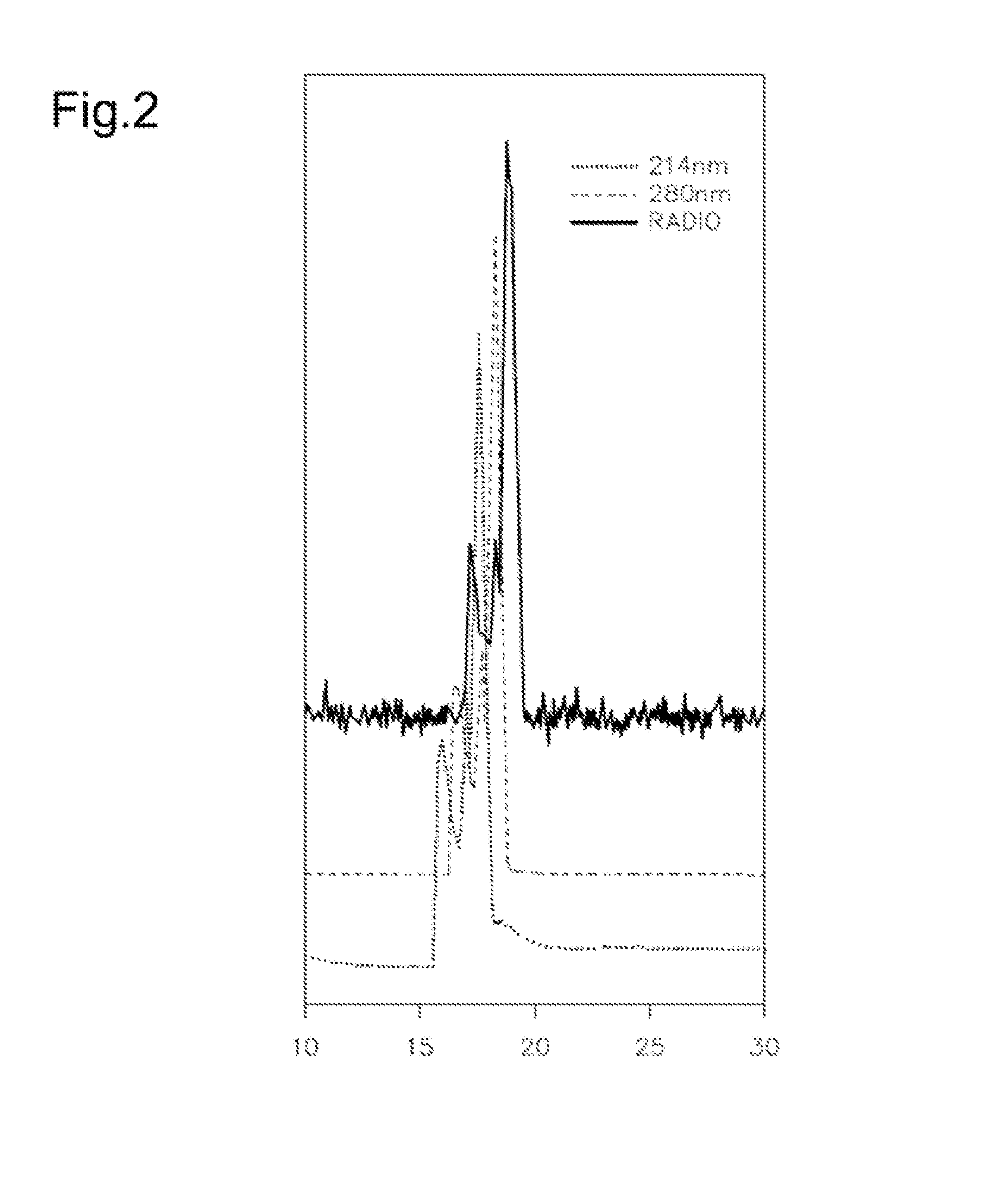
[0189]This Example illustrates HPLC data confirming synthesis and radiolabeling of [ENBDMP-3-isopropoxy-PI-67Ga]+. In these experiments, the 67Ga-labeled complex (1A) was synthesized and characterized via HPLC. FIG. 2 presents HPLC data for [ENBDMP-3-isopropoxy-PI-67Ga]+ 1A co-injected with unlabeled 1. In FIG. 2, peaks have been offset for visualization.

example 3

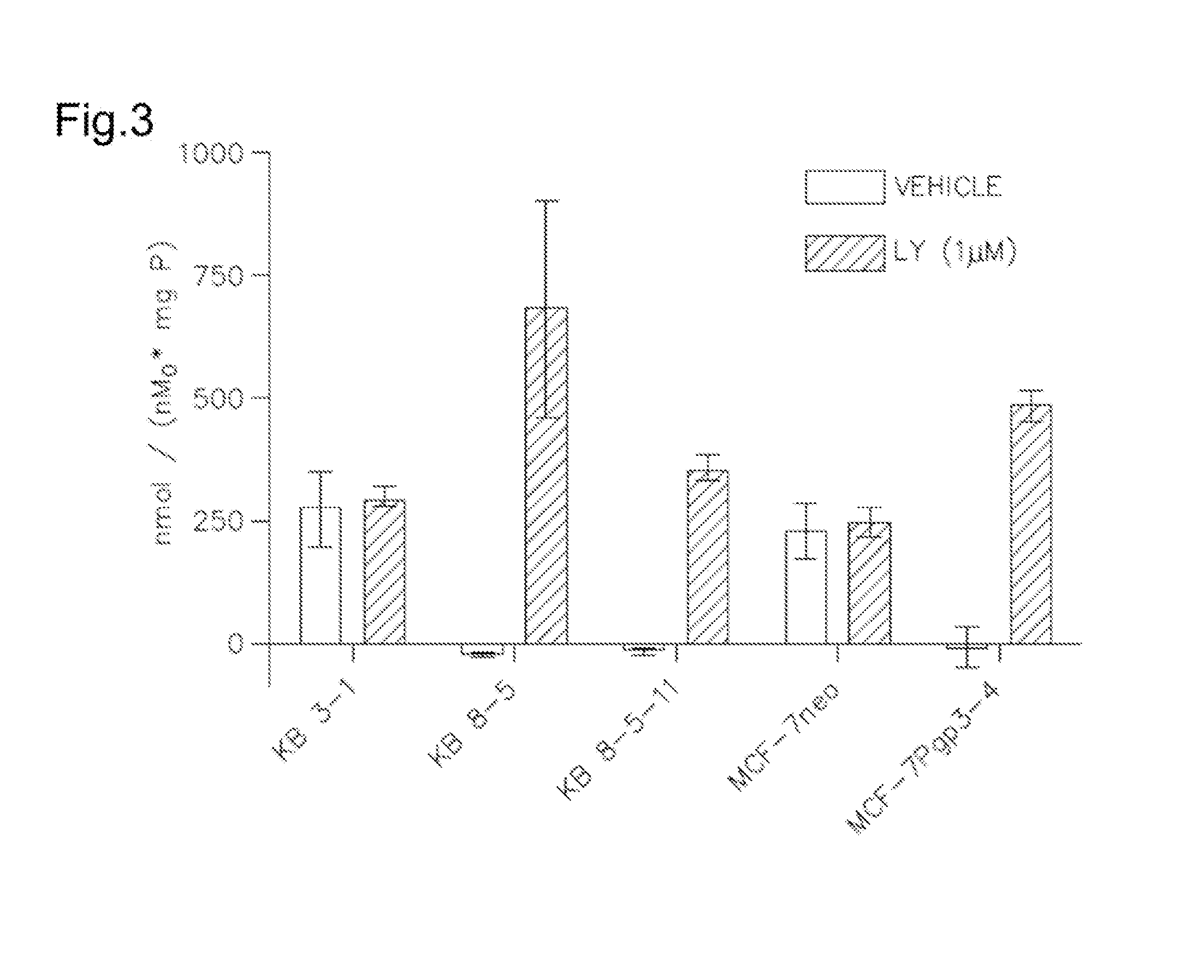
[0190]This Example illustrates characterization of [ENBDMP-3-isopropoxy-PI-67Ga]+ for 1A, FIG. 3 shows cellular accumulation of 1A in KB-3-1 cells (−Pgp). MCF-7 cells (−Pgp), MDR KB-8-5 (+Pgp), KB-8-5-11 (Pgp++) cells and stably transfected MCF-7 / MDR1 cells as indicated. Shown is net uptake at 90 minutes (fmol (mg protein)−1 (nM0)−1) using control buffer in the absence or presence of MDR1Pgp inhibitor LY335979 (1 μM). Each bar represents the mean of 4 determinations; line above the bar denotes +SEM.


PUM

PropertyMeasurementUnit
retention timeaaaaaaaaaa
pHaaaaaaaaaa
structureaaaaaaaaaa

Abstract

Tracers that can be used for PET or SPECT imaging of the distribution of Pgp are disclosed. The tracers are metalloprobes that can comprise a radioactive metal ion such as 67Ga or 68Ga. Methods of synthesizing the tracers, and methods of imaging heart and other tissues are also disclosed. The tracers can be used to obtain high signal-to-background ratios for imaging tissues in vivo such as heart or tumor tissue. In various embodiments, disclosed tracers can exhibit, a) enhanced first pass extraction into heart tissue compared to presently available probes, b) linearity with true blood flow, c) enhanced detection of myocardial viability compared to presently available probes, d) reduced liver retention compared to presently available probes, and e) more efficient clearance from non-cardiac and adjoining tissues compared to presently available probes.

Description

CROSS-REFERENCE TO RELATED APPLICATIONS[0001]The present application is a National Stage entry application of and claims the benefit of PCT / US12 / 024,752 international filing date 10 Feb. 2012 and claims the benefit of U.S. Provisional Patent Application Ser. No. 61 / 441,732 filed 11 Feb. 2011. These applications are incorporated by reference, each in its entirety.GOVERNMENT RIGHTS[0002]This invention was made with the support of Grants AG033328, P50 ADRC, and, P50 CA94056 from the National Institutes of Health. The government of the United States of America may have certain rights in this work.TECHNICAL FIELD[0003]The present disclosure is in the field of ligands and radioisotopic tracers that can be useful as imaging agents for in vivo positron emission tomography (PET) or single photon emission computed tomography (SPECT) imaging of tissues such as heart or tumors.BACKGROUND[0004]Coronary heart disease (CHD) is a leading cause of death in the United States. Annual costs of approxim...

Claims


Application Information

Patent Timeline
Patent Type & Authority Applications(United States)
IPC IPC(8): A61K51/04C07B59/00
CPCA61K51/0478C07B59/002C07B59/001C07C251/24C05D9/02C07F5/003
Inventor SHARMA, VIJAYSIVAPACKIAM, JOTHILINGAMPIWNICA-WORMS, DAVID
Owner WASHINGTON UNIV IN SAINT LOUIS
Learn More