
Liquid medium and sample vial for use in a method for detecting cancerous cells in a cell sample

a cell sample and liquid medium technology, applied in the field of early diagnosis of cancers, can solve the problems of loss of cell structure and information stored in the sample, time-consuming and cumbersome work, and requires well-trained specialists, and achieve the effect of fast and accurate prediction, without the need of cumbersome and laborious handling steps

Active Publication Date: 2014-11-06
OVIZIO IMAGING SYST NV SA
View PDF3 Cites 10 Cited by
  • Summary
  • Abstract
  • Description
  • Claims
  • Application Information

AI Technical Summary

Benefits of technology

The present invention is a method for predicting the presence of cancer cells in a liquid sample obtained from a person. The method is based on the analysis of morphological and biomarker data from the sample, which can be obtained simultaneously using a digital holographic microscopy system. The technical effect is a fast and accurate prediction, without the need for additional handling steps.

Problems solved by technology

This is a time consuming and cumbersome work, and requires well-trained specialists.
Moreover, as many of the solutions used to fix and stain the cells, this approach will inevitably lead to loss of cell structures and information stored therein.
This might thus interfere with the possibility of a reliable interpretation and diagnosis from the sample.
Inadequate processing of a sample may lead to an increased number of false negatives diagnoses.
For instance, of the over 50 million cervical cytological PAP smears, which are performed in the USA each year, a high false-negative interpretation rate of 20-40% has been described (Williams et al., 1998), frequently leading to fatal consequences.
Most of these false negatives are the result of inadequate sample processing.
However, most of these devices are not designed to assist diagnosis by supplying the calculable parameters to eliminate interpretation errors and inter-observer discrepancy.
In addition, it is not applicable for general cytological laboratory because of high cost and technical or linguistic gaps.
Thus, without a reproducible and quantitative tool, it is still an unsolved problem for a routine cytological laboratory to improve the diagnostic divergence caused by visual observation.
Therefore, the throughput is not desirable to accommodate the 60-70 million cervical cytology specimens obtained every year in the US and the 150+ million samples worldwide.
Further, molecular techniques performed on a slide are cumbersome and time consuming, characteristics that are not amenable to cervical cancer screening.

Method used


Image

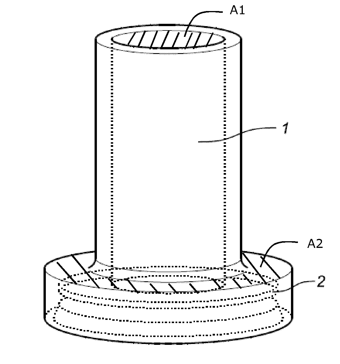
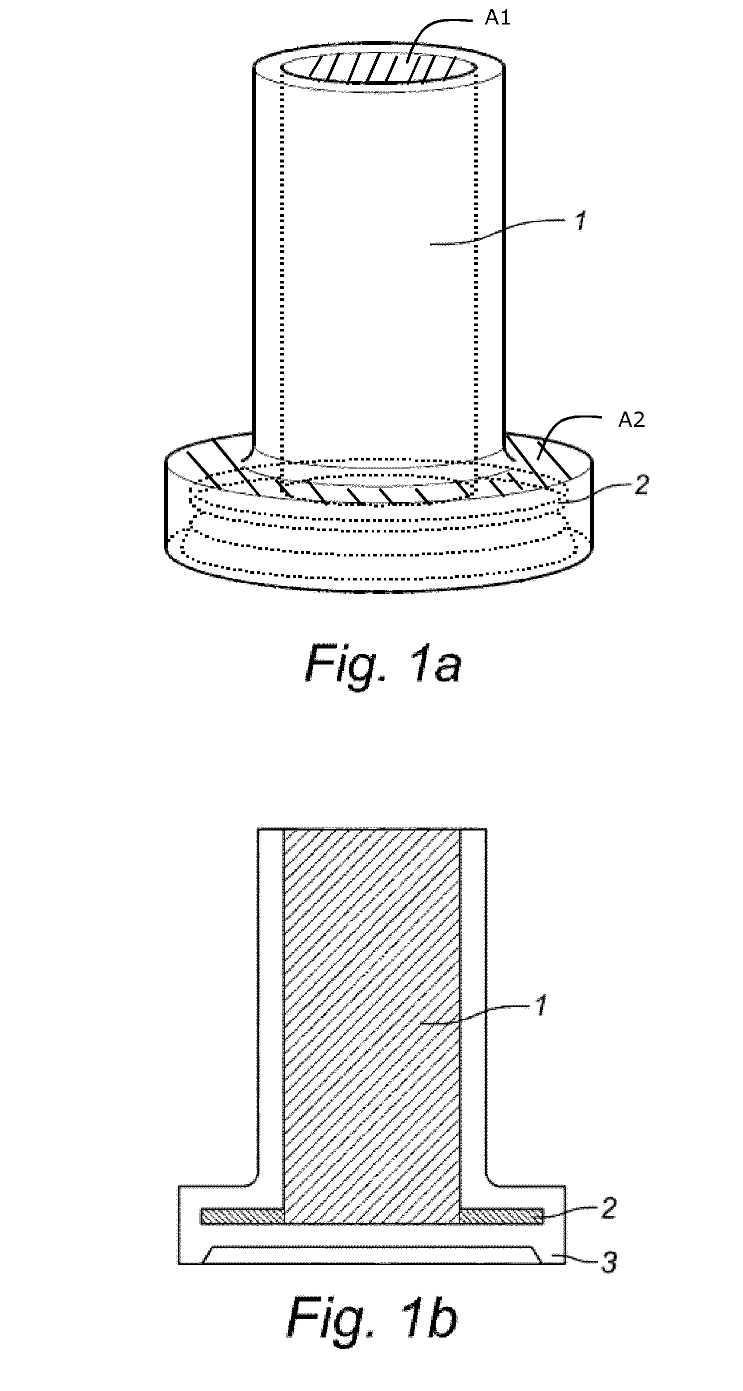
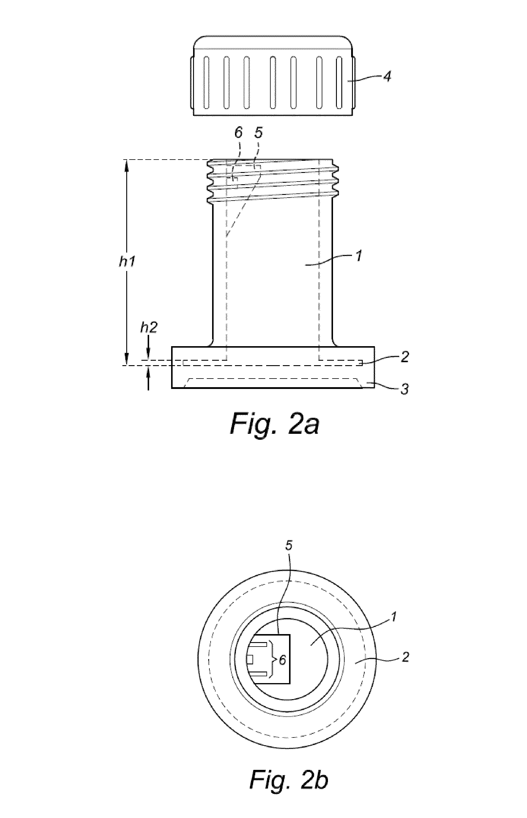
  • Liquid medium and sample vial for use in a method for detecting cancerous cells in a cell sample
  • Liquid medium and sample vial for use in a method for detecting cancerous cells in a cell sample
  • Liquid medium and sample vial for use in a method for detecting cancerous cells in a cell sample

Examples


experimental example 1

[0152]Samples of 16 selected patients previously diagnosed by the Thinprep® liquid based cytology confirmed by HPV Abott® assay or histology diagnosis for CIN2 / 3, were obtained and analyzed by the method of the current invention. Samples were collected by means of a cervical brush and immediately solubilized in a liquid medium of the current invention, comprising a labelled (fluorescent) biomarker against either HPV E6 or E7 protein (samples were divided in two groups of 8 samples, one group received the liquid medium with anti-E6, the other with anti-E 7).

[0153]DHM enables a quantitative multifocal phase contrast imaging that has been found suitable for quantitative and qualitative inspection, and for 3-dimensional cell imaging. Both biomarker data and morphological data was obtained.

[0154]188 cells were identified and measured in an automated way. Cells were in a first instance analyzed based on the presence of HPV specific proteins. In a second instance, information regarding the...

experimental example 2

[0158]In a similar set-up, cervical samples of 15 patients were obtained and solubilized in a liquid medium of the current invention, comprising a fluorescently labelled bio-marker against p16 (antibody). A linear correlation between cells found to be positive for p16 and predicted cancerous based on the obtained morphological data was shown.

experimental example 3

[0159]Experiment 1 and 2 were samples from the same patients, this time with biomarkers labelled with gold plated nanoparticles. Comparable results were obtained as in example 1 and 2.

[0160]The method of the current invention, using combined morphological and biomarker data (e.g. E6, E7, p16, p16INK4a, Ki-67 or annexin A5 overexpression) on a cell-by-cell basis performed on cells in suspension increased the sensitivity and specificity for detection of (pre)cancerous cells (e.g. a CIN2 an CIN3 lesion to more than 90%, compared to currently known single tests such as PAP smear and HPV DNA.


PUM

PropertyMeasurementUnit
concentrationaaaaaaaaaa
temperatureaaaaaaaaaa

Abstract

The current invention concerns a method for determining the presence of a (pre)cancerous cell in a liquid cell sample, comprising the steps of: —suspending and preserving a cell sample obtained from a subject in a sample vial comprising a liquid medium, said liquid medium comprises means for labeling cells or epitope(s) on or in said cells; —obtaining data from said labelled liquid cell sample; and —determining the presence of said (pre)cancerous cells based on said obtained data; characterized in that said data comprises morphological data and biomarker data. In a further aspect, the invention relates to a liquid medium for fixing, preserving and labeling cells in a cell sample and a sample vial specifically designed to be used in conjunction with the current method.

Description

TECHNICAL FIELD[0001]The invention pertains to the technical field of the early diagnosis of carcinomas as well as their preliminary stages, particularly carcinomas of the cervix and uterus.BACKGROUND[0002]To diagnose whether a patient is suffering from cancer or has a predisposition, cells need to be sampled from a patient and a thorough analysis of the cell sample is required in order to evaluate whether abnormal or aberrant cells are present. Mainly, a pathologist or other skilled medical personnel will base the diagnosis on specific characteristics of the cells in the sample, such as cell morphology, the presence of certain types of cells or proteins and more. These cytological tests are based on a two-dimensional presentation of the cells present in the sample and mostly require the fixation of cells on a substratum and the use of dyes or stainings to visualize specific features of the cells. This is a time consuming and cumbersome work, and requires well-trained specialists. M...

Claims


Application Information

Patent Timeline
Patent Type & Authority Applications(United States)
IPC IPC(8): G01N33/574A61B10/02
CPCG01N33/57411A61B10/0291A61B2010/0216G01N33/48778B01L3/502B01L3/5082B01L2200/0647B01L2200/0652B01L2300/023B01L2300/042B01L2300/0609B01L2300/0681B01L2300/0803B01L2300/0832B01L2300/0851B01L2300/0858B01L2400/086G01N1/405G01N21/03G03H1/0443G03H2001/005
Inventor MAGNIETTE, OLIVIER
Owner OVIZIO IMAGING SYST NV SA
Learn More