Use of g-csf dimer in preparation of medicament for treatment of neurodegenerative diseases
a neurodegenerative disease and dimer technology, applied in the field of biology and medicine, can solve the problems of affecting the living quality of patients, abnormal behavior and dysfunction of patients, premature death, etc., and achieve the effect of reducing the survival rate of pc12 cells, significantly improving the protective effect of gcsf dimer on pc12 cells, and improving the protective
- Summary
- Abstract
- Description
- Claims
- Application Information
AI Technical Summary
Benefits of technology
Problems solved by technology
Method used
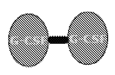
Image


Examples
example 1
Preparation of G-CSF Dimer
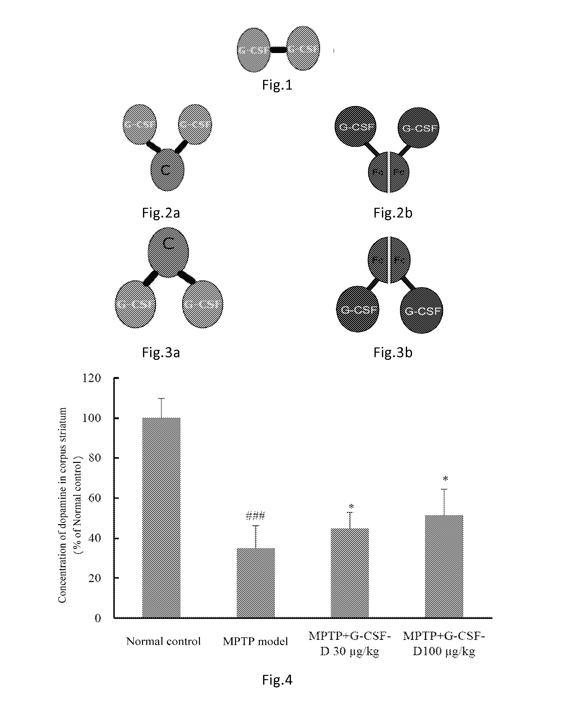
[0100]The G-CSF dimer of the present invention has an amino acid sequence of SEQ ID NO: 1 or comprises dimers as illustrated in FIGS. 1-3 comprising G CSF-Fc complexes with an amino acid sequence selected from SEQ ID NOs: 2-7. Preparation methods are described as follows:
a. Construction of a cell line expressing G-CSF dimer
[0101]The full length cDNA sequence of the G-CSF-Fc complexes (such as the sequence shown in SEQ ID NO: 10 or SEQ ID NO: 9) was synthesized. cDNA sequence of human G-CSF monomer was connected with cDNA sequence of Fc fragment of IgG2. cDNA sequences containing HindIII site, and expression elements required by mammalian cell such as Kozak sequence and signal peptide sequence were introduced at the 5′ end. cDNA sequence containing EcoRI site was introduced at the 3′ end. The full length cDNA sequence was cloned into pUC19 plasmid to obtain pG-CSF-Fc, which was used to transform E. coli TG1. The plasmid was digested with HindIII and EcoRI, a...
example 2
In Vivo Half-Life of G-CSF Dimer
[0109]Rats received a single dose of 100 μg / kg of G-CSF dimer consisting of two G-CSF-Fc complexes (SEQ ID NO: 3) by subcutaneous injection. The pharmacokinetic parameters were calculated and listed in Table 1 below (n=6). The half-life of G-CSF monomer in rats was approximately 2 hr.
TABLE 1Pharmacokinetic ParametersParameter (n = 6)UnitAverage ValueSDAUC(0−t)ng / mL * h4234.8640.3MRT(0−t)h21.61.4t(1 / 2)h7.71.2Clz / FL / h / kg0.0240.003Cmaxng / mL162.230.2
example 3
Pharmacokinetic Properties of G-CSF Dimer in Human Beings
[0110]24 healthy subjects were randomly divided into four dosage groups of 30, 60, 120, 240 μg / kg respectively receiving a single dose of 30, 60, 120, 240 μg / kg of G-CSF dimer (comprising two G-CSF-Fc monomers with sequence shown in SEQ ID NO: 6). Blood samples were collected at the 0.5, 1st, 2nd 4th, 8th, 16th 24th, 36th, 48th, 72nd, 96th hour, Day 6 (120 hours), 7, 9, 11, 13, and 15 after administration. Serum was separated and stored in −70oC freezer. The blood drug concentrations were measured by ELISA (ELISA, Quantikine human G-CSF ELISA kit, R&D System, Inc. Minneapolis, Min, Cat: PDCS50). The pharmacokinetic parameters were calculated using the non-compartmental analytical procedures (Software WinNonlin v 5.2, Pharsight Corporation, USA). The results were shown in Table 2.
TABLE 2Pharmacokinetic ParametersParameter (n = 6)30 μg / kg60 μg / kg120 μg / kg240 μg / kgCmax (ng / mL)21.3(10.3)44.6(17.7)219.9(76.6)759(160)Tmax (h, median...
PUM
| Property | Measurement | Unit |
|---|---|---|
| structure | aaaaa | aaaaa |
| affinity | aaaaa | aaaaa |
| crystalline structure | aaaaa | aaaaa |
Abstract
Description
Claims
Application Information
 - R&D
- Intellectual Property
- Life Sciences
- Materials
- Tech Scout
- Unparalleled Data Quality
- Higher Quality Content
- 60% Fewer Hallucinations
Browse by: Latest US Patents, China's latest patents, Technical Efficacy Thesaurus, Application Domain, Technology Topic, Popular Technical Reports.
© 2025 PatSnap. All rights reserved.Legal|Privacy policy|Modern Slavery Act Transparency Statement|Sitemap|About US| Contact US: help@patsnap.com