Carbon nanotubes for imaging and drug delivery

a technology of carbon nanotubes and drug delivery, which is applied in the field of imaging and drug delivery, can solve the problems of false positives as well as false negatives, difficult to specifically locate, and unnecessary invasive tests on a human subject, and achieve the effect of effective human use and delivery of carbon nanotubes

Inactive Publication Date: 2014-03-20
THE BOARD OF TRUSTEES OF THE LELAND STANFORD JUNIOR UNIV
View PDF2 Cites 5 Cited by
  • Summary
  • Abstract
  • Description
  • Claims
  • Application Information

AI Technical Summary

Problems solved by technology

In some of these procedures screening can lead to false positives as well as false negatives, depending on the test used.
It can also lead to unnecessary invasive tests on a human subject when a false positive exists.
However, it is often very difficult to specifically locate a tumor and such efforts at localization can be very invasive.
Coronary artery atherosclerosis can be fatal in both men and women due to unstable plaque growth and sudden atherothrombotic events due to thrombosis caused by unstable plaques.
Plaques, made of fat, cholesterol, calcium and other substances from the blood, build up in the blood vessels, resulting in narrowing, and ultimately blocking of these vessels that are needed to oxygenate and remove CO2 from the body's tissue.
More importantly, some of such plaques can be unstable, meaning that they are vulnerable to rupture at any moment causing, for example, a heart attack.
While this is a positive move toward early diagnosis of atherosclerosis, testing has so far only been in animals, and the process may not work in humans.
While the Robinson work shows the safety of using SWNTs in imaging and photothermal tumor treatment, it does not address the issue of locating a tumor or other tissue in order to diagnose the presence or absence of damaged tissue in a particular location.
Nor does Robinson teach monitoring or treatment of such tissue, as the Robinson techniques relied on knowing where a mouse tumor existed in order to treat it.

Method used


Image

  • Carbon nanotubes for imaging and drug delivery
  • Carbon nanotubes for imaging and drug delivery
  • Carbon nanotubes for imaging and drug delivery

Examples


example 1

Visualization of the Movement of Single-Walled Carbon Nanotubes (SWNTs)

[0089]This example shows the injection of various forms of nanotubes into the body of a mouse that has at least one tumor. Movement of the nanotubes is followed by the use of fluorescent dyes as visualized through intravital microscopy.

[0090]To observe nanotube targeting, fluorescence microscopy was used. This consists of the instrument shown in FIGS. 1a and 1b, with the lens juxtaposed to the mouse's body. The microscope employs four input lasers and three simultaneous output channels to dynamically image a tumor in living subjects. FIG. 1c shows the dorsal skinfold chamber which is surgically implanted into the mice; the device allows stabilization of mouse motion and provides a transparent window for optical microscopy.

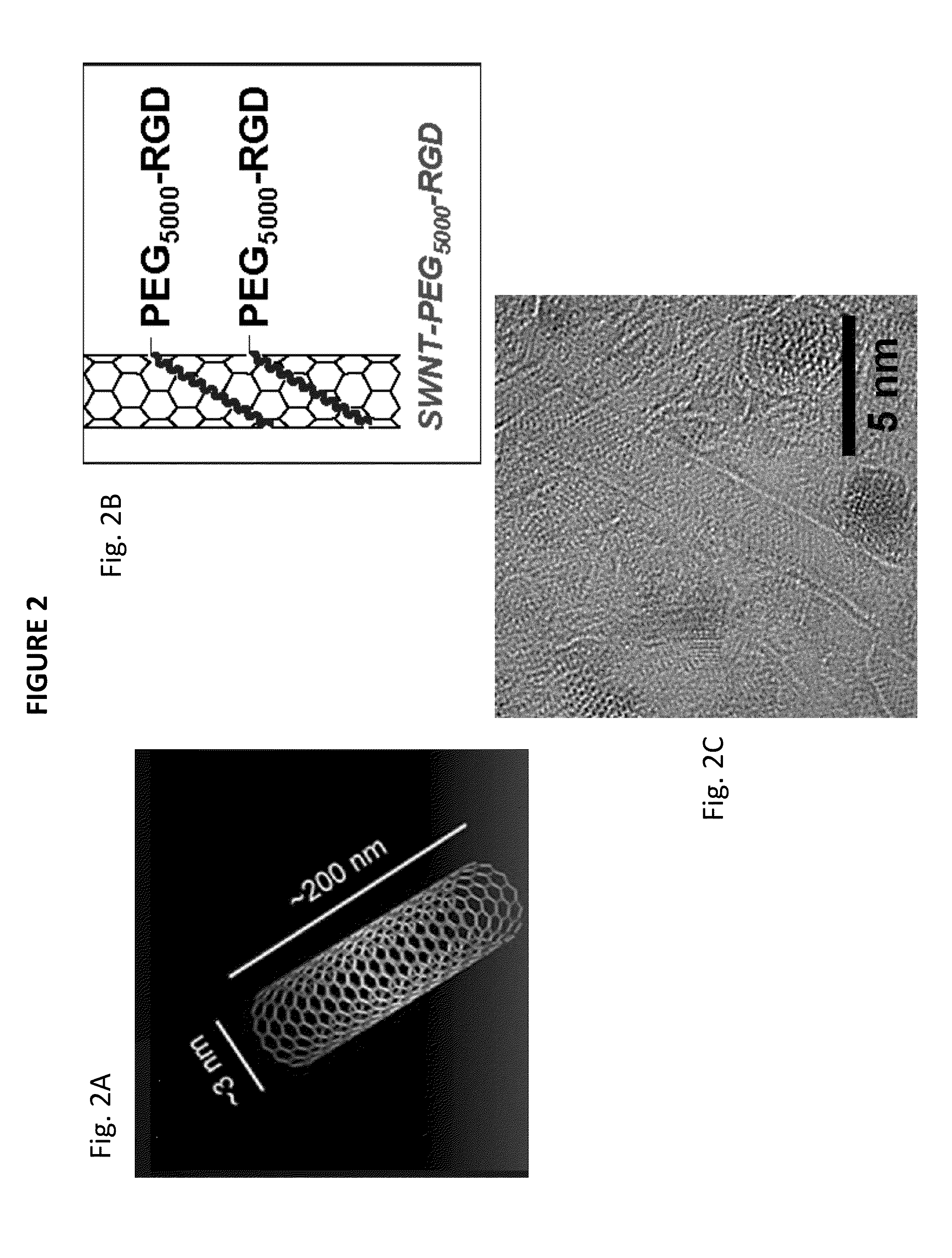
[0091]FIG. 2 shows nanotubes in a variety of forms. FIG. 2a shows the netting-type shape of the nanotubes used which have dimensions that range from approximately 3 nm to about 200 nm. FIG. 2b i...

example 2

SWNTs Functionalized with Peptides

[0092]For initial intravital microscopy, mice were injected into the tail with approximately 5×10⁵ EGFP-transfected U87MG tumor cells and the tumor was allowed to grow for about 10-14 days. EGFP is enhanced green fluorescent protein, first isolated from jellyfish, and then modified to enhance the green fluorescence. Cy5.5 was used to show the nanotubes, and a long-term dye was used to show the circulating blood.

[0093]For intravital microscopy of the SWNTs, 18 mice were injected with various experimental and control nanotubes: SWNTs with conjugated RGD, SWNTs with conjugated RAD, and plain SWNTs without attached peptides, as well as BSA without any SWNTs and other controls. SWNT behavior was visualized from injection into the mouse until about 4 hours post-injection, and then at designated time-points throughout the first day and first week post-injection. At each time point, 5-20 fields-of-view in the tumor were acquired to create a time series. Mor...

example 3

Peptide Dependency of the SWNT Uptake

[0096]FIG. 4 illustrates the uptake of SWNTs and shows that uptake is peptide dependent. Three types of nanotubes were compared: plain, i.e. non-conjugated, SWNTs; RGD-conjugated SWNTs, and RAD-conjugated SWNTs. Cells per minute per field of view were counted within 10 minutes of injection. As seen in FIG. 4, uptake of the SWNTs is clearly a function of peptide presence (p<0.001). While the plain SWNTs were taken up into circulating cells almost immediately after injection, uptake of RGD- or RAD-conjugated SWNTs into circulating cells was much slower. The kinetics of interaction between cells containing RGD- or RAD-SWNTs and the vasculature is shown by the amount of cells per minute. Clearly, plain SWNTs are taken up by circulating cells much faster than are RGD- or RAD-conjugated SWNTs. The same type of information is shown in FIG. 5, where in vitro experiments verified the peptide dependence of uptake of SWNTs into RAW cells. At even 10× lower ...


PUM

PropertyMeasurementUnit
timeaaaaaaaaaa
molecular weightaaaaaaaaaa
diameteraaaaaaaaaa

Abstract

The invention provides compositions and methods for visualizing particular tissues and delivering one or more therapeutics to that tissue using single-walled carbon nanotubes (SWNTs), which are taken up and delivered to target tissues by specific monocytes in the body. The delivery of SWNT to target tissues allows the visualization of the affected tissue for diagnostics and therapy in diseases where the specific monocyte is implicated in the disease pathogenesis. These nanotubes can be conjugated to a peptide, such as RGD, which helps direct the SWNT-containing monocytes to the vascular endothelium.

Description

CROSS-REFERENCE TO RELATED APPLICATION[0001]This application claims priority and other benefits from U.S. Provisional Patent Application Ser. No. 61 / 698,242 filed Sep. 7, 2012, entitled “Carbon nanotubes for imaging and drug delivery”. Its entire content is specifically incorporated herein by reference.STATEMENT OF GOVERNMENT SUPPORT[0002]This invention was made with Government support under NIH CA151459, CA119367 and CA160764 awarded by the National Institutes of Health. The Government has certain rights in the invention.TECHNICAL FIELD OF THE INVENTION[0003]The present invention relates to the field of imaging and drug delivery. In particular, it relates to the use of carbon nanotubes for visualizing particular tissues and delivering therapeutic treatment to that tissue.BACKGROUND[0004]The effort to determine the existence and location of a disease in a human subject has a long history. Recently, procedures have advanced for more precisely locating specific tissue in a body. Human...

Claims


Application Information

Patent Timeline
Patent Type & Authority Applications(United States)
IPC IPC(8): A61K49/00A61K47/48A61K49/22
CPCA61K49/0056A61K47/48976A61K49/222B82Y5/00A61K47/60A61K47/64A61K47/6949
Inventor SMITH, BRYAN R.GHOSN, ELIVER
Owner THE BOARD OF TRUSTEES OF THE LELAND STANFORD JUNIOR UNIV
Why Eureka
Try Eureka