
Covalently immobilized protein gradients in three-dimensional porous scaffolds

a technology of porous scaffolds and protein gradients, which is applied in the direction of peptides, biocides, peptide/protein ingredients, etc., can solve the problems of restricting the process to simple patterns, and limiting the scale and speed of such processes

Inactive Publication Date: 2013-09-05
TRUSTEES OF TUFTS COLLEGE +1
View PDF3 Cites 3 Cited by
  • Summary
  • Abstract
  • Description
  • Claims
  • Application Information

AI Technical Summary

Benefits of technology

The invention provides a method to create a gradual increase or decrease of a protein or other agent within a 3D scaffold. This is done by activating the surface of a scaffold made from a material like silk or collagen, and then adding the agent to the scaffold. The solution with the agent can be forced to flow through the scaffold to create a gradual change in the agent's concentration. This method can be used to create a scaffold that has a specific concentration gradient of the agent, which can help to attract or guide certain types of cells. The scaffold can also be made from a combination of different polymers or other materials, and multiple gradients can be created in different directions. This invention provides a way to create a highly customizable scaffold for use in regenerative medicine and tissue engineering.

Problems solved by technology

The disadvantage of the two-dimensional flow through microchannels is that it is restricted to simple patterns.
However, the scale and speed of such processes are limiting and the process is confined to two dimensional surfaces.

Method used


Image

  • Covalently immobilized protein gradients in three-dimensional porous scaffolds
  • Covalently immobilized protein gradients in three-dimensional porous scaffolds
  • Covalently immobilized protein gradients in three-dimensional porous scaffolds

Examples


example 1

Materials and Methods

Preparation of Silk Fibroin Scaffold

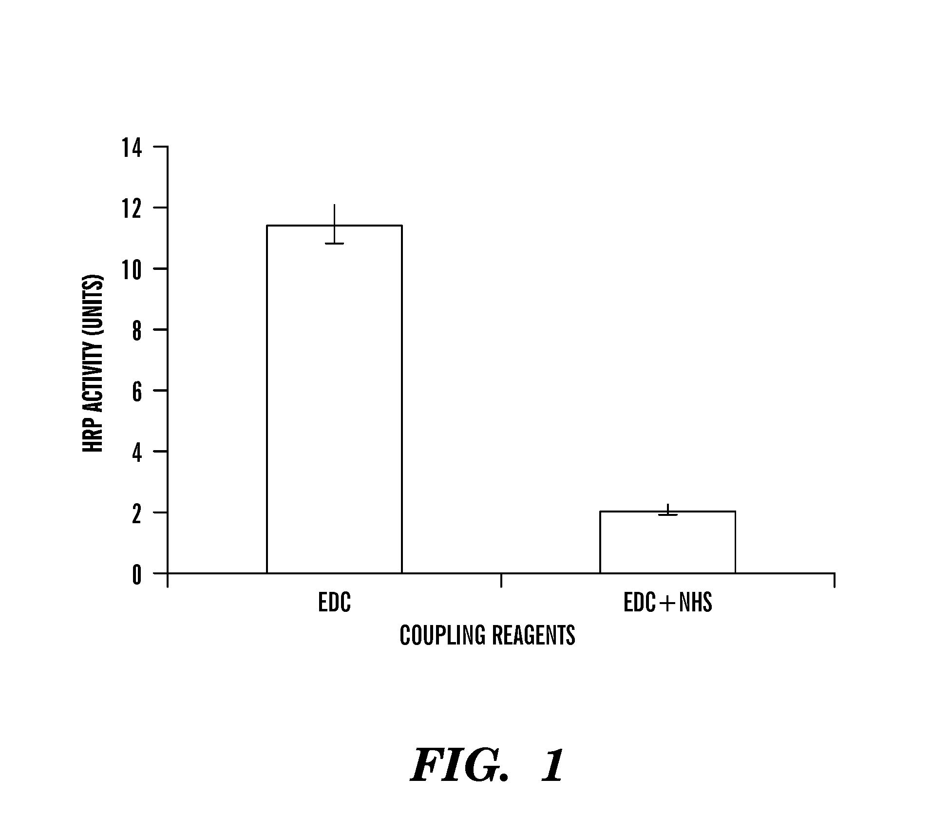
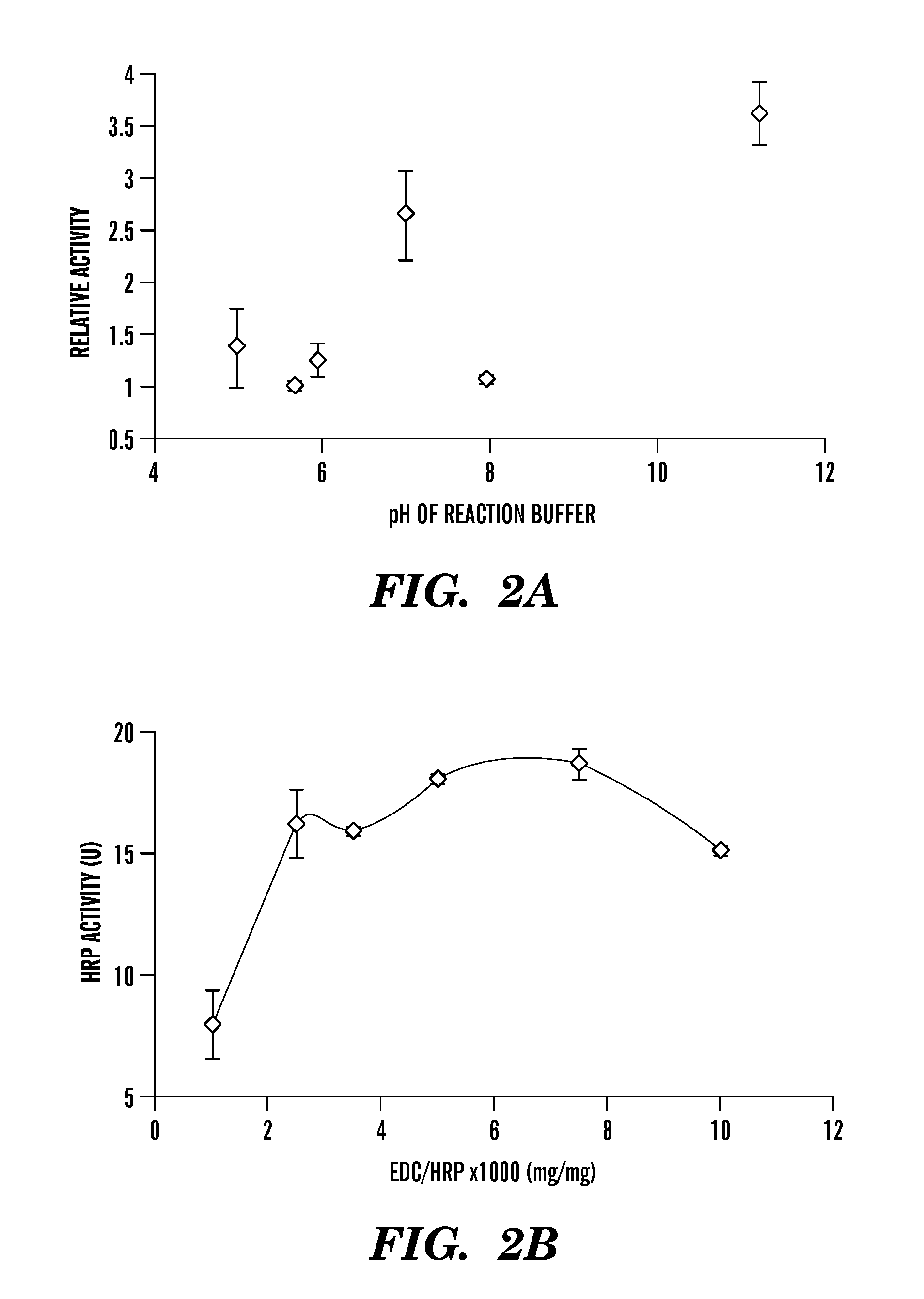
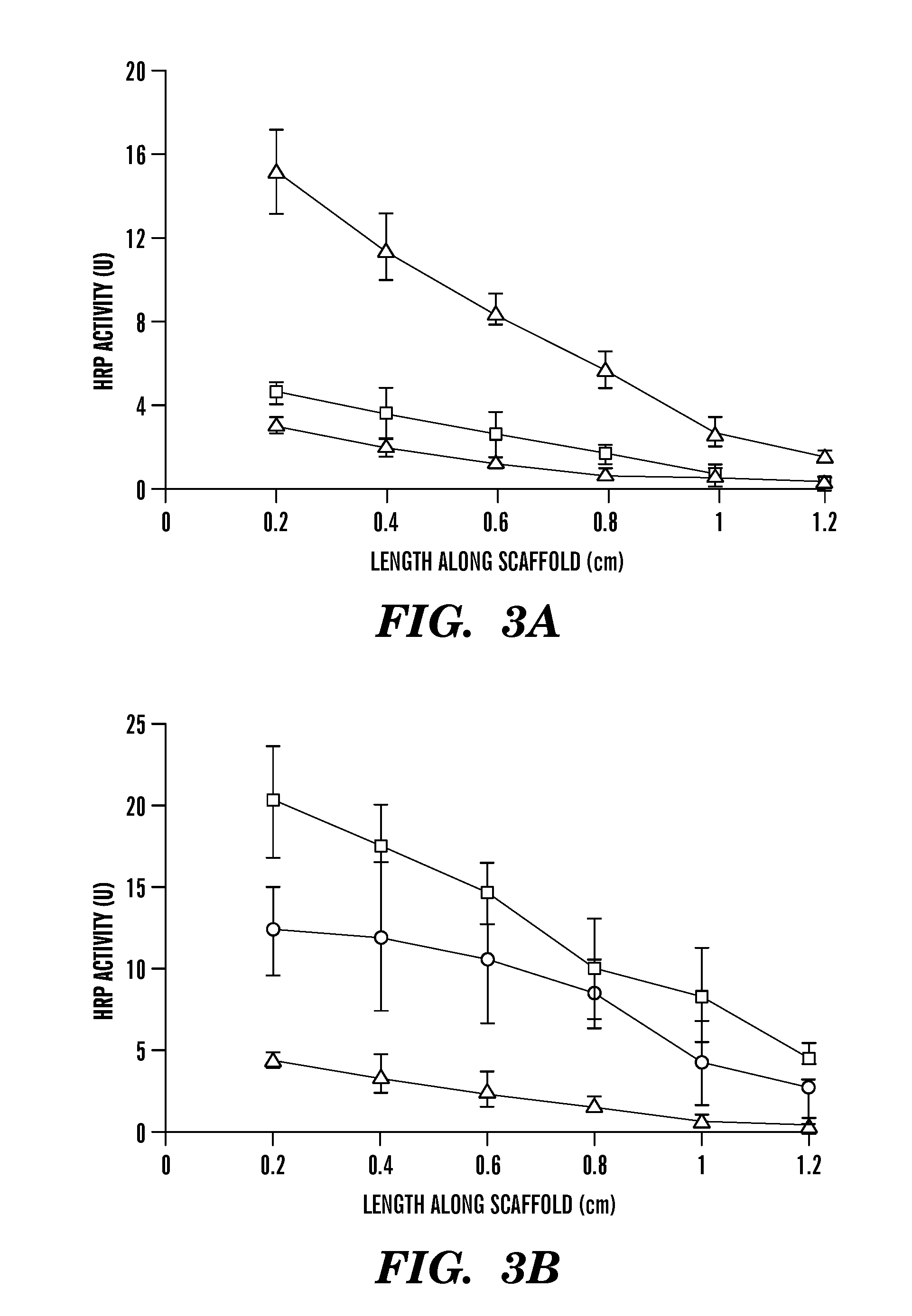
[0037]Cocoons from Bombyx mori (supplied by M. Tsukada, Institute of Sericulture, Tsukuda, Japan) were de-gummed for 1 hr in 0.02 M Na2CO3 (Fisher, Pittsburgh, Pa.) solution to extract sericin from fibroin. Silk fibroin was washed thoroughly to remove sericin, dried and dissolved in 9.3 M lithium bromide at 500 c to generate a 10% (w / v) solution. The solution was dialyzed in a Slide-A-Lyzer (3500MW cassette, Pierce, IL) against water for 3 days to remove lithium bromide. The solution was lyophilized and then dissolved in hexafluoro-2-propanol (Aldrich, Milwaukee, Wis.) to generate a 15% silk solution, a modification of our previously reported procedure [(Nazarov et al. 2004; Sofia et al. 2001).

[0038]Granular sodium chloride (crystal sizes from 150 to 700 μm, Sigma, Mo.) was used as a porogen (Nazarov et al, 2004). The 15% silk solution was poured into a Teflon hexagon dish (medium size, Fisher, Pittsburgh, Pa.) containing leve...

example 2

[0066]Recombinant Human BMP-2 (rhBMP-2) Gradients

[0067]Using methods similar to Example 1 to confirm our findings with HRP, rhBMP-2 gradients were produced. Silk fibroin scaffolds were activated using EDC / NHS for 15 minutes. Different volumes of rhBMP-2 were allowed to diffuse and create different immobilized gradients of rhBMP-2 within the silk fibroin scaffold (FIGS. 9A and 9B).

[0068]Bone marrow stromal cells were seeded into the silk fibroin scaffolds and supplied with osteogenic media. Scaffolds were grown in 6 well plates for 4-5 weeks and supplied with 50 μg / ml ascorbic acid and 3 mM B-glycerophosphate and 10−8 M dexamethasone. Calcium mineral assay showed a gradient response along the length of the scaffold.

[0069]Increase in the amount of BMP-2 (from 4 μL to 9 μL), results in an increase in the calcium mineral content showing a dose dependent increase in mineral deposition with increase in BMP-2. Immobilized BMP-2 shows control in the response of human bone marrow stromal cel...


PUM

PropertyMeasurementUnit
porosityaaaaaaaaaa
volumeaaaaaaaaaa
volumeaaaaaaaaaa

Abstract

The invention provides a method for forming an immobilized agent gradient within a 3-dimensional porous scaffold. A 3-dimensional scaffold formed from a biocompatible material is provided. The surface of the scaffold and / or the agent is activated so as to allow binding of the agent to the scaffold. The activated scaffold is contacted with a solution containing the agent. Contact with the solution is maintained for a sufficient period of time to allow diffusion of the solution through a portion of the scaffold, thereby forming a desired gradient of the agent through the 3-dimensional scaffold.

Description

CROSS-REFERENCE TO RELATED APPLICATIONS[0001]This application is a continuation application of U.S. patent application Ser. No. 11 / 407,373 filed on Apr. 19, 2006, which claims benefit under 35 U.S.C. §119(e) of U.S. Provisional Application No. 60 / 673,074 filed on Apr. 20, 2005, the contents of each of which are incorporated herein by reference in their entireties.[0002]This application claims the benefit under 35 U.S.C. §119(e) of the U.S. Provisional Application Ser. No. 60 / 673,074, filed on Apr. 20, 2005, the entire contents of which are incorporated herein by reference.GOVERNMENT SUPPORT[0003]This invention was made with government support under grant number P41 EB002520 awarded by the National Institutes of Health. The government has certain rights in this invention.BACKGROUND OF THE INVENTION[0004]Immobilized enzymes are utilized extensively in biosensor and related detection protocols in many technological applications. Immobilization generally offers improved enzyme stability...

Claims


Application Information

Patent Timeline
Patent Type & Authority Applications(United States)
IPC IPC(8): C07K17/02C12N11/02
CPCC07K17/04C12N11/04C12N11/02C12N11/10C07K17/02C12N11/06
Inventor VEPARI, CHARUKAPLAN, DAVID L.VUNJAK-NOVAKOVIC, GORDANA
Owner TRUSTEES OF TUFTS COLLEGE
Learn More