
Functionalization of gold nanoparticles with oriented proteins, application to the high-density labeling of cell membranes

a technology of oriented proteins and gold nanoparticles, applied in the field of nanoparticles, can solve the problems of direct adsorption approach, partial denaturation, loss of biological properties,

Inactive Publication Date: 2013-08-22
BRISSON ALAIN +1
View PDF5 Cites 9 Cited by
  • Summary
  • Abstract
  • Description
  • Claims
  • Application Information

AI Technical Summary

Benefits of technology

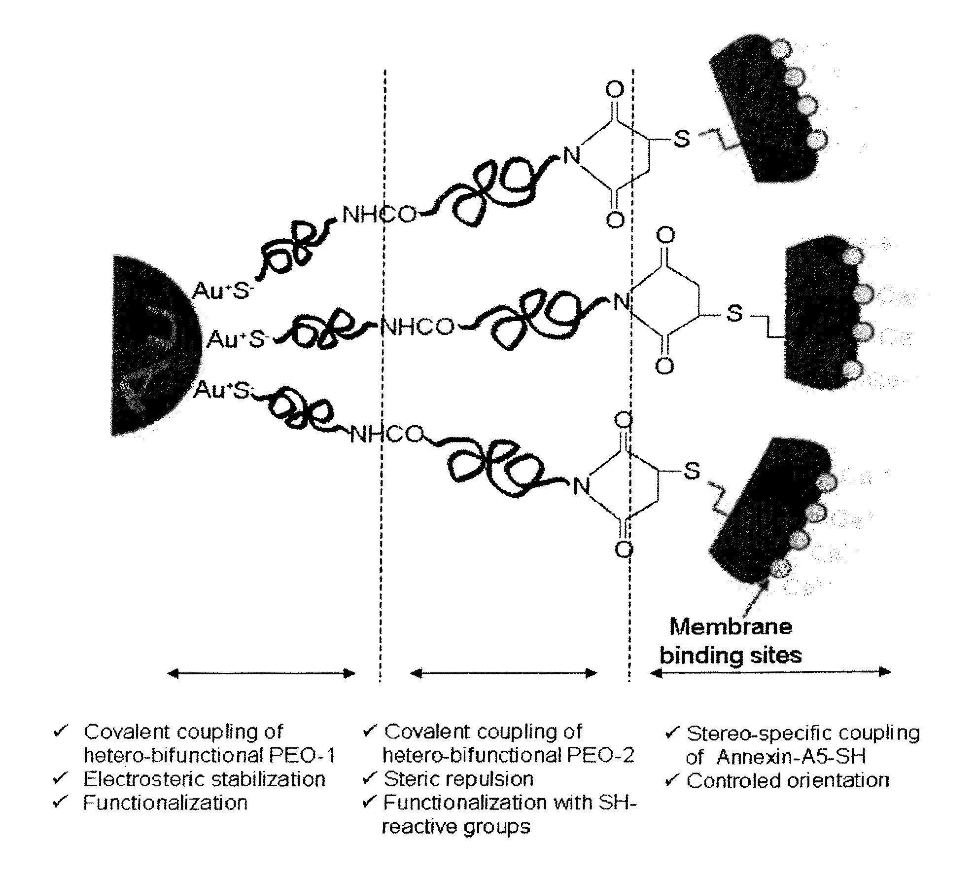
The patent describes a method for creating stable protein-gold nanoparticle assemblies that can be used for various detection methods. The method allows for the preservation of the structure and function of the protein, as well as the colloidal stability of the nanoparticles without the need for additional stabilizing agents. The high concentrations and large quantities of the suspensions of nanoparticles functionalized by covalent and stereo-specific coupling of proteins make them ideal for studying the distribution, localization, or quantification of target elements present in biological materials. The presence of PEO molecules on the surface of the nanoparticles ensures colloidal stability in physiological medium. The strategy of bio-functionalization of the gold nanoparticles is valid for any protein, peptide, or molecule presenting an accessible sulfhydryl group. The nanoparticles have several properties that make them suitable for various detection methods, including high electron scattering cross section, optical and spectroscopic properties, and interaction with X-rays or other types of radiation. The density of antibodies linked to the nanoparticles can be controlled by adjusting the respective concentrations of nanoparticles and antibodies. The use of different sizes of functionalized gold nanoparticles with the same biological sample can be advantageous for multiple detection of several target molecules.

Problems solved by technology

However, physical adsorption presents severe limitations as the interactions involved may lead to molecular rearrangements, which may result in a partial denaturation in the case of proteins and in a loss of their biological properties.
The direct adsorption approach presents several other severe limitations, principally the absence of binding specificity, as in theory any molecule can bind, and the lack of control of the orientation of molecules bound to the surface.
In the case of gold nanoparticles, or other colloidal particles, the question of the colloidal stability of the particles constitutes an additional issue.
Indeed, bare nanoparticles are in general unstable in physiological solutions.
The physical adsorption of proteins is in general not sufficient to stabilize nanoparticles.
The presence of these additives is however problematic as they may interfere with the molecular processes investigated or perturb the integrity of the studied cellular structures, as it is well proven for surfactants.
In addition, the physical adsorption of proteins on gold nanoparticles is irreproducible, inefficient and tedious, as it must be optimized for every new protein to be coupled.
Therefore this approach is costly in time and costly in products, as it requires large amounts of proteins, in general of high value.
The multiple limitations associated with the physical adsorption approach explain the development of alternative approaches for coupling biological molecules to solid supports in a controlled manner.
In addition, amine groups often participate in active sites or ligand-binding sites, and their modification may lead to loss of recognition properties.
This approach has limited application, because only few proteins present accessible SH-groups, even after disulfide reduction.
In addition, the production of Fab′-SH proteins is not straightforward, requiring experts in the art, and has low yield.
In conclusion, no reliable strategy has yet been proposed for controlling the orientation of proteins linked to gold particles, in such a way that the sites complementary to the target molecule of interest are properly exposed to the aqueous environment.
Said nanoparticles have the limitations of physical adsorption reported above: lack of colloidal stability, necessary presence of stabilizing agents, protein denaturation, no control of protein orientation, and low concentrations of functionalized nanoparticles.

Method used


Image

  • Functionalization of gold nanoparticles with oriented proteins, application to the high-density labeling of cell membranes
  • Functionalization of gold nanoparticles with oriented proteins, application to the high-density labeling of cell membranes
  • Functionalization of gold nanoparticles with oriented proteins, application to the high-density labeling of cell membranes

Examples


example 1

Synthesis of Aqueous Suspensions of 10 nm-Diameter Gold Nanoparticles, Functionalized Covalently and Stereo-Specifically with Proteins

[0106]1.1 Preparation of 10 nm-Diameter Gold Nanoparticles (solA).

[0107]Gold nanoparticles are prepared according to a method derived from the protocol of Turkevich et al. (25) in which tetrachloroaurate salts (HAuCl4, KAuCl4) are reduced by citrates, leading to the formation of suspensions (called sol hereunder) of 10 nm-diameter gold nanoparticles.

[0108]Typically, for preparing 550 mL of aqueous sol of gold nanoparticles of 10 nm-diameter (FIG. 33) and for a concentration equal to 32.62 nM of particles (1.964.1016 particles / L, namely 10−3 M Au): a volume of 400 mL of ultrapure water (4 (99.999%, Aldrich) are added. The reaction medium is carried to the water reflux (110° C.). The reduction of auric salts occurs upon addition of 50 mL of 3.4 mM sodium citrate dihydrate solution (99%, Aldrich). The reaction is left 30 minutes to the water reflux and t...

example 2

UV-Visible Absorption Spectroscopy of Suspensions of Gold Nanoparticles

[0126]Measurements of UV-visible absorption spectra of gold nanoparticle suspensions give access to the concentration of gold nanoparticles of SolA, SolAPN, SolAPPmal, SolAPP-A5 and to the quality of these dispersions with respect to their colloidal stability.

[0127]The UV-visible spectrum of 10 nm-diameter gold nanoparticles presents an absorption band at a wavelength λ=520 nm attributed to the plasmon resonance band of gold nanoparticles (FIG. 4). For a given particle size, the optical density is directly proportional to the particle concentration, which is determined by the Beer-Lambert law: O.D.=εp.Cp.l, where O.D. is the optical density, εp the molar extinction coefficient of 10 nm-diameter gold particles (εp=1.086×10⁸ (mole of particules)−1.L·cm), Cp the concentration in mole of particles, L the path length (1 cm).

example 3

Binding of solAPP-A5 Gold Nanoparticles to Supported Lipid Bilayers, by Quartz Crystal Microbalance with Dissipation Monitoring (QCM-D)

[0128]The binding of Anx5-functionalized gold nanoparticles (solAPP-A5), prepared according to example 1.4, to supported lipid bilayers containing PS was measured in a quantitative manner by the QCM-D method (37), according to reference measurements established for Anx5 (17).

[0129]FIG. 5 shows 1) that the kinetics of binding of the solAPP-A5 particles (blue curve) to a (PC / PS, 4:1) supported lipid bilayers saturates, 2) that Anx5-gold nanoparticles bound to a supported lipid bilayer are able to bind PS-containing liposomes, demonstrating that several molecules of Anx5 are bound per solAPP-A5 nanoparticle, 3) that the binding of solAPP-A5 particles is PS-specific, as their binding is only reversed by addition of the calcium chelating agent ethylene-bis(oxyethylenenitrilo)tetraacetic acid (EGTA).

[0130]The mass of Anx5-coupled gold nanoparticles bound t...


PUM

PropertyMeasurementUnit
sizeaaaaaaaaaa
sizeaaaaaaaaaa
sizeaaaaaaaaaa

Abstract

The present invention relates to nanoparticles the surface of which is modified by deposition of proteins. The invention further relates to a method for producing said nanoparticles and to their use in biological research and in the biomedical field (for example labelling and diagnosis).

Description

FIELD OF THE INVENTION[0001]The present invention relates to nanoparticles the surface of which is modified by deposition of proteins. The invention further relates to a method for producing said nanoparticles and to their use in biological research and in the biomedical field (for example labelling and diagnosis).BACKGROUND OF THE INVENTION[0002]Colloidal gold particles of small size, from 1 to 20 nm in diameter, have been used for several decades as specific labels in cell ultrastructure research by Transmission Electron Microscopy (TEM) (1-2). Indeed gold nanoparticles functionalized with antibodies, or other types of biological molecules, allow characterizing the number and the localization of cellular antigens, or other types of complementary elements, in thin sections or in homogenized suspensions of cells or tissues, according to classical techniques of TEM. Classical methods for immobilizing antibodies—or other types of proteins or molecules—on gold particles are based on th...

Claims


Application Information

Patent Timeline
Patent Type & Authority Applications(United States)
IPC IPC(8): G01N33/58G01N33/92
CPCG01N33/54346G01N33/92G01N33/587
Inventor BRISSON, ALAINMORNET, STEPHANE
Owner BRISSON ALAIN
Learn More