Composition for regenerating normal tissue from fibrotic tissue

Active Publication Date: 2013-07-04
NITTO DENKO CORP
View PDF2 Cites 7 Cited by
  • Summary
  • Abstract
  • Description
  • Claims
  • Application Information

AI Technical Summary

Benefits of technology

The present invention has shown that normal tissue can be regenerated from fibrotic tissue, which was previously thought to be unable to do so. This means that therapy can be developed for fibrotic diseases where there was previously no effective treatment. The invention also means that fibrotic tissue can be treated even when it is constantly exposed to a fibrotic stimulus. This contributes to the development of new treatments for fibrotic diseases and makes an impact on medical and veterinary treatment.

Problems solved by technology

When tissue is damaged by a stimulus such as oxidative stress, hypoxia, inflammation, or apoptosis, damaged tissue is repaired by replacement with extracellular matrix, but in the case of the damage being serious or in the case of such stimulation becoming chronic, the accumulation of extracellular matrix becomes excessive, and the tissue cannot perform its function sufficiently.
However, there have been no detailed reports regarding what is specifically happening in the tissue after pathological accumulation of extracellular matrix decreases, and it has been completely unknown until now for regeneration of normal tissue to occur in such fibrotic tissue or for regeneration of normal tissue to be possible.
Furthermore, the fibrosis of tissue not only includes fibroses for which the cause of the disease is clear and can be removed, such as fibrosis derived from viral infection, drinking alcohol, drugs, etc., but also includes fibroses for which the direct cause of the disease is unclear, such as for example cryptogenic cirrhosis, idiopathic pulmonary fibrosis, or idiopathic myelofibrosis, and those for which the direct cause of the disease is known but the origin of the cause of the disease is unclear or is difficult to remove, such as for example primary biliary cirrhosis, nonalcoholic steatohepatitis (NASH)-derived hepatic fibrosis, and primary sclerosing cholangitis.

Method used


Image

  • Composition for regenerating normal tissue from fibrotic tissue
  • Composition for regenerating normal tissue from fibrotic tissue
  • Composition for regenerating normal tissue from fibrotic tissue

Examples


example 1

Preparation of VA-lip siRNA

[0092](1) Preparation of siRNA

[0093]As a sense chain and an antisense chain of siRNA (Hokkaido System Science Co., Ltd., Sapporo, Japan) targeted to the base sequence of gp46 (GenBank Accession No. M69246), which is the rat homologue of human HSP47, a molecular chaperone common to collagens (types I to IV), those below were used.

A:GUUCCACCAUAAGAUGGUAGACAACAG(sense chain siRNA starting fromthe 757th base on the gp46 basesequence, SEQ ID NO: 1)B:GUUGUCUACCAUCUUAUGGUGGAACAU(antisense chain siRNA, SEQ ID NO: 2)

[0094]As siRNA random (also called siRNAscramble), those below were used.

C:CGAUUCGCUAGACCGGCUUCAUUGCAG(sense chain siRNA, SEQ ID NO: 3)D:GCAAUGAAGCCGGUCUAGCGAAUCGAU(antisense chain siRNA, SEQ ID NO: 4)

[0095]In some experiments, sense chains having 6′-carboxyfluorescein (6-FAM) or fluorescein isothiocyanate (FITC) conjugated to the 5′ terminal were used. It was confirmed by a BLAST search that these sequences did not have homology with other known rat mRN...

example 2

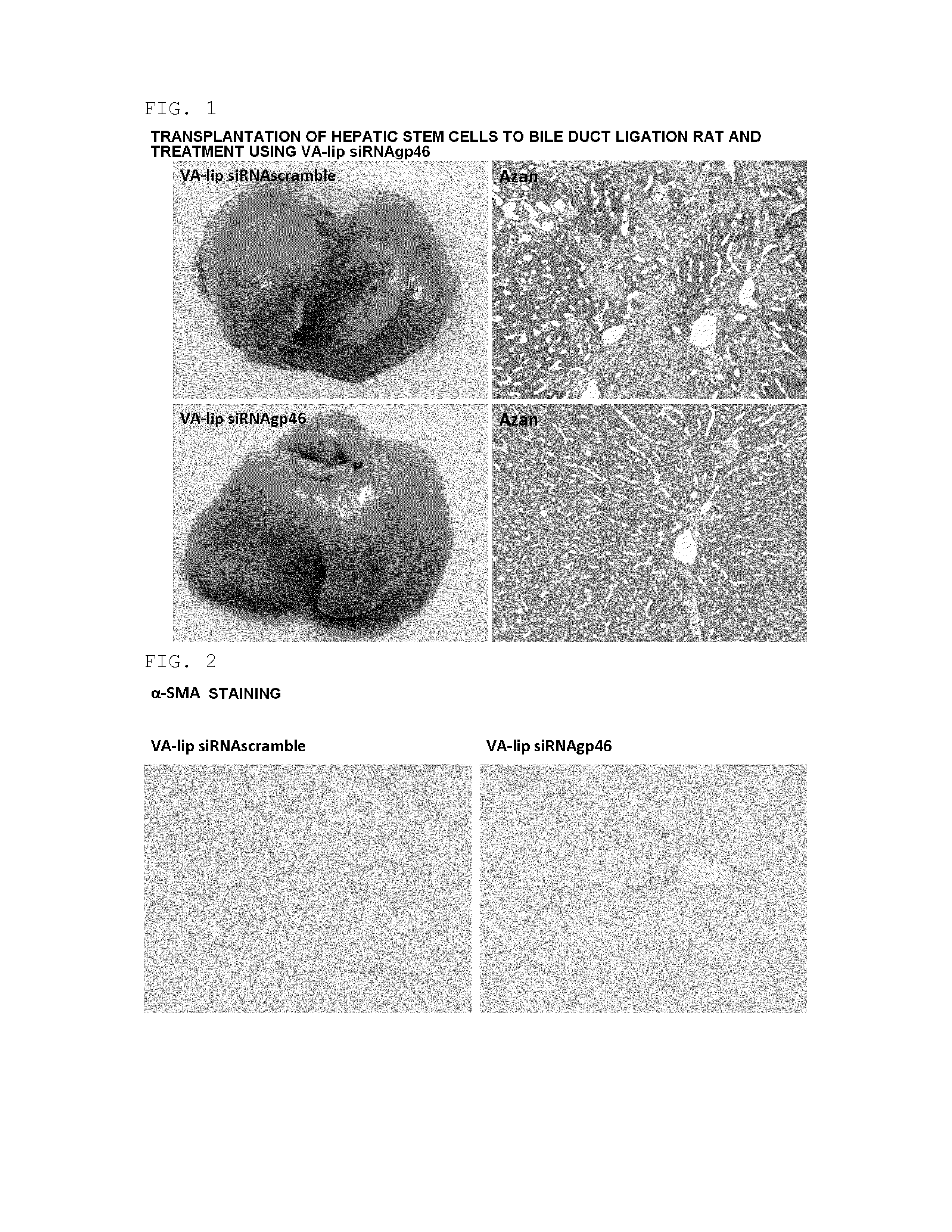
Regenerative Therapy Experiment Using Hepatic Fibrosis Model Rat

(1) Preparation of Hepatic Fibrosis Model Rat

[0097]A hepatic fibrosis model rat was prepared by subjecting a male SD rat (body weight 150 to 200 g) (Slc Japan, Shizuoka, Japan) to common bile duct ligation, and an individual on the 28th day after ligation was subjected to the present experiment. The present model rat was in a state in which cholestasis was caused by the common bile duct ligation and the liver tissue was continually exposed to a fibrotic stimulus.

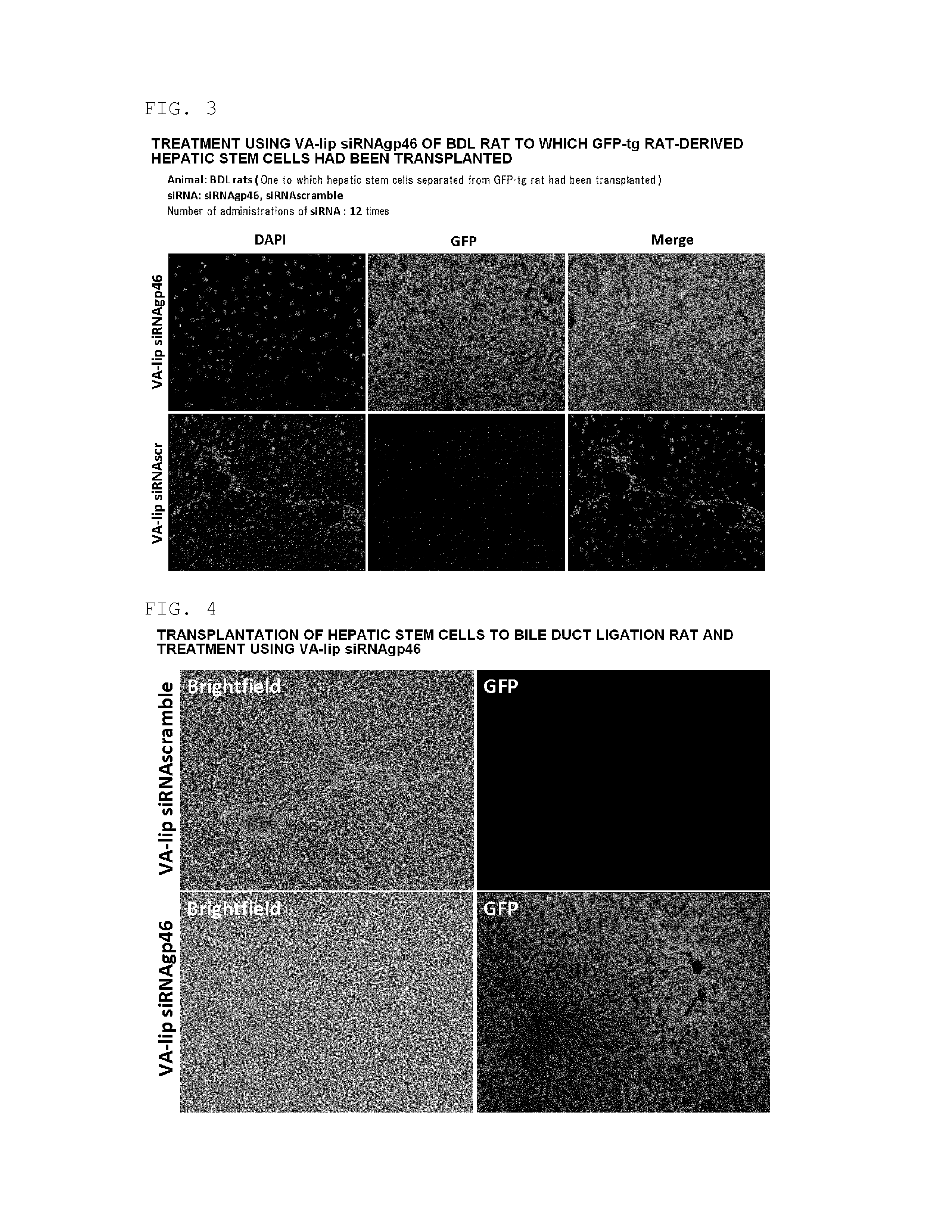
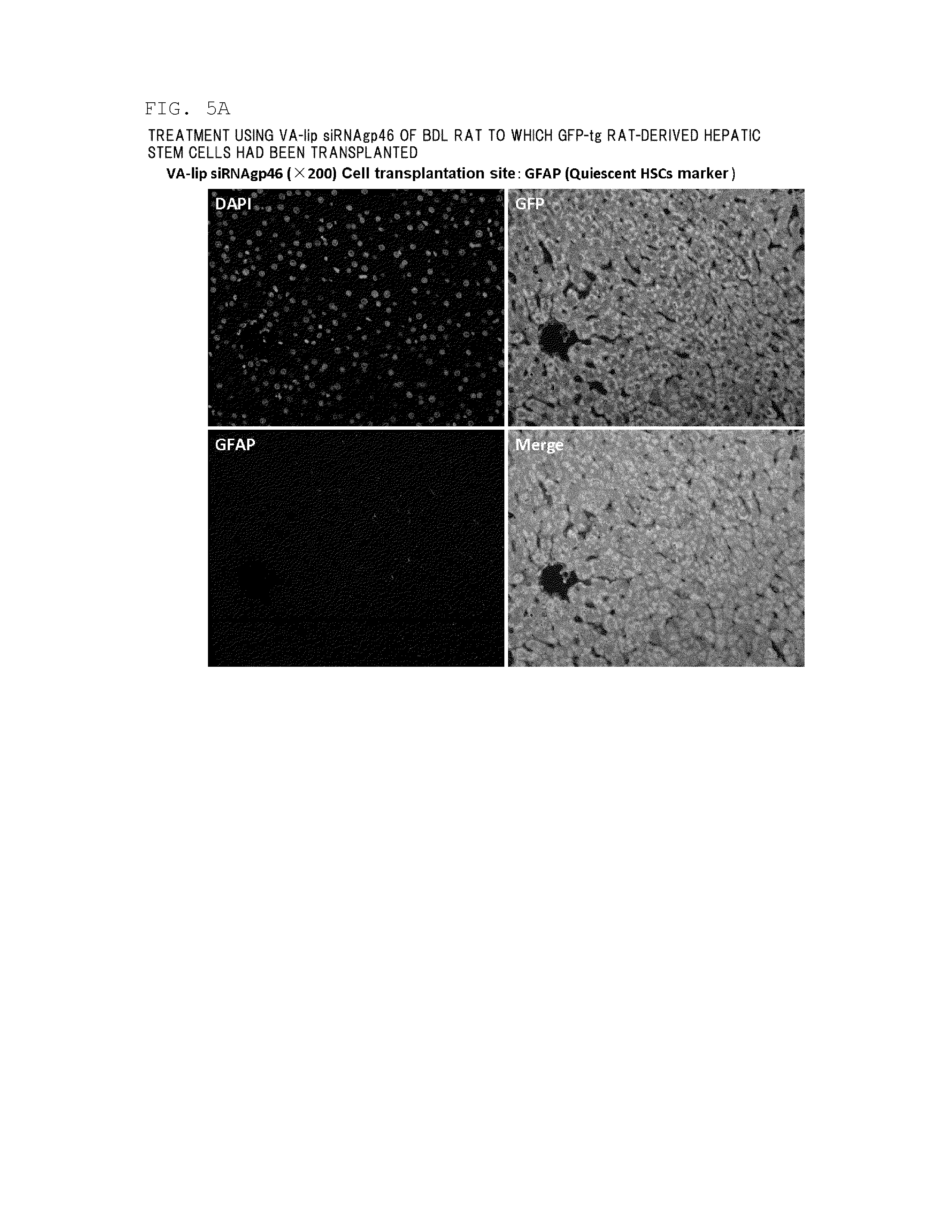
(2) Preparation of GFP-Labeled Rat Hepatic Stem Cells

[0098]GFP-labeled rat hepatic stem cells were harvested from the liver of a 4 week old GFP transgenic rat (Slc Japan). First, an EGTA solution and a collagenase solution were perfused through the GFP transgenic rat, the liver was then harvested, and the harvested liver was finely cut and then filtered using a cell strainer (pore diameter 100 μm). Hank's balanced salt solution (HBSS)+0.25% bovine serum albumin ...

example 3

Stellate Cell-Specific Delivery by Means of VA

(1) Isolation of Rat Pancreatic Stellate Cells (PSC)

[0113]Rat pancreatic stellate cells (PSC) were isolated using a density gradient centrifugation method in accordance with a previous report (Apte et al. Gut 1998; 43: 128-133). Purity was assayed by microscopic examination, autofluorescence of endogenous VA, and an immunocytochemical method using a monoclonal antibody (1:25, Dako) for desmin, which is a muscle actin crosslinking protein. The viability of cells was assayed by trypan blue exclusion. Both the cell purity and the viability exceeded 95%. The cells were cultured in Iscove's modified Dulbecco's medium (Iscove's modified Dulbecco's medium: IMDM) supplemented with 10% fetal bovine serum (FBS) at 37° C. with 95% air / 5% CO2 under a humidified environment.

(2) Intracellular Distribution Analysis of VA-lip siRNAgp46-FAM

[0114]Rat pPSCs (primary pancreatic stellate cells, primary PSC) were sown so that there were 1×10⁴ cells per chambe...


PUM

PropertyMeasurementUnit
Massaaaaaaaaaa
Massaaaaaaaaaa
Massaaaaaaaaaa

Abstract

The present invention relates to a pharmaceutical composition and a method for regenerating normal tissue from fibrotic tissue, the pharmaceutical composition and the method employing a collagen-reducing substance. In accordance with the present invention, normal tissue can be therapeutically regenerated from fibrotic tissue.

Description

TECHNICAL FIELD[0001]The present invention relates to a composition and method for regenerating normal tissue from fibrotic tissue.BACKGROUND ART[0002]Fibrosis of tissue is caused by the excessive production and accumulation in tissue of extracellular matrix, which is mainly collagen. When tissue is damaged by a stimulus such as oxidative stress, hypoxia, inflammation, or apoptosis, damaged tissue is repaired by replacement with extracellular matrix, but in the case of the damage being serious or in the case of such stimulation becoming chronic, the accumulation of extracellular matrix becomes excessive, and the tissue cannot perform its function sufficiently. Fibrosis is seen in various types of organs, such as the liver, pancreas, lung, kidney, bone marrow, and heart, and it is thought that collagen-producing cells such as myofibroblasts are related to a disease state. Conventionally, it is though that fibrosis is an irreversible phenomenon and that once tissue has become fibrotic...

Claims


Application Information

Patent Timeline
IPC IPC(8): A61K31/713
CPCA61K31/713A61K9/0019A61K9/1272A61K31/41A61K31/55A61K45/06A61P1/16A61P1/18A61P11/00A61P17/02A61P43/00A61K2300/00C12N15/113C12N2310/14C12N2320/31
Inventor NIITSU, YOSHIROYONEDA, AKIHIROISHIWATARI, HIROTOSHI
Owner NITTO DENKO CORP
Why Eureka
Try Eureka