
Compositions and Methods for Quantitative Histology, Calibration of Images in Fluorescence Microscopy, and ddTUNEL Analyses

Inactive Publication Date: 2013-06-20
THE METHODIST HOSPITAL RES INST
View PDF1 Cites 13 Cited by
  • Summary
  • Abstract
  • Description
  • Claims
  • Application Information

AI Technical Summary

Benefits of technology

The present invention provides new and useful compositions of tissue analogs and histological phantoms that can be used as reference standards in fluorescence microoscopic analysis. These compositions use a gelatin-based solution that has an intrinsically low absorbance in the ultraviolet spectral region. This allows for effective labeling with fluorescent dyes and provides accurate and reliable results. The invention also provides methods for calibrating and quantifying images using these new compositions.

Problems solved by technology

The inherent variability of fluorescence characteristics, however, has meant that such methods have, to date, only been qualitative in nature, and not quantitative.
This limitation has been problematic for a number of reasons: Quantification and calibration of images in fluorescence microscopy is notoriously difficult (see e.g., Swedlow, 2007 and Wolf, 2007).
These types of standards have greatly aided the quantification of fluorophore signals, but are not easy to prepare, are not robust and only quantify the levels of the fluorophore, not any probe to which the fluorophore may be attached.
They therefore have limited utility in the calibration of biologically relevant samples (Zwier et al., 2004).
Similarly, the quantitative analysis of nucleic acids using optical microscopy methods has also been limited.
A major problem with TUNEL is the use of dUTP as a substrate, as Tdt generates —(U)n—OH 3′ polymers at each of the free DNA-OH 3′ ends; the generation of these poly-U-OH 3′ polymers is very good for signal amplification, but makes the TUNEL assay intrinsically unquantifiable.
Likewise, the ability to perform quantitative analysis of nucleic acids in situ has been constrained by a lack of ligase-based assays that are suitable for the selective detection of specific markers of necrosis and / or apoptosis in tissue sections.
However, either these procedures are laborious or the starting materials are difficult to obtain.
However, this cannot be used in a quantitative manner in either fixed cells or tissues, due to the interference of aldehydes / ketones present in other biomolecules (Kow and Dare, 2000).

Method used


Image

  • Compositions and Methods for Quantitative Histology, Calibration of Images in Fluorescence Microscopy, and ddTUNEL Analyses
  • Compositions and Methods for Quantitative Histology, Calibration of Images in Fluorescence Microscopy, and ddTUNEL Analyses
  • Compositions and Methods for Quantitative Histology, Calibration of Images in Fluorescence Microscopy, and ddTUNEL Analyses

Examples


example 1

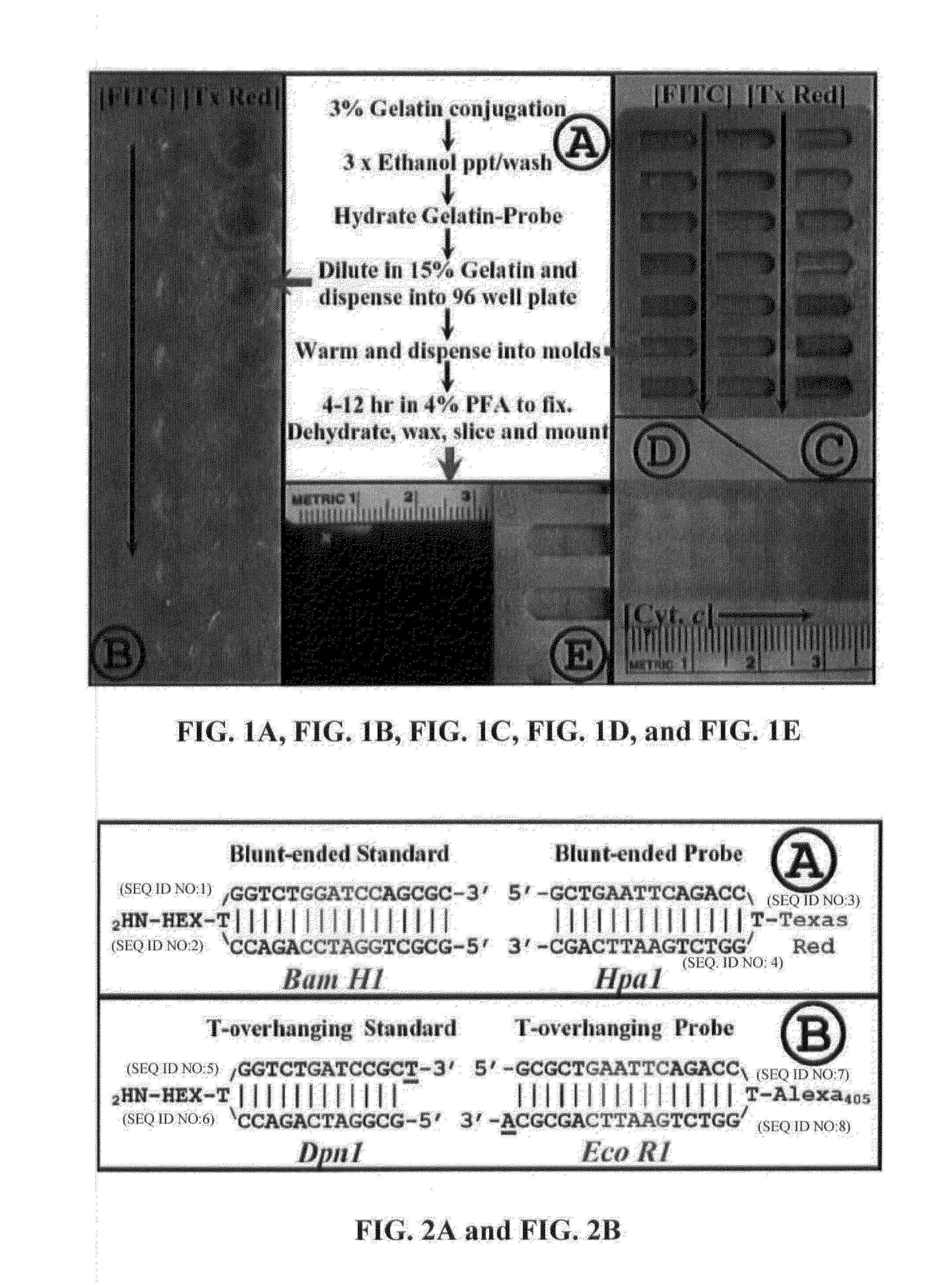
Preparation of FITC-Labeled Gelatin Phantoms; Identification and Calibration of Images in Fluorescence Microscopy

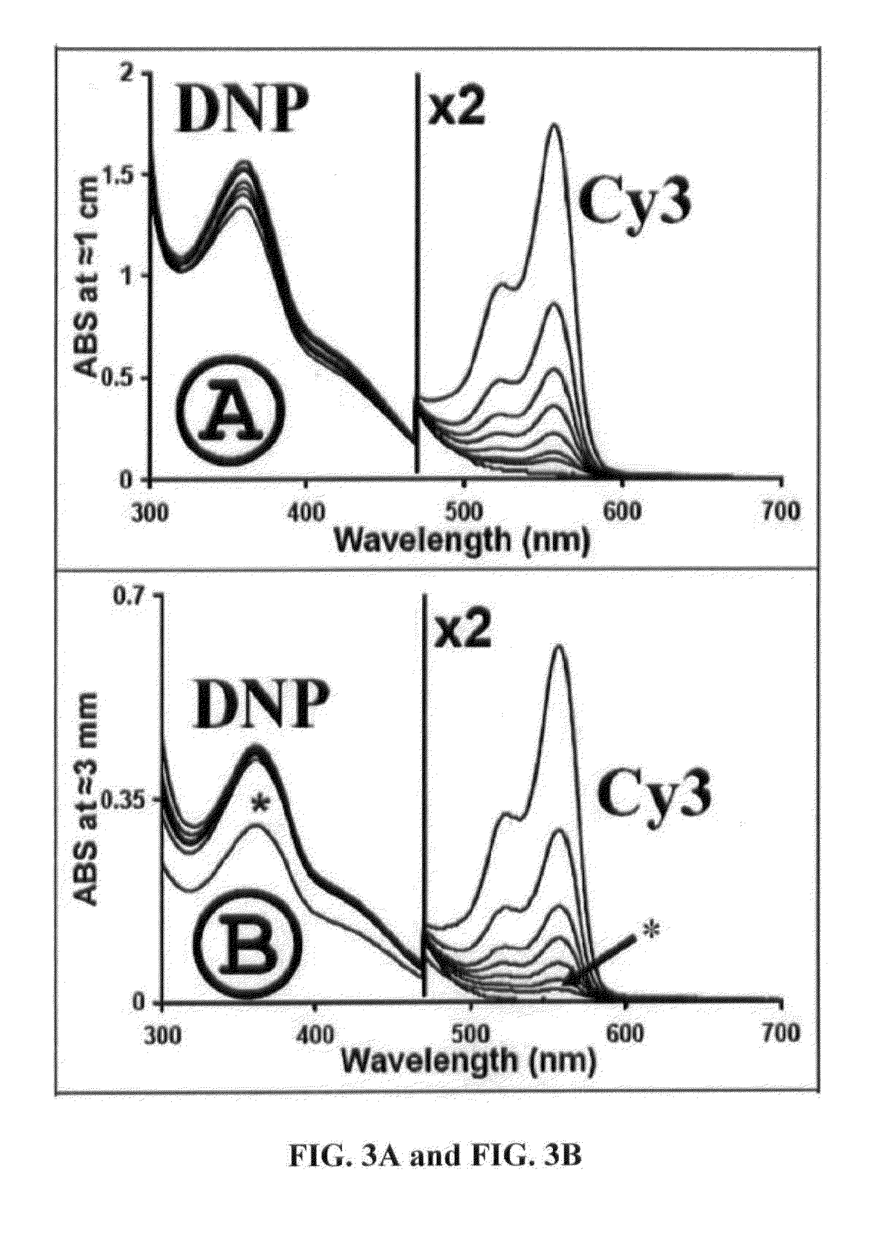
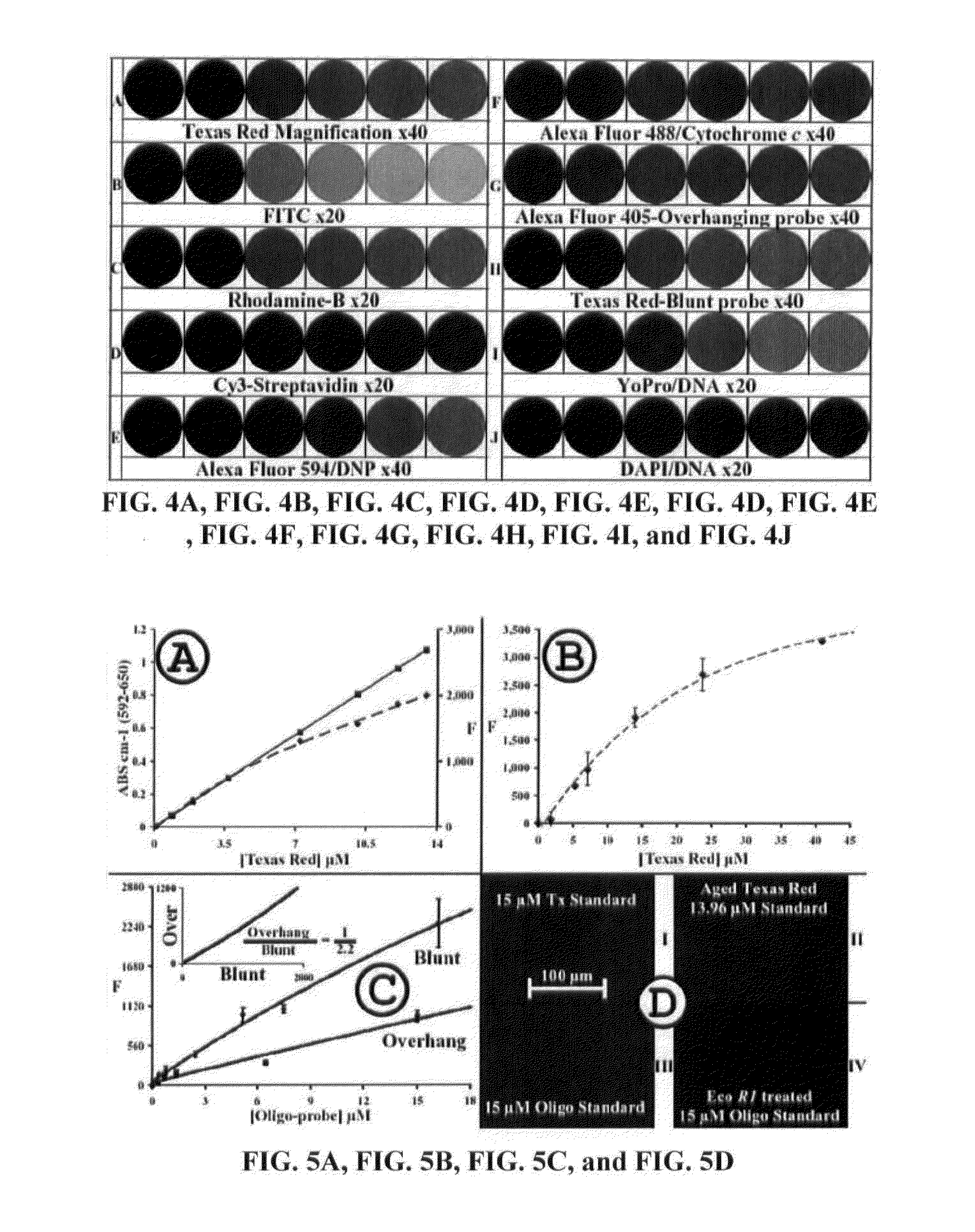
[0061]Fluorescently-labeled, oligonucleotide probes have been developed, which can be used, inter alia, to study programmed cell death, or for visualizing the presence of different types of DNA breaks (see, e.g., Didenko et al., 2006; Didenko et al., 2004; Baskin et al., 2003; Didenko et al., 2003; Didenko et al., 2002; Didenko, Ngo and Baskin, 2002; Didenko et al., 1999) in a biological sample. This methodology was facilitated by the development of tissue “phantoms” that contain known amounts of: 1) one or more chromophores or fluorophores; 2) one or more nucleic acids (e.g., a DNA, an RNA, or any combination thereof); 3) one or more peptides, proteins, polypeptides, enzymes, and / or antibodies (either labeled or unlabeled), or any combination thereof; 4) one or more oligonucleotide standards (either DNA, RNA, or any combination thereof); or 5) any combination of one or m...

example 2

Direct and Quantitative Measurement of Blunt and Overhanging DNA Ends

[0135]The present example describes a method of quantifying the levels of blunt- and overhanging-DNA ends using a method that employs oligonucleotide standards bound to gelatin slices. Using these tissue ‘phantom’ standards, a ligating efficiency of essentially 100% and a background staining level of 2O2 and Paraquat), or to one of the three chemotherapeutic agents routinely used for treating this disease (carmustine [BCNU], temozolomide, or irinotecan) have also been characterized.

[0136]The present ligase-based assay for selective detection of specific markers of necrosis / apoptosis in tissue sections utilizing oligonucleotide probes employs biotinylated, oligonucleotide, hairpin probes to detect the products of internucleosomal enzymatic DNA cleavage. Both blunt- and overhanging-DNA 3′-OH ends were detectable, and the typical products of DNase I type activity using in situ labeling of double-stranded DNA breaks. T...

example 3

Quantification of DNase Type I and II Ends and Oxidized / Acylated Bases

[0165]In the present example, a quantitative assay has been developed by substituting labeled ddUTP in place of dUPT in the conventional TUNEL assay, and a protocol developed to permit, for the first time, a TUNEL-based assay to be used in a quantifiable manner. The inventors are also the first to demonstrate how such a ddTUNEL assay can be combined with phosphatase treatment to detect and specifically quantitate the levels of DNase Type II activity in a single sample.

[0166]The ddTUNEL assay described herein has been combined with the base-modification repair enzyme, formamidopyrimidine-DNA glycosylase (Fpg), to interrogate the levels of modified DNA in tissues or in fixed, cultured cells. Using rat mammary gland, from Days 1 and 7 of involution, the inventors have validated the new methodology's ability to label apoptotic nuclei and apoptotic inclusion bodies. In addition, the types of DNA damage and modification...


PUM

PropertyMeasurementUnit
Fractionaaaaaaaaaa
Fractionaaaaaaaaaa
Concentrationaaaaaaaaaa

Abstract

Disclosed are compositions and methods for quantitation and calibration of images in fluorescence microscopy. Also provided are tissue phantoms that contain known amount(s) of fluorophore standard(s), as well as components and diagnostic kits containing the same for use in various histological analyses. In certain embodiments, three distinct nucleic-acid based assays provide improvements over conventional TUNEL methods to facilitate precise quantitation of a variety of nucleic acids obtained from a biological sample.

Description

CROSS-REFERENCE TO RELATED APPLICATIONS[0001]The present application claims priority to U.S. Provisional Patent Appl. No. 61 / 492,331, filed Jun. 1, 2011, the entire contents of which is specifically incorporated herein in its entirety by express reference thereto.STATEMENT REGARDING FEDERALLY SPONSORED RESEARCH OR DEVELOPMENT[0002]Not Applicable.NAMES OF THE PARTIES TO A JOINT RESEARCH AGREEMENT[0003]Not Applicable.BACKGROUND OF THE INVENTION[0004]1. Field of the Invention[0005]The present invention relates generally to the fields of optical microscopy and molecular biology. More particularly, it concerns compositions and methods for facilitating quantitative analysis of images in fluorescence / epifluorescence-based microscopic analysis, and for analyzing and directly quantitating nucleic acids including blunt and overhanging DNA ends. In certain embodiments, the invention provides tissue phantoms containing known amounts of chromo / fluorophores to serve as analytical markers for the ...

Claims


Application Information

Patent Timeline
IPC IPC(8): G01N21/64G01N33/53C12Q1/68
CPCG01N21/6428C12Q1/68G01N33/567G01N21/6458G01N33/53
Inventor SHARPE, MARTYN ALUNBASKIN, DAVID S.
Owner THE METHODIST HOSPITAL RES INST
Learn More