
Method and device for determining the location of an endoscope

a technology of endoscopy and location, which is applied in the field of image-guided endoscopy, can solve the problems of electromagnetic signal noise, enb system suffer from patient motion (breathing, coughing, etc., and different skill levels

Inactive Publication Date: 2012-08-09
PENN STATE RES FOUND
View PDF33 Cites 142 Cited by
  • Summary
  • Abstract
  • Description
  • Claims
  • Application Information

AI Technical Summary

Benefits of technology

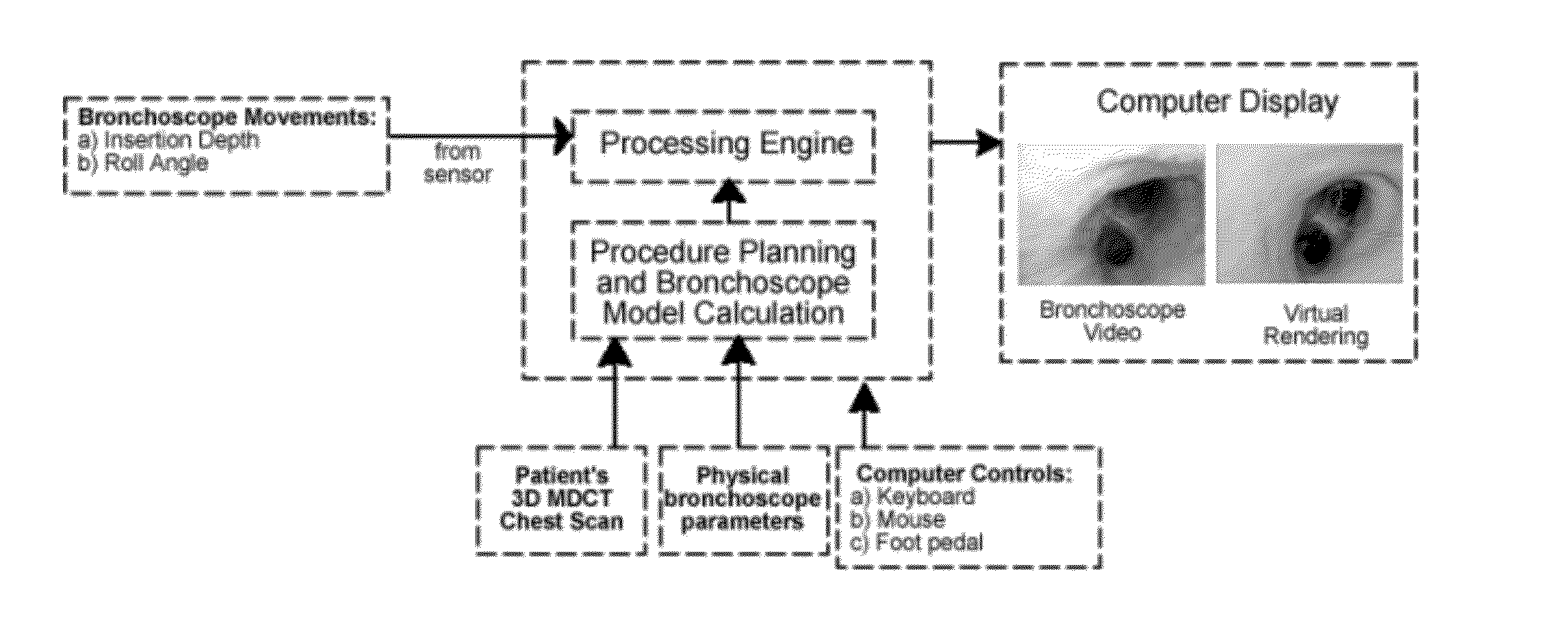
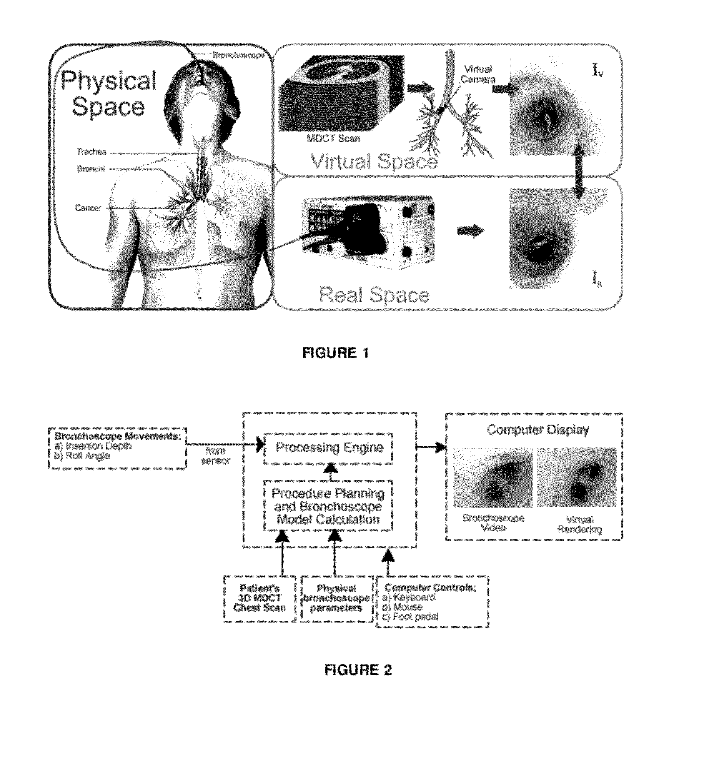
[0009]This invention overcomes the drawbacks of electromagnetic navigation bronchoscopy (ENB) and image-based bronchoscopy systems by comparing real-time measurements of actual instrument movements to precomputed insertion depth values provided by shape models. The preferred methods implement this comparison in real-time, providing continuous prediction of the instrument's tip location and orientation. In this way, the invention enables technician-free guidance and continuous procedure guidance irrespective of adverse events.
[0010]A method of determining the location of an endoscope within a body lumen according to the invention comprises the step of precomputing a virtual model of an endoscope that approximates insertion depths at a plurality of view sites along a predefined path to a region of interest (ROI). A “real” endoscope is provided with a device such as an optical sensor to observe actual insertion depths during a live procedure. The observed insertion depths are compared in real time to the precomputed insertion depths at each view site along the predefined path, enabling the location of the endoscope relative to the virtual model to be predicted at each view site by selecting the view site with the precomputed insertion depth that is closest to the observed insertion depth. An endoluminal rendering may then be generated providing navigational instructions based upon the predicted locations. The lumen may form part of an airway tree, and the endoscope may be a bronchoscope.

Problems solved by technology

Because of the challenges in standard bronchoscopy, physician skill levels vary greatly, and navigation errors occur as early as the second airway generation [6, 31].
ENB systems suffer from patient motion (breathing, coughing, etc.), electromagnetic signal noise, and require expensive equipment.
Often times, video frames lack enough structural information to allow for image-based registration or tracking.
Because registration and tracking techniques are not robust to these events, an attending technician is required to operate the system.

Method used


Image

  • Method and device for determining the location of an endoscope
  • Method and device for determining the location of an endoscope
  • Method and device for determining the location of an endoscope

Examples


Embodiment Construction

[0023]To overcome the drawbacks of ENB and image-based bronchoscopy systems, we propose a fundamentally different method. Our method compares real-time measurements of the bronchoscope movement to precomputed insertion depth values in the lungs provided by MDCT-image-based bronchoscope-shape models. Our method uses this comparison to provide a real-time, continuous prediction of the bronchoscope tip's location and orientation. In this way, our method then enables continuous procedure guidance irrespective of adverse events. It also enables technician-free guidance.

Branching Organ Representation

[0024]Let M be a 3D MDCT scan of the patient's airway tree N. While we focus on bronchoscopy, the invention is applicable to any procedure requiring guidance though a tubular structure, such as the colon or vasculature.

[0025]A virtual N is segmented from M using the method of Graham et al. [10]. This results in a binary-valued volume:

v(x,y,z)={1,iflisinsideN0,otherwise(1)

representing a set of ...


PUM


Abstract

A technician-free strategy enables real-time guidance of bronchoscopy. The approach uses measurements of the bronchoscope's movement to predict its position in 3D virtual space. To achieve this, a bronchoscope model, defining the device's shape in the airway tree to a given point p, provides an insertion depth to p. In real time, the invention compares an observed bronchoscope insertion depth and roll angle, measured by an optical sensor, to precalculated insertion depths along a predefined route in the virtual airway tree to predict a bronchoscope's location and orientation.

Description

REFERENCE TO RELATED APPLICATION[0001]This application claims priority from U.S. Provisional Patent Application Ser. No. 61 / 439,529, filed Feb. 4, 2011, the entire content of which is incorporated herein by reference.GOVERNMENT SPONSORSHIP[0002]This invention was made with government support under NM Grant Nos. R01-CA074325 and R01-CA151433 awarded by the National Cancer Institute. The government has certain rights in the invention.FIELD OF THE INVENTION[0003]This invention relates generally to image-guided endoscopy and, in particular, to a system and method wherein real-time measurements of actual instrument movements are compared in real-time to precomputed insertion depth values based upon shape models, thereby providing continuous prediction of the instrument's location and orientation and technician-free guidance irrespective of adverse events.BACKGROUND OF THE INVENTION[0004]Bronchoscopy is a procedure whereby a flexible instrument with a camera on the end, called a bronchosc...

Claims


Application Information

Patent Timeline
IPC IPC(8): A61B1/267A61B1/00
CPCA61B1/00006A61B1/00133A61B1/2676A61B5/066A61B6/12A61B6/5247G06K9/6289A61B2019/462A61B2019/502A61B2019/5246A61B2019/5261G06K9/00208G06K9/00214A61B19/5244A61B2034/102A61B2034/2046A61B34/20A61B2090/062A61B2034/2061G06V20/647G06V20/653G06V10/803G06F18/251
Inventor HIGGINS, WILLIAM E.GIBBS, JASON D.CORNISH, DUANE C.
Owner PENN STATE RES FOUND
Learn More