
Compositions and methods to generate pilosebaceous units

a technology of pilosebaceous cells and compositions, applied in the field of compositions and methods to generate pilosebaceous cells, can solve the problems of morbidity rather than mortality, lifelong suffering, and the inability of grafted skin to completely restore normal skin function

Inactive Publication Date: 2011-12-29
UNIV OF SOUTHERN CALIFORNIA
View PDF1 Cites 14 Cited by
  • Summary
  • Abstract
  • Description
  • Claims
  • Application Information

AI Technical Summary

Benefits of technology

[0013]This invention provides a new procedure that allows multipotential skin precursor cells to form a large number of new hair follicles which are arranged in a physiological plane with a cosmetically acceptable appearance. This procedure can be performed efficiently, reproducibly and on a large scale so as to be appropriate for clinical applications.

Problems solved by technology

While most survive, the deformities and scarring can cause lifelong suffering.
Now, morbidity rather than mortality is the main issue in burn care.
However, even with the new advances in skin substitutes, grafted skin is unable to completely restore normal skin function.
Burns can cause lasting appearance and functional defects to visible areas on the skin.
Current technology has improved the mortality rate of burns tremendously, but improvement of the morbidity rate of burn injuries has a long way to go.
While one may find these to be ancillary, lack of any or all of these causes significant suffering in a patient with scars.
The current gold standard of treatment for hair loss is hair transplantation, which is a laborious and expensive procedure.
Patients who do not have enough hair as a result of previous injury (such as burn), cannot spare extra hair to transplant.
There is just no way to increase the total number of hairs.
If one accepts the fact that hair-follicle development involves many molecular and cellular events embedded in discrete morphogenetic steps, then it would not be so surprising to encounter incomplete and imperfect structures as the science advances to the engineering of hair follicles—it is hard to get every step right in this dawn of bioengineering.
Failure of any of these events will lead to disrupted hair-follicle structures, resulting in various degrees of incomplete hair-follicle formation (Chuong et al.
However, the procedure is time consuming and cumbersome.
The method is only good for laboratory purposes on animal research.
However, due to the fact that the environment in which the growth of these hairs is trapped, the hair grows on the underside and cannot cycle and thus is not practical for clinical use.

Method used


Image

  • Compositions and methods to generate pilosebaceous units
  • Compositions and methods to generate pilosebaceous units
  • Compositions and methods to generate pilosebaceous units

Examples


experimental examples

Example 1

A Simplified Planar Hair Forming Protocol for High Throughput Assay Clinical Use

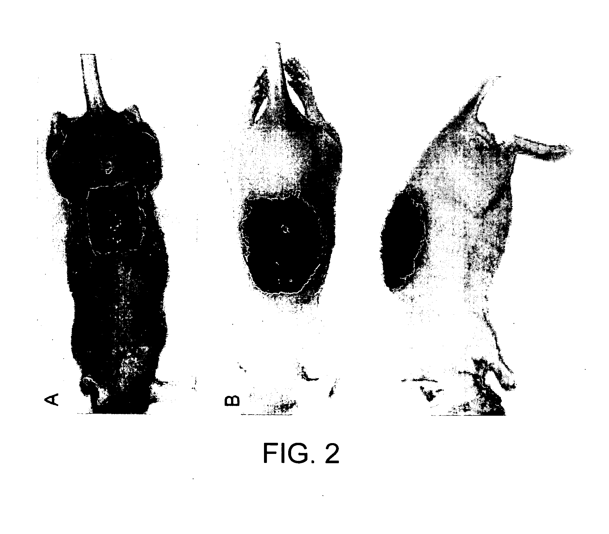
[0136]This example shows a method in which a large number of pilosebaceous units, including hair, are able to be grown from a dissociated cell suspension in vivo. While methods for hair generation have been described in the past, this invention is an improvement over prior art methods by making the process more user friendly for the clinician as well as more comfortable for the patient or animal. Additionally, by seeding these cells in a scaffold-like scaffold, it was unexpectedly shown that the cells will reorganize themselves in a way such that proper orientation of hair growth is obtained in a cosmetically acceptable manner. This protocol can be useful for high throughput screening of molecules or drugs important for hair formation. It may also have future clinical benefits for hair regeneration after severe wound injury and the treatment of alopecia.

Material and Methods:

[0137]Cell Isolation:...

example 2

Prepare Skin Stem Cells and Their Environment for Delivery

[0143]Given one million skin cells capable of forming hairs, 10 big hairs or 1000 small hairs can be formed. Big hairs are generally the preferred outcome. The regulatory patterning of this process has been studied by the inventors (Maini et al. (2006) Science 314(5804):1397-1398). In vitro studies showed that Turing reaction-diffusion plays a critical role in periodic patterning a homogenous population of stem cells (Jung et al. (1998) Dev Biol. 196(1):11-23; Jiang et al. (1999) Development 126(22):4997-5009). This suggests that the system can self-organize and cells respond to their local environment to assemble organs with a specific architecture based on cells ability to use their intrinsic (e.g. growth factor receptors, adhesion molecules) and extrinsic (e.g. growth factors, extracellular matrix molecules, etc.) properties. Before explant culture, dissociated multi-potential skin precusor cells are pre-plated at high cel...

example 3

A Simplified Planar Hair Forming Protocol for High Throughput Assay Clinical Use

[0147]In an extension of the experiment described in Example 1, Applicant provides the following Example 3.

Material and Methods:

[0148]Cell Isolation: Multipotential skin precursor cells are currently obtained from neonatal mice using techniques from previously published work. Briefly, neonatal mice are harvested shortly after birth (within the first 24 hours) and euthanized. The truncal skin is then dissected off with sharp forceps. Epidermis and dermis are separated by floating in cold 0.25% trypsin overnight. Epidermal cells are then dissociated into a cell suspension by cutting into fine pieces and manual tituration with a serological pipet. Single epithelial cells are separated through a 70 μm cell strainer to exclude cells of the stratum corneum. The dermal cells are individually dissociated using warm 0.35% collagenase for 40-50 minutes at 37° C. DNase I is then added for 5 minutes at RT before man...


PUM


Abstract

The invention provides compositions and methods to generate pilosebaceous units. In one aspect, the invention comprises a biocompatible scaffold and an effective amount of dermal and epidermal precursor cells.

Description

CROSS-REFERENCE TO RELATED APPLICATIONS[0001]This application claims the benefit under 35 U.S.C. §119(e) of U.S. Provisional Application Ser. No. 61 / 116,620, filed Nov. 20, 2008, the content of which is incorporated by reference into the present disclosure in its entirety.STATEMENT OF FEDERAL SUPPORT[0002]This invention was supported by grants from the National Institutes of Health (Grant Nos. AR 047364 and F32 GM08019). The government has rights in this application.BACKGROUND[0003]The skin consists of two layers, an epidermis layer derived from the ectoderm, and a dermis layer derived from mesoderm underneath the epidermis layer. Dermis is subdivided into two strata, the superficial papillary layer and the reticular layer. The epidermis is a stratified squamous epithelium. The epidermis is the outer layer of the skin and acts as a protective film against external insults. The majority of epidermal cells undergo keratinization and form the dead superficial layers of the skin. The th...

Claims


Application Information

Patent Timeline
IPC IPC(8): A01K67/027A61K35/36A61K49/00C12N5/071
CPCC12N5/0628C12N2533/54C12N2500/25C12N2500/99C12N2501/11C12N2501/115C12N2501/135C12N2501/155C12N2501/165C12N2501/385C12N2501/999C12N2502/092C12N2502/094C12N2502/1323C12N2503/06C12N5/0698C12N2500/90
Inventor LEE, LILYCHUONG, CHENG MINGGARNER, WARRENJIANG, TING XIN
Owner UNIV OF SOUTHERN CALIFORNIA
Learn More