Eureka AIR delivers breakthrough ideas for toughest innovation challenges, trusted by R&D personnel around the world.

Flow sound in x-ray examination

a flow sound and x-ray technology, applied in the field of representation of blood flow related information, can solve the problems of insufficient derivable information, difficult or even impossible to retrieve qualitative and quantitative information about blood flow, and insufficient information, etc., to achieve the effect of easy detection

Inactive Publication Date: 2011-12-29
KONINKLIJKE PHILIPS ELECTRONICS NV
View PDF14 Cites 8 Cited by
  • Summary
  • Abstract
  • Description
  • Claims
  • Application Information

AI Technical Summary

Benefits of technology

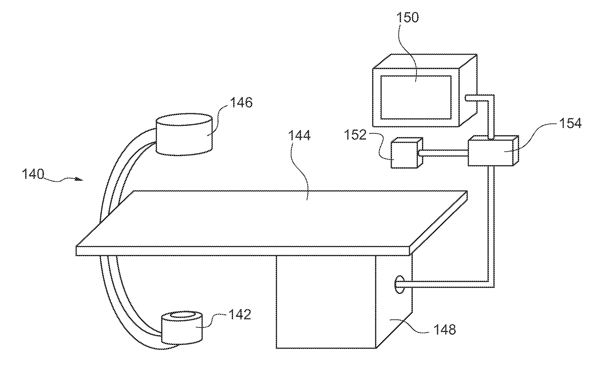
[0006]According to an exemplary embodiment of the invention, the image sequences are used to get vessel maps for neurological intervention and the visualization of aneurysm structure. Then, flow maps are produced using space time filtering and, for example, Optical Flow methods. These flow maps provide complex visual information about the actual flow characteristics. However, to comprehend this information, the user's visual attention is required. According to the invention, knowing the spatial distribution of blood velocity, it is possible to artificially synthesize a sound defined by the visual spectrum determined by the velocity distribution. Providing the information to the acoustic sense, the user, e.g. the surgeon, is relieved in such a way that his visual attentiveness can be addressed to other aspects. Hence, providing a sound signal representing the blood flow will help clinicians to appreciate and qualify the vascular flow characteristics. It is also recognized that the sensibility of the ear is able to detect subtle variations, due to flow disturbances, or irregularities, more than any imaging representation. That means that by providing an acoustic representation of the blood flow, it is possible to provide more detailed information compared to a visual representation. Another advantage is that detailed blood flow information can be provided for regions of interest regardless of their situation within the body of patient, for example for neurological vessels or for obtaining information about coronary flows. Providing sound information based on X-ray imaging technique allows cardiologists and neurologists, for example, an enhanced way of evaluating vascular lesions and a better control on quality of resulting blood flow.
[0011]By adapting the sound characteristics to the sound impression of Ultrasound Doppler flow sound, a Doppler Spectrum analysis comparable to the known Ultrasound Doppler Flow Exam is provided. Indeed, Ultrasound Doppler exam is a commonly used reference exam for vascular evaluation. Hence, by providing a sound signal with the characteristics of an Ultrasound Doppler Flow sound, the clinician can use his skills from Ultrasound exams to interpret the sound and spectrum provided by the X-ray imaging system. Thus, the sound representation is beneficial for interpreting the X-ray results. By providing Doppler like sound information, the need for an interpretation of these new images by the user is avoided, which by the way would require huge clinical validations. In other words, reproducing results in a known manner, wherein the results are usually obtained with a different imaging modality, will enhance the acceptance and interpretation of X-ray base flow measure, because the clinician can appreciate the flow quality using the same skills he developed with ultrasound.
[0021]This allows presenting additional information that is included in the X-ray image, which results in a very significant image with high information density. The effect of the superimposition is that coloured pixel representing certain flow characteristics appear within the X-ray image that is traditionally only in black and white, i.e. in greyscale. This superimposition of coloured pixels can be accomplished by a covering overlaying, i.e. an opaque colour application over the greyscale image or by a semitransparent overlaying, i.e. where the greyscale image is still visible to a certain degree. Hence, as an effect the greyscale image partly appears coloured at least in some pixels.
[0030]These artificial patterns mimic an intrinsic aliasing effect, which occurs for high velocities in Ultrasound CFI keeping a good colour dynamics for normal flows and enhancing the high flows that are typically associated to tight stenoses. To produce this aliasing phenomenon if velocity values are abnormally high, in one embodiment the threshold is corresponding to normal maximum blood flow velocities.
[0036]It has shown that the contrast phase within an artery submitted to the arterial pulsed pressure creates a periodic time modulation of the contrast density. With the blood flow, this modulation is transported in the arterial network, creating a kind of “contrast wave” pattern. By tuning the characteristics of a dedicated time filter to the cardiac period it is possible to enhance the contrast wave pattern. This sequence is an attractive visualization of the flowing contrast and may be the input for the velocity vector field estimation step.

Problems solved by technology

But it has shown that with common techniques in X-ray procedures, blood flow images are produced in such a way that the qualitative and quantitative information about the blood flow is difficult or even impossible to retrieve.
For example, these images themselves do not provide the desired information for a detailed vessel analysis to the surgeon, for example.
Even by combining several images to a sequence, for example representing a cardiac cycle, the derivable information is not sufficient.

Method used


Image

  • Flow sound in x-ray examination
  • Flow sound in x-ray examination
  • Flow sound in x-ray examination

Examples


Embodiment Construction

[0057]In order to produce a flow sound, in an exemplary embodiment the following steps are provided: First, a first sequence of images is acquired by an X-ray image acquisition device. Further, a second sequence of images is acquired with injected contrast agent. Then, the contrast signal is extracted through a DSA operation. Further, the vessel structure is registered to cancel the motion of the vessels. Then, in one exemplary embodiment, time filtering of the sequence is used to enhance the moving components of the contrast. The images are then used for generating vector velocity fields which are represented by contrast motion within the vessels. This extraction is achieved with an optical flow (OF) method. The output of the OF operation is then used to produce velocity distribution curves. This graphical information is then converted into a synthetic sound signal.

[0058]In a preferred embodiment, the sound signal is adapted to the acoustic Doppler effect.

[0059]Hence, an acoustic s...


PUM


Abstract

The invention relates to the representation of blood flow-related information using an X-ray image acquisition device. In order to provide an improved method in an X-ray imaging system for representing the blood flow, for vessel analysis purposes for example, a device and a method are provided where X-ray images are used to determine the spatial distribution of blood velocity. Using this information it is possible to artificially synthesize a sound defined by the visual spectrum determined by the velocity distribution. Providing sound information based on X-ray imaging technique allows cardiologists and neurologists, for example, an enhanced way of evaluating vascular lesions and a better control on quality of resulting blood flow.

Description

FIELD OF THE INVENTION[0001]The invention relates to the representation of blood flow-related information. In particularly, the invention relates to a method for representing blood flow-related information using an X-ray image acquisition device and to an apparatus with an X-ray image acquisition device for representing blood flow-related information.BACKGROUND OF THE INVENTION[0002]Information concerning the blood flow is needed for different reasons. For example, vessel maps are commonly used for vessel analysis, neurological interventions or for the visualization of aneurysm structures. For example, information on the blood flow quality is needed before an intervention, such as a stent placement for instance, for planning purposes and after the intervention, e.g. the stenting, for an outcome control. But the recovery of satisfactory physiological blood flow cannot be verified. Usually, echography is used to get quantitative flow information. Indeed, Ultrasound is used for the exa...

Claims


Application Information

Patent Timeline
IPC IPC(8): G06K9/00G08B3/00G06K9/32
CPCA61B6/463A61B6/481A61B6/504G06T7/0024A61B6/507G06T2207/20224G06T2207/30104G10H1/0008G10H2220/371G06T2207/10116G06T7/30
Inventor BONNEFOUS, ODILEFLORENT, RAOULAUVRAY, VINCENT MAURICE ANDRE
Owner KONINKLIJKE PHILIPS ELECTRONICS NV
Why Eureka
Eureka Blog
Learn More