
Encapsulated agent guided imaging and therapies

a technology of encapsulated agents and imaging, applied in the direction of powder delivery, chemical/physical/physicochemical processes, viruses, etc., can solve the problems of short vascular circulation time, limited true potential of non-toxic and clinically proven optical probes, and difficulty in optical imaging and phototherapy of various diseases, etc., to achieve the effect of improving the circulation tim

Inactive Publication Date: 2011-12-22
RGT UNIV OF CALIFORNIA
View PDF1 Cites 7 Cited by
  • Summary
  • Abstract
  • Description
  • Claims
  • Application Information

AI Technical Summary

Benefits of technology

[0017]The invention may employ a virus, viral capsid, virus-resembling structure, or artificial virus. The invention utilizes viral coat proteins capable of forming a capsid or other structures which can enclose, or partially enclose the delivered agent such that it is protected or targeted to specific tissues, or preferentially accumulates in certain tissues. One aspect of the invention is the incorporation of a selected contrast agent into the virus, or a virus-resembling structures, or artificial viruses for both imaging and therapy. The advantages of this system as compared to other contrast agents including polymer-based capsules known in the art lies in biocompatibility, appropriate size dimensions, high level of monodiversity, and specific binding or distribution to a selected target site.
[0027]A further aspect of the invention provides imaging agents and methods of use that utilize coating materials, ligands or receptors that are attached to the reassembled virus capsid that provide or improve circulation time, half life and target specificity. One embodiment the viral capsid does not have viral activity and target specificity is provided by the ligand.

Problems solved by technology

For example, development of highly sensitive probes with high specificity for the targeted structure to enable diagnosis of pre-malignant lesions and early stage tumors remains as one of the great challenges of oncology.
Despite its clinical usage, ICG in its current formulation of being freely dissolved in solution has several major drawbacks consisting of optical instability at physiologically relevant conditions, short vascular circulation time with plasma clearance half-life on the order of 3-4 minutes, and nearly exclusive uptake by the liver.
Therefore, the true potentials of this non-toxic and clinically proven optical probe, for both optical imaging and phototherapy of various diseases remains limited.
However, there remain significant issues related to toxicity, physical stability, chemical flexibility and synthesis difficulty.
First, synthesis of these nano-capsules is often difficult.
For example, depending on the construct type, synthesis steps can include complicated procedures such as cycles of freeze-vacuum-thaw and polymerization, several heating and cooling steps, filtration and drying steps and complex evaporation procedures.
Another deficiency found with the use of synthetic constructs is that the synthetic polymeric formulations do not provide the appropriate size dimension and level of monodispersity required for appropriate circulation time, biodistribution, and availability within the body.
These types of nano-capsules are poly-dispersed in size, influencing their intended biodistribution, they often aggregate in solution, compromising their effectiveness for imaging purposes.

Method used


Image

  • Encapsulated agent guided imaging and therapies
  • Encapsulated agent guided imaging and therapies
  • Encapsulated agent guided imaging and therapies

Examples


example 1

[0062]In order to demonstrate the functionality of the encapsulation methods, a new type of an optical nano-probe, based on genome-depleted plant Brome Mosaic Virus (BMV) whose interior is doped with indocyanine green (ICG), an FDA-approved near infrared (NIR) fluorescent dye was engineered. The nano-probes, derived from naturally occurring building blocks, are referred to as optical viral ghosts (OVG's) since the viral particles no longer contain the genomic machinery for replication. Only the capsid protein (outer shell) of the native virus remains intact, and provides the encapsulating structure for ICG.

[0063]BMV is a member of the family Bromoviridae of plant RNA viruses. It is a non-enveloped icosahedral virus whose shell is composed of 180 subunits of a 20 kDa capsid protein (cp). The genome of BMV is divided among four RNAs. Viral replication is dependent on interaction between non-structural replicase proteins, encoded by genomic RNA1 and RNA2. The capsid protein gene is enc...

example 2

[0079]To demonstrate the viability of optical viral ghosts (OVG's) in cellular imaging, normal human bronchial epithelial cells (LHS-9) as model cells were used since certain types of lung cancers are derived from the bronchial epithelium. The cells were cultured for 24 hours in serum free culture medium supplied by the commercial vendor. OVG's were then delivered into cell culture medium and incubated for two hours. An optical filter that allowed transmission of NIR light greater than 780 nm was used to capture the fluorescent signal onto a CCD camera. Tungsten light filtered between 730 nm and 775 nm was used as the excitation source.

[0080]False-color confocal fluorescent images of the human bronchial epithelial cells at 2 hr post-incubation with OVG's or free ICG (control) were obtained from a plane across the cells. The concentration of free ICG and that used in the construction of OVG's was 10 μg / ml. While free ICG remains mostly localized to the “rim” of the cells, OVG's are a...

example 3

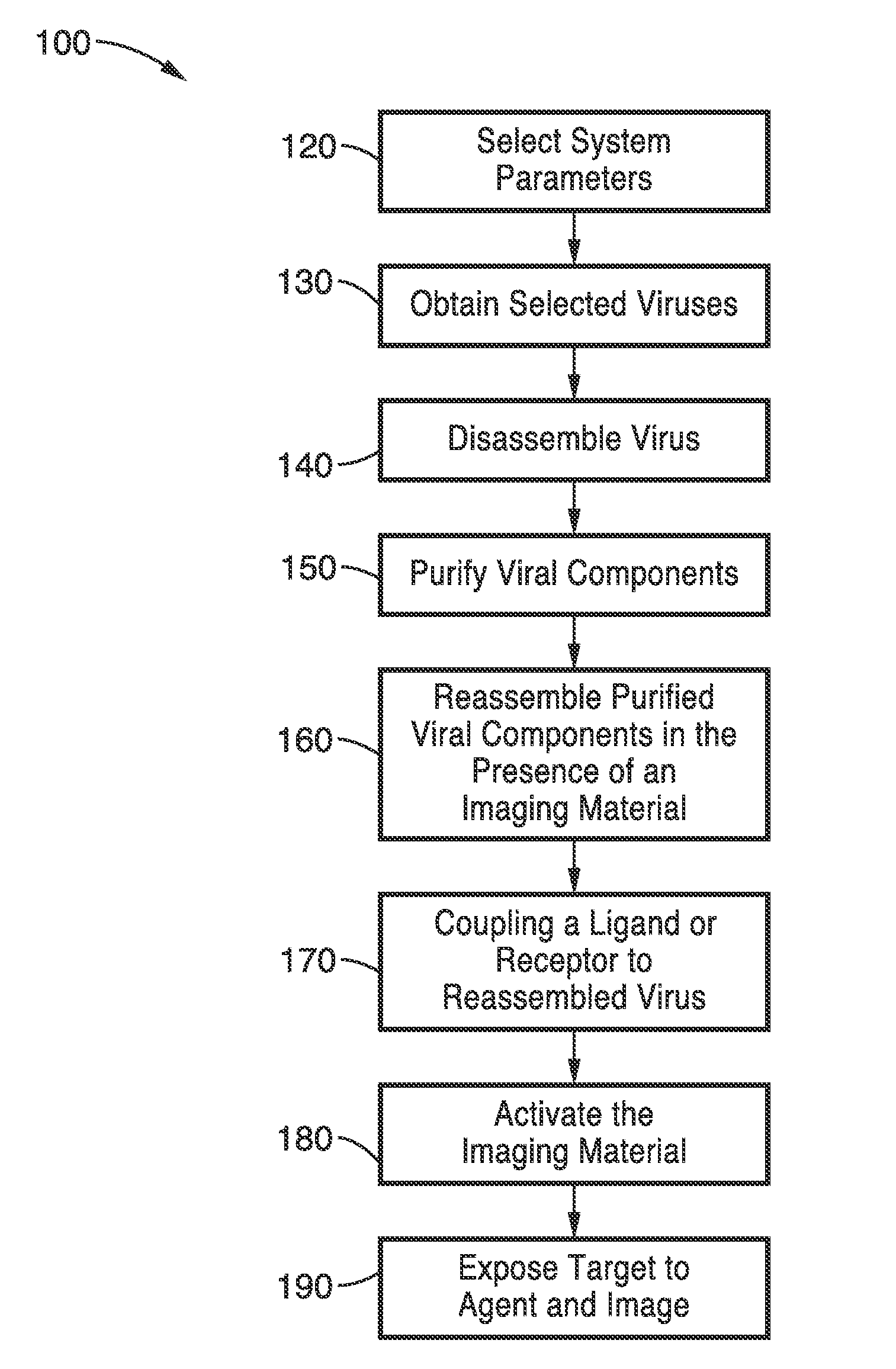
[0083]To demonstrate the utility of the encapsulation methods with imaging compounds other than ICG, the methods were used with Alexa 488-Dextran complex. The encapsulation process used the following steps: (1) disassociation / disassembly of the virus capsid; and (2) re-association / reassembly of the virus capsid in the presence of the encapsulation agent were used with Alexa Fluor 488 in this example.

[0084]To dissociate the virus capsid, the pH of the buffer solution containing the viral particles was raised to about pH 7.5 with 0.5 CaCl2 and 50 mM Tris-HCl. Additional reagents included 1 mM DTT to stabilize the solution, and 1 mM EDTA and 0.5 mM PMSF as protease inhibitors. A period of 24 hours for the disassociation process was allowed to eliminate the nucleotides of the virus (RNA in the case of BMV) and only preserve the capsid for the final reassociation / reassembly. Other reagents that increase the pH or alter the ionic strengths of the solution to levels that cause disassociati...


PUM

PropertyMeasurementUnit
Therapeuticaaaaaaaaaa
Energyaaaaaaaaaa
Biostabilityaaaaaaaaaa

Abstract

A nano-capsule construct for imaging and therapeutic uses and method for production are provided. One nano-probe embodiment based on genome-depleted plant brome mosaic virus (BMV) whose interior is doped with indocyanine green (ICG), an FDA-approved near infrared fluorescent dye, is used to illustrate the invention. The material encapsulated in viral shell components may be coated with functionalized coatings such as branched, dendritic polymer coatings to improve longevity and distribution in the body as well as antibody conjugation for increased target specificity. The constructs can also be coated with ferromagnetic iron oxide nanoparticles, enabling the ICG-containing capsules to be used as nano-probes with the capability of being detected in both optical and magnetic resonance imaging. The capsules may be produced by purifying a plant or animal viruses and disassembling the viruses to provide virus shell components. The virus shell components are reassembled in the presence of a material for encapsulation thereby encapsulating said material within the core of the construct in one embodiment.

Description

CROSS-REFERENCE TO RELATED APPLICATIONS[0001]This application a 35 U.S.C. §111(a) continuation of PCT international application Ser. No. PCT / US2009 / 067396 filed on Dec. 9, 2009, incorporated herein by reference in its entirety, which is a nonprovisional of U.S. provisional patent application Ser. No. 61 / 121,186 filed on Dec. 9, 2008, incorporated herein by reference in its entirety. Priority is claimed to each of the foregoing applications.[0002]The above-referenced PCT international application was published as PCT International Publication No. WO 2010 / 068705 published on Jun. 17, 2010, and is incorporated herein by reference in its entirety.STATEMENT REGARDING FEDERALLY SPONSORED RESEARCH OR DEVELOPMENT[0003]Not ApplicableINCORPORATION-BY-REFERENCE OF MATERIAL SUBMITTED ON A COMPACT DISC[0004]Not ApplicableBACKGROUND OF THE INVENTION[0005]1. Field of the Invention[0006]This invention pertains generally to imaging agents and delivery methods and more particularly to an imaging cons...

Claims


Application Information

Patent Timeline
IPC IPC(8): A61K49/00A61K31/403A61K31/409A61K31/197B01J19/12A61K49/22A61K49/04A61K49/18A61K9/14A61K9/00A61K31/221
CPCA61K47/48776C12N2770/14042C12N2770/14023C12N7/00A61K47/6901
Inventor ANVARI, BAHMAN
Owner RGT UNIV OF CALIFORNIA
Learn More