Contrast agents in porous particles
a technology of contrast agents and porous particles, applied in the field of contrast agents in porous particles, can solve the problems of insufficient contrast generation and potential toxicities, low circulation time, and various limitations of the use of cas for imaging
- Summary
- Abstract
- Description
- Claims
- Application Information
AI Technical Summary
Benefits of technology
Problems solved by technology
Method used
Image

Examples
example 1
Characterization of Gd-Based Contrast Agents
[0087]In this work, the enhanced efficiency of Gd-based CAs (Gd-CAs) has been demonstrated by confining them within the nanoporous structure of intravascularly injectable silicon particles (SiMPs)8. Enhanced efficiency was shown for two different Gd-CAs, namely Magnevist (MAG) and gadonanotubes (GNTs).
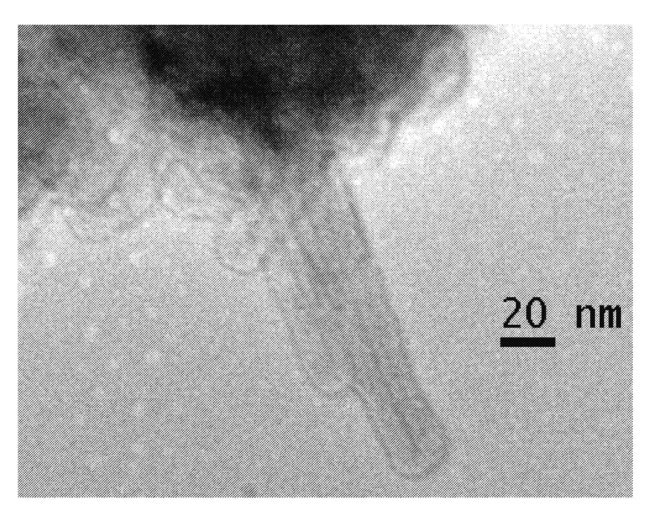
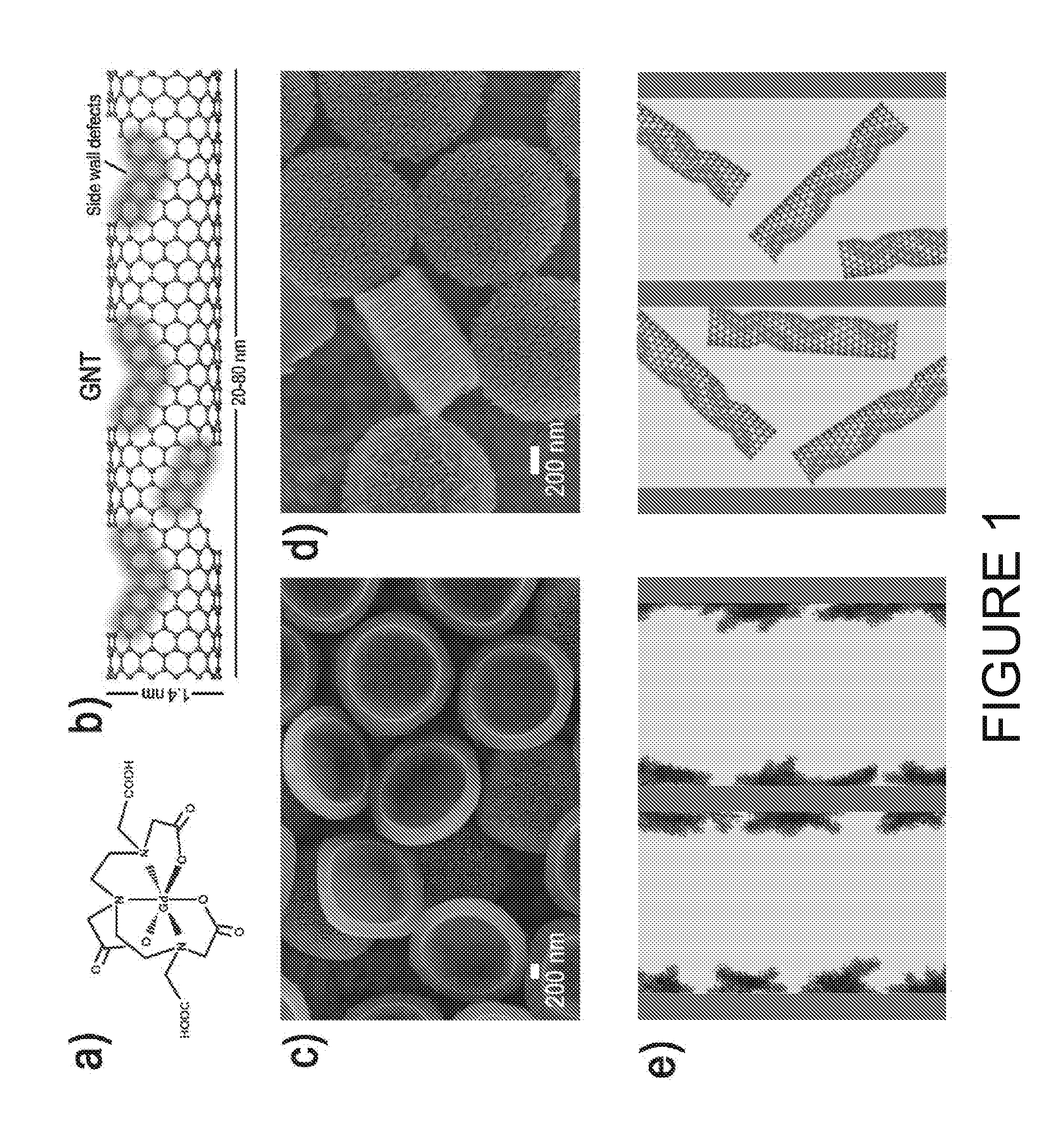
[0088]Magnevist (Gd-DTPA) is an example of gadolinium polyamino carboxylate complexes (FIG. 1A), widely used in the clinic as T1-weighted MRI contrast agents6. GNTs are carbon nanostructure-based lipophilic contrast agents showing great promise in MRI. As shown in FIG. 1B, GNTs are nanoscale carbon capsules (derived from full-length single-walled carbon nanotubes) with a length of 20-80 nm and a diameter of about 1.4 nm, which are internally loaded with Gd3+ ion clusters. Within the GNTs, the Gd3+ ions are present in the form of clusters (3+ ions per cluster), and each GNT contains approximately 50 to 100 Gd3+ ions. The Gd3+ clusters are stab...
example 2
Supplemental Information for Example 1
[0120]Mathematical Model for Estimating the Longitudinal Relaxivity of MRI CAs
[0121]The ability of MRI CAs to shorten the longitudinal T1 relaxation time of water (T1=3000 ms) is reflected by their r1 relaxivity. This is given by summing the contributions of the so-called inner-sphere rIS and outer-sphere rOS relaxivities. The inner-sphere contribution comes from the nuclear spin residing in the water molecules entering the first coordination shell of the metal ion of interest, whilst the water molecules outside of this shell contribute to the outer-sphere portion. The classical theory for estimating the inner-sphere contribution is due to the work of the SBM theory.3 This simplified theory provides a close form expression for rIS and can be effectively used to interpret experimental data on the relaxivity of several complexes. In particular it has been shown that the SBM theory can accurately predict and reproduce NMRD profiles in the high fiel...
example 3
Optimized Loading of Magnevist into Mesoporous Silicon Particles
[0171]Discoidal mesoporous silicon particles with a diameter of 1000 nm and a thickness of 400 nm were loaded with Magnevist (MAG). As set forth previously, MAG is a clinically available Gd3+ ion-based contrast agent for T1 weighted MRI. It has been show that by reducing the size of the pores in which MAG is loaded, the longitudinal relaxivity r1 of the nanoconstructs can be enhanced even more than what was demonstrated by using the HP particles. See Examples 1-2 and Nature Nanotechnology 5:815-821 (24 Oct. 2010).
[0172]As shown in FIG. 17, a longitudinal relaxivity r1 of about 10 (mM sec)−1 was demonstrated for the HP particles (pore size 30-40 nm in diameter). With the SP particles (pore size 5-10 nm in diameter), the longitudinal relaxivity r1 was boosted up to about 25 (mM sec)-1.
PUM
| Property | Measurement | Unit |
|---|---|---|
| pore size | aaaaa | aaaaa |
| pore size | aaaaa | aaaaa |
| pore size | aaaaa | aaaaa |
Abstract
Description
Claims
Application Information
 - R&D
- Intellectual Property
- Life Sciences
- Materials
- Tech Scout
- Unparalleled Data Quality
- Higher Quality Content
- 60% Fewer Hallucinations
Browse by: Latest US Patents, China's latest patents, Technical Efficacy Thesaurus, Application Domain, Technology Topic, Popular Technical Reports.
© 2025 PatSnap. All rights reserved.Legal|Privacy policy|Modern Slavery Act Transparency Statement|Sitemap|About US| Contact US: help@patsnap.com