Method for detecting dysplasia
a dysplasia and imaging technology, applied in the field of imaging, can solve the problems of oesophageal cancer, rare curable disease, lack of precise pre-operative staging, etc., and achieve the effect of facilitating the diagnosis of “
- Summary
- Abstract
- Description
- Claims
- Application Information
AI Technical Summary
Benefits of technology
Problems solved by technology
Method used
Image
Examples
example 1
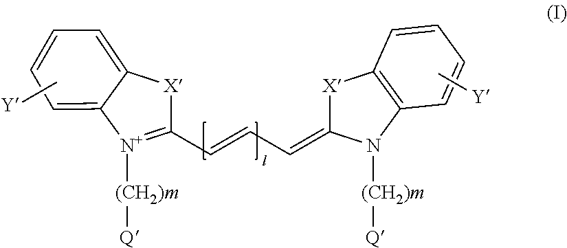
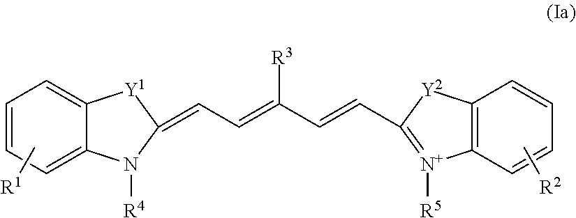
Synthesis of the Cyanine Dye 2-{(1E,3E,5E)-5-[1-(5-carboxypentyl)-3,3-dimethyl-5-sulfo-1,3-dihydro-2H-indol-2-yliden]penta-1,3-dienyl}-3-methyl-1,3-bis(4-sulfobutyl)-3H-indolium-5-sulfonate (Cy5**)
[0256]
(1a) 5-Methyl-6-oxoheptane-1-sulfonic Acid
[0257]
[0258]Ethyl 2-methylacetoacetate (50 g) in DMF (25 ml) was added to a suspension of sodium hydride (12.0 g of 60% NaH in mineral oil) in DMF (100 ml), dropwise with ice-bath cooling over 1 hour, (internal temperature 0-4° C.). This mixture was allowed to warm to ambient temperature for 45 mins with stirring before re-cooling. A solution of 1,4-butanesultone (45 g) in DMF (25 ml) was then added dropwise over 15 minutes. The final mixture was heated at 60° C. for 18 hours. The solvent was removed by rotary evaporation and the residue partitioned between water and diethyl ether. The aqueous layer was collected, washed with fresh diethyl ether and rotary evaporated to yield a sticky foam. This intermediate was dissolved in water (100 ml) an...
example 2
Synthesis of 2-[(1E,3E,5E)-5-(1-{6-[(2,5-dioxopyrrolidin-1-yl)oxy]-6-oxohexyl}-3,3-dimethyl-5-sulfo-1,3-dihydro-2H-indol-2-ylidene)penta-1,3-dienyl]-3-methyl-1,3-bis(4-sulfobutyl)-3H-indolium-5-sulfonate, diisopropylethylamine Salt (NHS Ester of Cy5**)
[0264]
[0265]Cy5** (Example 1, 10 mg) was dissolved in anhydrous DMSO (3 ml); to this were added HSPyU (20 mg) and N,N′-diisopropylethylamine (80 μl). The resulting solution was mixed for 3 hours, whereupon TLC (RPC18. Water / MeCN) revealed complete reaction. The dye was isolated by precipitation in ethyl acetate / diethyl ether, filtered, washed with ethyl acetate and dried under vacuum. UV / Vis (Water) 650 nm. MS (MALDI-TOF) MH+983.5. MH+═C42H53N3O16S4 requires m / z 984.16.
example 3
Synthesis of Compounds 1-11
[0266]a) Peptide Synthesis
[0267]The peptidyl resin corresponding to the sequences of Compounds 1-11 in Table 2 were assembled by standard solid-phase peptide chemistry [Barmy, Int. J. Peptide Protein Res., 30, 705-739 (1987)] on either a Rink Amide MBHA resin (from NovaBiochem, typical loading 0.72 mmol / g, synthesis of 1, 2, 3, 5, 6, 7, 9, 10 and 11), a Fmoc-Ala-Wang resin (from NovaBiochem, loading 0.52 mmol / g, synthesis of 4) or on a Fmoc-Cys(Trt)-Sasrin resin (from Bachem, loading 0.45 mmol / g, synthesis of 8). Fmoc-Cys(Trt)-OH was loaded manually onto the Rink Amide MBHA resin using PyAOP / collidine activation in 50% DCM:DMF (synthesis of 6 and 10). All other amino acids were assembled manually on the solid phase using a microwave assisted peptide synthesizer (CEM Liberty). The residues (from the carboxyl terminus) were coupled on a 0.1 mmol scale typically using single coupling cycles (5 min at 75° C.) of 0.5 mmol Fmoc-amino acids (0.2 M solution in NMP...
PUM
| Property | Measurement | Unit |
|---|---|---|
| of wavelength | aaaaa | aaaaa |
| near-infrared wavelength | aaaaa | aaaaa |
| vivo imaging | aaaaa | aaaaa |
Abstract
Description
Claims
Application Information
 - R&D Engineer
- R&D Manager
- IP Professional
- Industry Leading Data Capabilities
- Powerful AI technology
- Patent DNA Extraction
Browse by: Latest US Patents, China's latest patents, Technical Efficacy Thesaurus, Application Domain, Technology Topic, Popular Technical Reports.
© 2024 PatSnap. All rights reserved.Legal|Privacy policy|Modern Slavery Act Transparency Statement|Sitemap|About US| Contact US: help@patsnap.com


