METHOD OF MEASURING CYTOKERATIN 19 mRNA

a cytokeratin and cytokeratin technology, applied in the field of cytokeratin 19 mrna measurement, can solve the problems of difficult to distinguish between cancer cells, inability to detect cancer cells, and complexity of the procedure, and the risk of secondary infection

Inactive Publication Date: 2011-09-22
TOSOH CORP
View PDF2 Cites 0 Cited by
  • Summary
  • Abstract
  • Description
  • Claims
  • Application Information

AI Technical Summary

Benefits of technology

[0021]An object of the present invention is to provide a method of rapidly measuring cytokeratin 19 mRNA with a procedure carried out at a constant temperature and in a single step on a sample obtained from human cells or tissue and the like.

Problems solved by technology

However, sometime it is difficult to distinguish between cancer cells and normal cells in the case of HE staining, resulting in the discovery of cancer cells being overlooked.
(and it may be necessary to separately carry out an additional detection step depending on the case), complexity of the procedure and the risk of secondary infection are suggested.
In addition, since it normally requires 2 hours or more to carry out the two steps, there have been problems with respect to poor reproducibility attributable to carrying out multiple steps as well as reducing large-volume specimen processing and testing costs.
Although a One Step RT-PCR method has been developed that enables both steps to be carried out simultaneously in order to shorten reaction time, this method has been indicated as having lower detection sensitivity and resulting in the production of non-specific amplification products in comparison with RT-PCR methods in which each step is performed separately.
Moreover, since amplification by PCR involves amplification of double-stranded DNA, there is concern over the possibility of amplifying contaminating chromosomal DNA, thereby resulting in the need to completely remove all chromosomal DNA by digesting with DNase and the like in order to precisely analyze mRNA expression.
Consequently, this results in greater complexity of the procedure and poorer reproducibility.
Moreover, since the PCR method requires the reaction temperature to be rapidly raised and lowered, it serves as an obstacle to labor saving and cost reduction of reaction devices at the time of automation.
However, this method requires a large number of primers to be designed, and there are numerous restrictions on the design of each primer thereby making design extremely complex.
Since the Tm value, GC content and the like of each primer is related to reactivity at this time, those domains for which primers can be designed are limited considerably.
Thus, not only is it difficult to design a large number of primers in this manner, but this method is also disadvantageous in terms of production cost.
Since the method used to detect turbidity does not involve direct detection of amplified nucleic acids, not only can amplified nucleic acids not be detected quantitatively in the case non-specific amplification products are produced, but it is also not possible to determine whether or not non-specific amplification products have been produced based on the measurement results.
In addition, since the LAMP method is characterized by a large number of primers being involved in the reaction, it is difficult to completely inhibit the production of non-specific amplification products.
Thus, this method is not suited to simultaneous detection and amplification of multiple tumor markers as in Multiplex PCR as reported for some PCR methods.
In addition, although the temperature during amplification in the LAMP method is constant at 65° C., since high-temperature treatment is typically required before and after the reaction, this treatment is an obstacle to saving on energy and reducing costs of the reaction apparatus.
Although these RNA amplification methods are suitable for easily measuring RNA since they amplify only RNA at a constant temperature and in a single step, since the hybridization procedure and the like requires a complex procedure, not only are they not suitable for large-volume specimen processing and automation, they also have the shortcomings of poor reproducibility and the potential for secondary contamination by amplified nucleic acids as a result thereof.
In addition, it normally takes 90 minutes or more to obtain results for both NASBA and TMA, thus preventing results from being obtained rapidly.
However, a primer sequence for amplification of CK19 mRNA suitable for this nucleic acid amplification method or combinations thereof, or an oligonucleotide probe sequence for detection, are currently not known.
In other words, typical single-stranded RNA in the manner of mRNA is known to easily form a high-dimensional structure, and under reaction conditions like those of this nucleic acid amplification method, the target mRNA forms a high-dimensional structure, and since this is thought to impair binding of the primer and probe, it is necessary to design an optimum primer and probe in a region that does not have a high-dimensional structure.
Although it is possible to calculate and estimate secondary structure from nucleic acid sequences using secondary structure analytical software as an indicator of RNA high-dimensional structure, it is extremely difficult to estimate actual high-dimensional structure from a calculated secondary structure.
However, since commonly used primer design techniques are premised on containing a step involving denaturation at a high temperature (e.g., PCR), it is difficult to design primers suitable for this nucleic acid amplification method using known primer design techniques.

Method used


Image

  • METHOD OF MEASURING CYTOKERATIN 19 mRNA

Examples


example 1

Preparation of Reference RNA

[0110]CK19 RNA used in the following examples (to be referred to as standard RNA) was prepared using the methods indicated (1) to (2) below.

[0111](1) Double-stranded DNA was cloned consisting of nucleotides from positions 160 to 1353 (1194 bases) of a CK19 base sequence registered with GenBank (GenBank Accession No. NM—002276, 1490 bases).

[0112](2) In vitro transcription was carried out using the double-stranded DNA prepared in (1) as a template. Continuing, RNA was prepared after completely digesting the double-stranded DNA by treating with DNase I and purifying. The RNA was quantified by measuring absorbance at 260 nm.

example 2

Preparation of Intercalating Fluorescent Dye-Labeled Oligonucleotide Probe

[0113]An oligonucleotide probe was prepared that was labeled with an intercalating fluorescent dye. Amino groups were introduced using Label-ON Reagents (Clontech) at the location of the 9th T from the 5′-end of the sequence listed as SEQ ID NO: 39, the 12th T from the 5′-end of the sequence listed as SEQ ID NO: 40, the 13th A from the 5′-end of the sequence described in SEQ ID NO: 41, the 13th C from the 5′-end of the sequence listed as SEQ ID NO: 42, the 7th T from the 5′-end of the sequence listed as SEQ ID NO: 43, the 11th G from the 5′-end of the sequence listed as SEQ ID NO: 44, the 10th C from the 5′-end of the sequence listed as SEQ ID NO: 45, and the 9th G from the 5′-end of the sequence listed as SEQ ID NO: 46, followed by further modifying the 3′-end with biotin. Oxazole yellow serving as intercalating fluorescent dye was labeled to the amino groups to prepare oxazole yellow-labeled oligonucleotide ...

example 3

Measurement of CK19 RNA (Part 1)

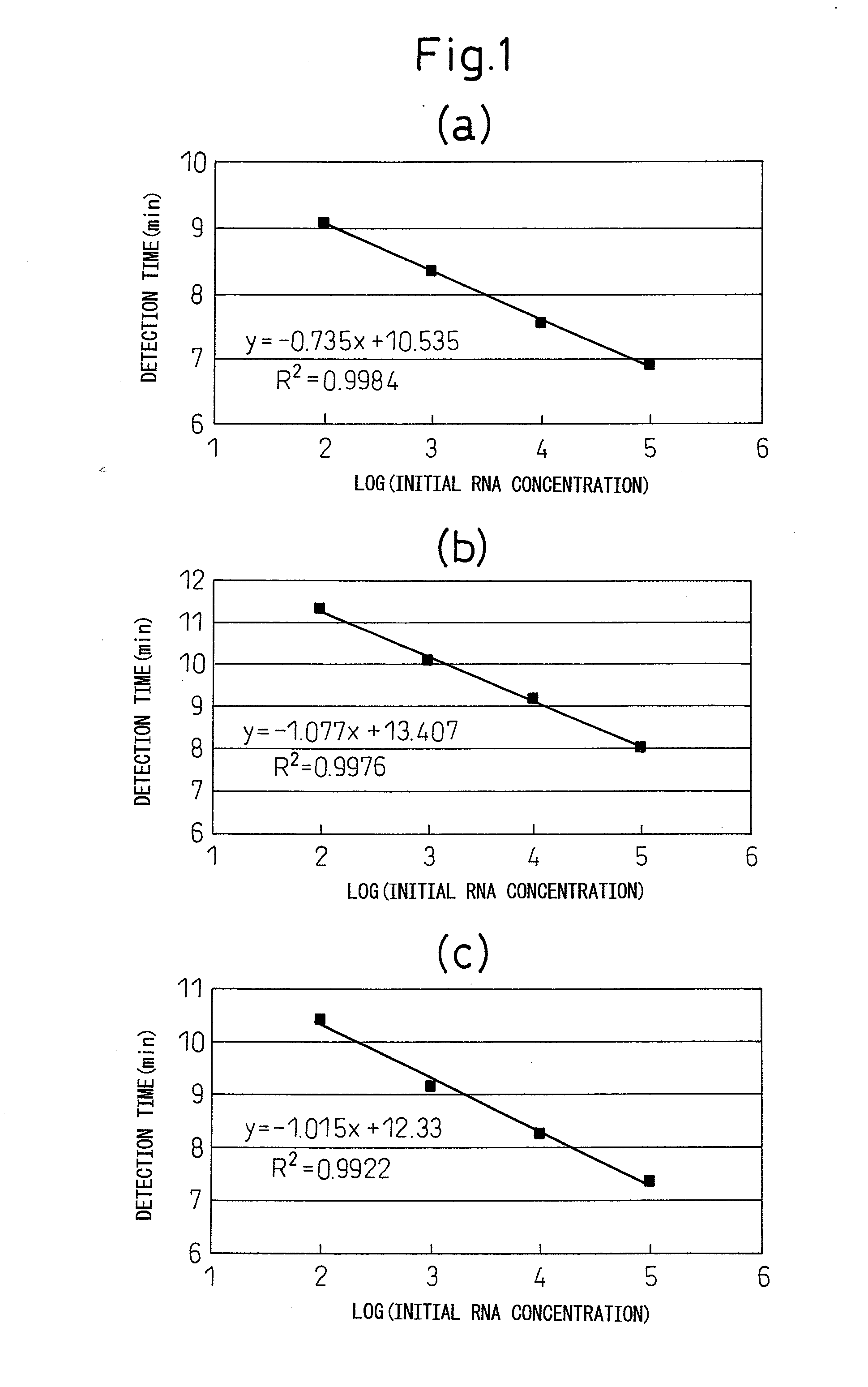
[0114]Standard RNA was measured according to the method indicated in (1) to (4) using the cleaving oligonucleotides, first primers, second primers and intercalating fluorescent dye-labeled oligonucleotide probes (to be referred to as “INAF probes”) indicated in the combinations of [1] to [28] shown in Table 1. Furthermore, in Table 1, SEQ ID NO: 19 to 22 constitute partial sequences of SEQ ID NO: 1, SEQ ID NO: 23 and 24 constitute partial sequences of SEQ ID NO: 2, SEQ ID NO: 25 and 26 constitute partial sequences of SEQ ID NO: 3, SEQ ID NO: 27 and 28 constitute partial sequences of SEQ ID NO: 4, SEQ ID NO: 29 to 32 constitute partial sequences of SEQ ID NO: 5, SEQ ID NO: 33 and 34 constitute partial sequences of SEQ ID NO: 6, SEQ ID NO: 35 and 36 constitute partial sequences of SEQ ID NO: 7, and SEQ ID NO: 37 and 38 constitute partial sequences of SEQ ID NO: 8.

TABLE 1Oligonucleotides Used (SEQ ID NO)OligonucleotideCleavingFirstSecondINAFcombinationol...


PUM

PropertyMeasurementUnit
temperatureaaaaaaaaaa
constant temperatureaaaaaaaaaa

Abstract

Disclosed is a method of amplifying and detecting cytokeratin 19 mRNA in RNA amplification process, comprising: a step for forming a double-stranded DNA containing a promoter sequence with a reverse transcriptase by use of a combination of oligonucleotides consisting of a first primer having a sequence homologous to a portion of cytokeratin 19 mRNA and a second primer having a complementary sequence, wherein the promoter sequence is added to the 5′-end of either the first primer or the second primer, forming an RNA transcription product by use of an RNA polymerase with using the double-stranded DNA as template, and forming the double-stranded DNA by use of a reverse transcriptase by continuing to use the RNA transcription product as a template of DNA synthesis, by measuring an amount of amplified RNA produced over time with an oligonucleotide probe designed so that signal properties change with the formation of a complementary double strand with the amplified RNA.

Description

TECHNICAL FIELD[0001]The present invention relates to a method of measuring cytokeratin 19 mRNA using a nucleic acid amplification method. More precisely, the present invention provides an oligonucleotide suitable for amplification and detection of mRNA at a constant temperature (40 to 60° C., and preferably 43° C.), and enables cytokeratin 19 mRNA present in a sample to be measured easily and rapidly. In addition, the present invention also provides a reagent for measuring cytokeratin 19 mRNA, and is useful for research, diagnosis and treatment in the fields of molecular biology, biochemistry and medicine and the like.BACKGROUND ART[0002]Cytokeratin is an intermediate-diameter filamentous protein that forms the cytoskeleton, and is characteristically present in epithelial tissue. More than 20 types of cytokeratin have currently been identified, and are classified based on their protein charge, with acidic cytokeratin classified as type I and neutral to basic cytokeratin classified ...

Claims


Application Information

Patent Timeline
Patent Type & Authority Applications(United States)
IPC IPC(8): C12Q1/68C07H21/04
CPCC12Q1/6853C12Q1/6865C12Q2525/143C12Q2545/114C12Q2561/113C12Q2531/143C12Q2521/107
Inventor OMOTO, DAISUKESAITO, JUICHIOONAKA, SATORUHAYASHI, TOSHINORI
Owner TOSOH CORP
Why Eureka
Try Eureka