
Method, apparatus, and computer program product for acquiring medical image data

Inactive Publication Date: 2011-09-22
KONINKLIJKE PHILIPS ELECTRONICS NV
View PDF9 Cites 33 Cited by
  • Summary
  • Abstract
  • Description
  • Claims
  • Application Information

AI Technical Summary

Benefits of technology

[0012]Embodiments of the invention overcome the aforementioned limitations by combining the advantages manual delineation of regions of interest and the advantages of using algorithms for automating the identification of regions of interest. Segmentation algorithms can be used for automating the identification of regions of interest. In other words, embodiments of the invention may mitigate the disadvantages of both:
[0035]In another aspect, embodiments of the invention provide for a computer program product comprising a set of computer executable instructions for performing the method of any of the previously described embodiments of the method. This has the advantage that the method can be performed more rapidly than a human could.

Problems solved by technology

manual delineation of regions of interest, which is tedious and prone to inconsistency

Method used


Image

  • Method, apparatus, and computer program product for acquiring medical image data
  • Method, apparatus, and computer program product for acquiring medical image data
  • Method, apparatus, and computer program product for acquiring medical image data

Examples


Embodiment Construction

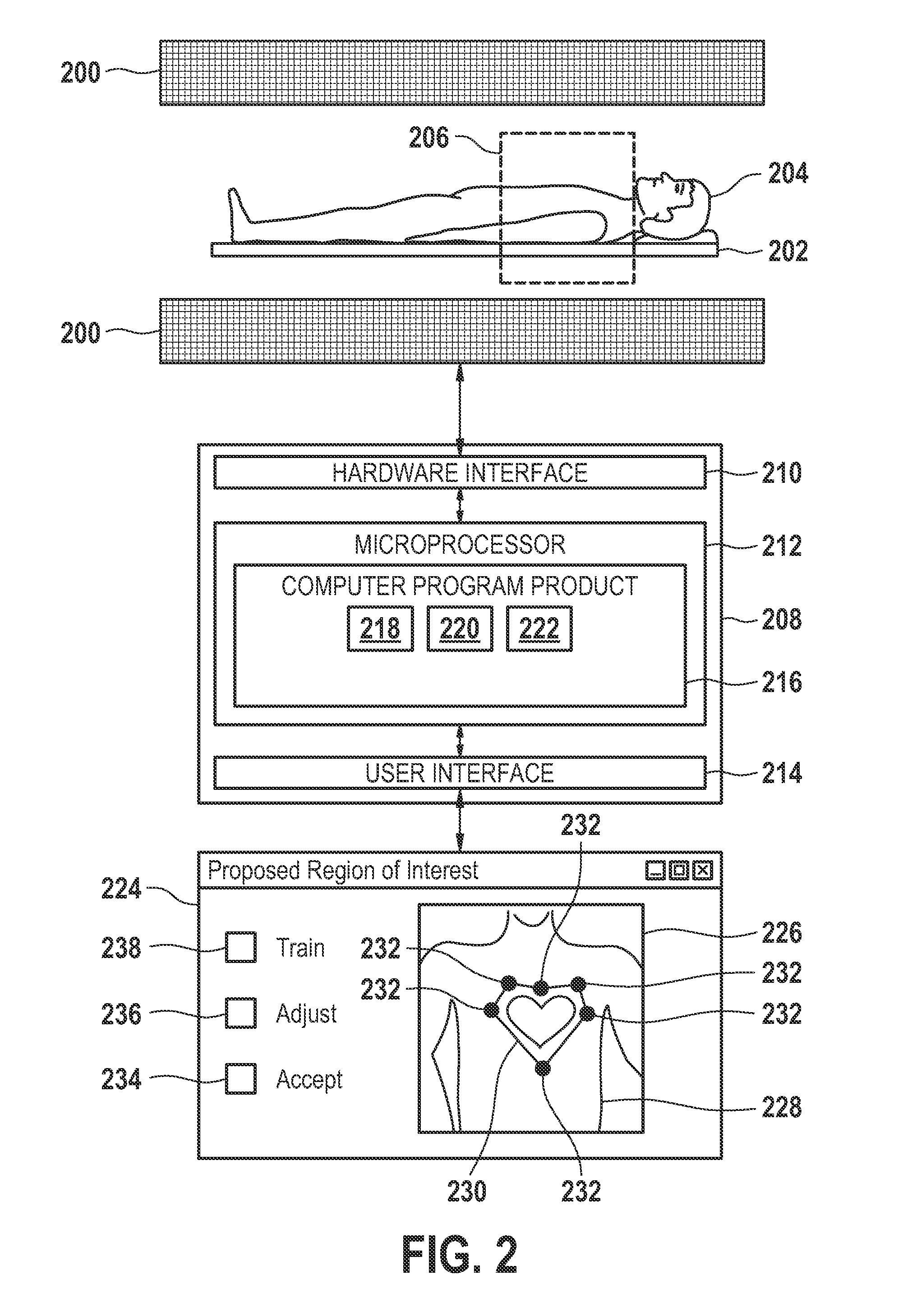
[0049]FIG. 1 shows an embodiment of a method of a acquiring medical image data with at least one region of interest with a predefined freely shaped geometry. The method comprises acquiring a first set of medical image data 100, identifying at least one anatomical landmark 102, determining the at least one region of interest with the trained pattern recognition module using the at least one anatomical landmark 104, acquiring a second set of medical image data 106, adjusting the location and / or shape of the at least one region of interest using the at least one anatomical landmark and the second set of image data 108, displaying the at least one region of interest graphically 110, receiving region of interest modifications from an operator 112, and using the modifications to perform a training step for the trained pattern recognition module 114. The first set of medical image data can be acquired using a variety of different image modalities such as magnetic resonance imaging, positro...


PUM


Abstract

A method of acquiring medical image data with at least one region of interest with a predefined freely shaped geometry comprising the following steps: acquiring a first set of medical image data, identifying at least one anatomical landmark in the first set of image data, determining the at least one region of interest with a trained pattern recognition module using the at least one anatomical landmark.

Description

FIELD OF THE INVENTION[0001]The invention relates to medical imaging, in particular it relates to the automatic identification of regions of interest within medical images.BACKGROUND OF THE INVENTION[0002]Medical imaging technologies such as Magnetic Resonance Imaging (MRI), Computed Tomography (CT), or Positron Emission Tomography (PET) are able to acquire information about the physical structure of a patient and construct two dimensional or three dimensional data sets. These data sets can then be used to construct visualizations of the data sets in a form which is useful for diagnosis by a physician.[0003]For example, a magnetic field is used by MRI scanners to align the nuclear spins of atoms as part of the procedure for producing images within the body of a patient. This magnetic field is referred to as the B0 field. During an MRI scan, Radio Frequency (RF) pulses generated by a transmitter coil cause perturbations to the local magnetic field and can be used to manipulate the or...

Claims


Application Information

Patent Timeline
IPC IPC(8): G06K9/00G06V10/25
CPCG06K9/3233G06T2207/20092G06T7/0081G06K2209/051G06T7/11G06V10/25G06V2201/031
Inventor DEN HARDER, JOHAN MICHIELVAN MUISWINKEL, ARIANNEYOUNG, STEWART
Owner KONINKLIJKE PHILIPS ELECTRONICS NV
Learn More