Monitoring skin metabolism products for evaluating burn injury

a technology of skin metabolism and products, applied in the direction of disease diagnosis, material testing goods, instruments, etc., can solve the problems of only affecting the epidermis, redness of the skin, damage to the cells of living tissue, etc., and achieve the effect of accurate evaluation of severity, rapid and cost-effectiv

Inactive Publication Date: 2011-06-23
AMIR ABRAHAM
View PDF38 Cites 4 Cited by
  • Summary
  • Abstract
  • Description
  • Claims
  • Application Information

AI Technical Summary

Benefits of technology

[0023]The present invention provides a rapid and cost-effective method and kit for accurate evaluation of the severity of a burn soon after it is sustained. The invention is based on measurement of the level of skin metabolism products in a clinical sample, particularly blood, using protein detection methods.

Problems solved by technology

Heating of the skin, however brief, can cause damage to the cells of its living tissue.
First degree burns cause redness of the skin and affect only the epidermis.
Such burns heal quickly, but the damaged skin may peel away after a day or two.
A second degree burn damages the skin more deeply, extending into the dermis and usually causes blistering.
Experience has shown that second degree superficial dermal (or partial-thickness) burns will normally heal spontaneously within two weeks with minimal scarring, whereas deep dermal second degree and third degree (or full-thickness) burns usually result in necrotic skin and in very slow wound healing.
However, it is usually almost impossible to assess the exact degree of the burn in the first 48-72 hours after the burn has been sustained.
Clinical criteria utilized to distinguish burn depth include sensitivity to pin prick, visual appearance, and viable cutaneous circulation, but these criteria are not objective and they do not constitute a practical method for evaluating burn injury.
Imaging techniques such as passive infrared thermography, laser Doppler, false-positive images, and high-frequency ultrasound are expensive and not sufficiently informative.
Intravenous administration of fluorescent drugs such as indocyanine green and fluorescein, followed by detection with a fiber-optic instrument, has been attempted with limited success.
Such techniques are hampered by the period of time required for the fluorescent drugs to reach the burn site and / or the time required for their clearance from the burn site, thus limiting the frequency of testing.
Further, intravenous administration is counter-indicated in many burn cases since burn patients frequently suffer from vasoconstriction at the early stages.
The prior art does not teach or suggest that measurement of collagen or basement membrane degradation products may be used for the evaluation of burn severity.

Method used


Image

  • Monitoring skin metabolism products for evaluating burn injury
  • Monitoring skin metabolism products for evaluating burn injury

Examples


example 1

C-telopeptide Concentration in Control and Post-Burn samples

[0169]A guinea pig model of burn injury is used. The animals are exposed to heat trauma using the following protocol: A guinea pig is shaved (electrical shaver) twenty-four hours prior the experiment. After shaving, the animal is anesthetized by 15-30 mg / kg pentobarbital i.p., placed in insulating fixed area shield, dorsum exposed, and immersed in a water bath of 80° C. for 30 sec. This produces a full-thickness burn, of about 25% of body surface area. Animal are then immediately resuscitated post-burn with 3 ml i.p. of sterile normal saline.

[0170]Blood samples are obtained from the anesthetized animals before the exposure to heat (time 0, control) and at 15, 30, 60 and 120 min post-burn. At least four samples for each time point are taken. Concentration of C-telopeptide is assessed using radio-immunoassay and anti-C-telopeptide antibody (Orion Diagnostica (Espoo, Finland). This model shows that the CTP concentration in a b...

example 2

Use of the Concentration of CTP-I in Blood Samples for Assessment of Burn Injury Severity

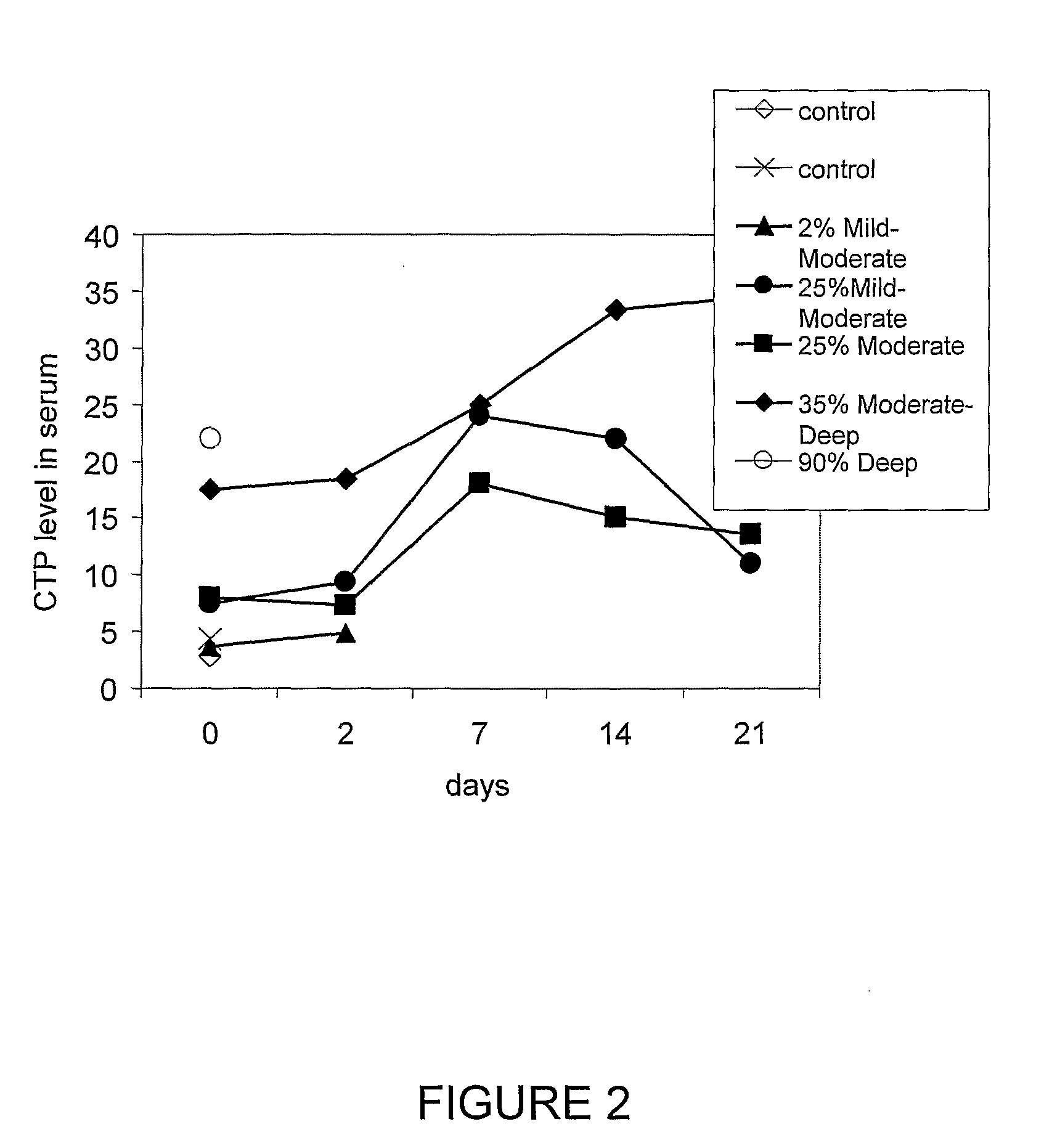
[0171]This preliminary study included 5 burn patients, each of which had a differing degree of burn severity and percentage of body area affected as follows: patient 1: mild, 2% body area; patient 2: mild-moderate, 25% body area; patient 3: moderate, 25% body area; patient 4: moderate-deep, 35% body area , and patient 5: deep, 90% body area). Four healthy individuals served as a control group. Blood samples were taken from patients on arrival to the hospital emergency room, or within 3 hours of arrival. CTP-I serum levels were measured using ELISA and anti-C-telopeptide antibody (UniQ). As shown in FIG. 1, the CTP-I levels of patients 1-5 were respectively 4.8 (empty bars); 6.95 (horizontally striped bars); 8.02 (diagonally striped bars); 18.48 (stippled bars); and 23.49 (vertically striped bars) ng per L, respectively. The CTP-I levels of the control group (black bars) were: 2.85, 4.42, 3.74, a...

example 3

Measuring Dynamics of CTP-I Serum Levels for Monitoring of Burn Patients

[0173]The patients described in Example 2 were continually monitored at periodic intervals (corresponding to 2, 7, 14 and 21 days following burn injury) for CTP-I levels in blood. The results, shown in FIG. 2, indicate that in some patients, the level of CTP-I declined as the burn injury healing process proceeded.

[0174]The foregoing description of the specific embodiments will so fully reveal the general nature of the invention that others can, by applying current knowledge, readily modify and / or adapt for various applications such specific embodiments without undue experimentation and without departing from the generic concept, and, therefore, such adaptations and modifications should and are intended to be comprehended within the meaning and range of equivalents of the disclosed embodiments. It is to be understood that the phraseology or terminology employed herein is for the purpose of description and not of ...


PUM

PropertyMeasurementUnit
Timeaaaaaaaaaa
Timeaaaaaaaaaa
Timeaaaaaaaaaa

Abstract

The present invention relates to methods and kits for evaluating the severity of a burn injury, which are based on the detection in a clinical fluid sample of skin metabolism products, such as collagen peptides which are released upon collagen degradation or synthesis.

Description

FIELD OF THE INVENTION[0001]The present invention relates to methods and kits for evaluating the severity of a burn injury, which are based on the detection in a clinical fluid sample of skin metabolism products, such as collagen peptides which are released upon collagen degradation or synthesis.BACKGROUND OF THE INVENTION[0002]Skin is composed of a thin outer layer, the epidermis, and a thicker inner layer,the dermis, which are separated by the basement membrane. Subcutaneous tissue is located beneath the dermis, which contains mostly fat, and beneath that fasciae and muscles are found. The epidermis forms a tough, waterproof protective coating that contains dead cells on its outside surface and living cells on its inner side. The living cells replace the dead cells that are continually worn away. The dermis is a living tissue containing blood vessels, nerves, sweat glands and hair follicles.[0003]Collagen is the main protein of connective tissue in animals, and its fibrillar form ...

Claims


Application Information

Patent Timeline
IPC IPC(8): G01N33/53C07K16/18
CPCG01N33/6881G01N2800/40G01N2333/78
Inventor AMIR, ABRAHAM
Owner AMIR ABRAHAM
Why Eureka
Try Eureka