Medical image analysis system using n-way belief propagation for anatomical images subject to deformation and related methods

a medical image and anatomical image technology, applied in image analysis, image enhancement, instruments, etc., can solve the problems of comparatively less computing resources, and achieve the effect of accurately determining the deformation between

Inactive Publication Date: 2011-04-07
HARRIS CORP
View PDF43 Cites 14 Cited by
  • Summary
  • Abstract
  • Description
  • Claims
  • Application Information

AI Technical Summary

Benefits of technology

[0013]In view of the foregoing background, it is therefore an object of the present invention to provide a medical image anal

Problems solved by technology

Twenty-six-way belief propagation, for example, may provide the most accurate results, at the expense of computing resources,

Method used


Image

  • Medical image analysis system using n-way belief propagation for anatomical images subject to deformation and related methods
  • Medical image analysis system using n-way belief propagation for anatomical images subject to deformation and related methods
  • Medical image analysis system using n-way belief propagation for anatomical images subject to deformation and related methods

Examples


Embodiment Construction

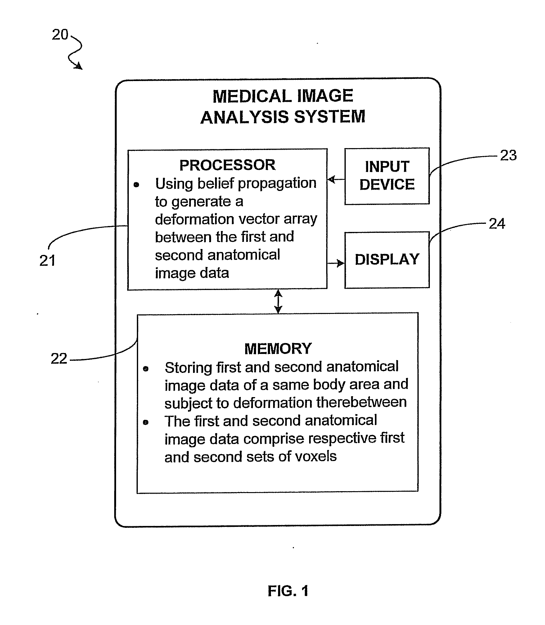
[0044]The present invention will now be described more fully hereinafter with reference to the accompanying drawings, in which preferred embodiments of the invention are shown. This invention may, however, be embodied in many different forms and should not be construed as limited to the embodiments set forth herein. Rather, these embodiments are provided so that this disclosure will be thorough and complete, and will fully convey the scope of the invention to those skilled in the art. Like numbers refer to like elements throughout.

[0045]Referring initially to FIG. 1, a medical image analysis system 20 is now described. The medical image analysis system 20 includes a processor 21. A memory 22, an input device 23, and a display 24 are coupled to the processor. The processor 21, memory 22, and display 24 may be any suitable devices known to those of skill in the art. The input device 23 may be a keyboard, mouse, or trackball, for example.

[0046]The memory 22 stores first and second anat...


PUM


Abstract

A medical image analysis system is for first and second anatomical image data of a same body area and subject to deformation, with the first and second anatomical image data including respective first and second sets of voxels. The medical image analysis system may include a processor cooperating with a memory and configured to generate a plurality of cost arrays, with each cost array based upon probabilities of a subset of voxels of the second anatomical image data matching voxels of the first anatomical image data. The processor may also solve each cost array using three-dimensional, N-way belief propagation to thereby generate a deformation vector array between the first and second anatomical image data, where N is an integer greater than or equal to six.

Description

FIELD OF THE INVENTION[0001]The present invention relates to the field of image analysis, and, more particularly, to a medical image analysis system and related methods.BACKGROUND OF THE INVENTION[0002]Medical imaging technologies provide medical practitioners detailed information useful for differentiating, diagnosing, or monitoring the condition, structure, and / or extent of various types of tissue within a patient's body. In general, medical imaging technologies detect and record manners in which tissues respond in the presence of applied signals and / or injected or ingested substances, and generate visual representations indicative of such responses.[0003]A variety of medical imaging technologies exist, including Computed Tomography (CT), Positron Emission Tomography (PET), Single Photon Emission Computed Tomography (SPECT), and Magnetic Resonance Imaging (MRI). Various medical imaging technologies are suitable for differentiating between specific types of tissues. A contrast agen...

Claims


Application Information

Patent Timeline
IPC IPC(8): G06K9/00G09G5/00
CPCG06T7/0034G06T2207/30004G06T2207/20076G06T2207/10072G06T7/35
Inventor BELL, DAVID M.BURRELL, LAUREN S.JACKSON, JEREMY D.CULP, TIMOTHY R.
Owner HARRIS CORP
Why Eureka
Try Eureka