
Method for detecting cancer cell caused by hpv, method for determining whether or not tissue is at stage of high-grade dysplasia or more severe stage, and primer set and kit used therefor

a cancer cell and hpv technology, applied in the field of detecting can solve the problems of difficult to detect a cancer cell caused by hpv in the uterine, high probability of invasive cancer in the lesion, and high accuracy, and achieve high accuracy, high accuracy, and high accuracy

Inactive Publication Date: 2011-01-27
SYSMEX CORP
View PDF2 Cites 6 Cited by
  • Summary
  • Abstract
  • Description
  • Claims
  • Application Information

AI Technical Summary

Benefits of technology

[0011]An object of the present invention is to provide a method for detecting a cancer cell caused by HPV easily with high accuracy. Another object of the present invention is to provide a method for determining whether or not a tissue is at a stage of high-grade dysplasia or a more severe stage easily with high accuracy. Still another object of the present invention is to provide a primer set capable of detecting a cancer cell caused by HPV and determining whether or not a tissue is at a stage of high-grade dysplasia or a more severe stage easily with high accuracy. Yet still another object of the present invention is to provide a kit capable of diagnosing cancer caused by HPV easily with high accuracy. Yet still another object of the present invention is to provide a kit capable of diagnosing the stage of dysplasia.

Problems solved by technology

However, in 5 to 10% of HPV infections, there are cases where HPV is not disappeared and the infection turns into a persistent infection causing uterine cervical cancer.
However, if a precursor lesion of high-grade dysplasia is left untreated, the lesion is highly likely to progress to invasive cancer.
However, there are cases where unmethylated E6 region and unmethylated LCR are detected in a cell other than a uterine cervical cancer cell.
Thus, it is difficult to detect a cancer cell caused by HPV in uterine cervical cells of a patient with high accuracy only by confirming the methylation state of E6 region and LCR.
However, confirming methylation of DNA by sequencing is time-consuming and involves cumbersome operations.
Thus, it is difficult to detect a cancer cell caused by HPV in a sample collected from a subject in a simple manner.

Method used


Image

  • Method for detecting cancer cell caused by hpv, method for determining whether or not tissue is at stage of high-grade dysplasia or more severe stage, and primer set and kit used therefor
  • Method for detecting cancer cell caused by hpv, method for determining whether or not tissue is at stage of high-grade dysplasia or more severe stage, and primer set and kit used therefor
  • Method for detecting cancer cell caused by hpv, method for determining whether or not tissue is at stage of high-grade dysplasia or more severe stage, and primer set and kit used therefor

Examples


experimental example 1

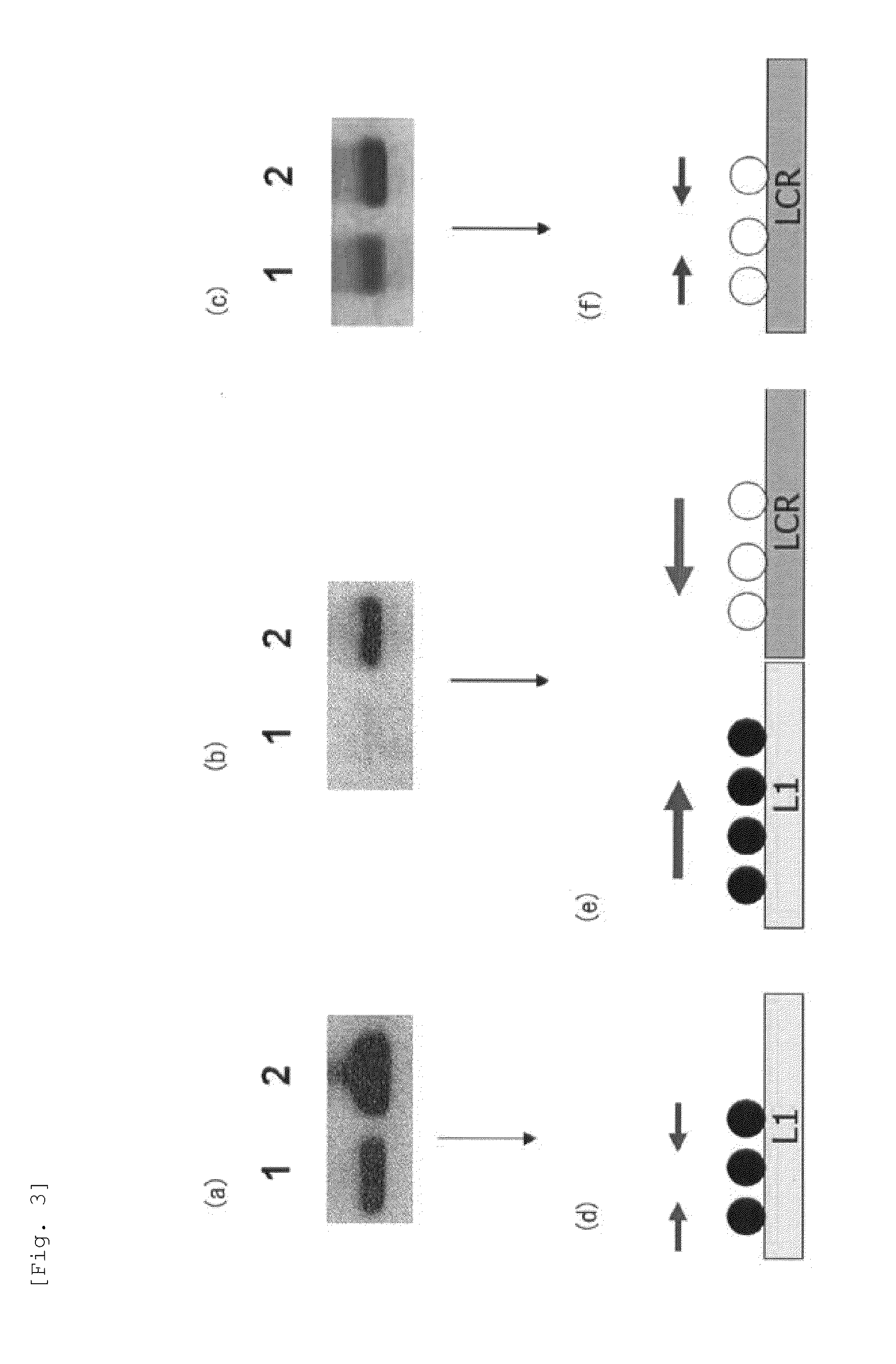
[0119]To 1 μg of genomic DNA of a SiHa cell, which is a cell line derived from uterine cervical cancer having HPV16 genome integrated into its chromosome, 300 μL of 0.3 M sodium hydroxide solution was added, followed by incubation at 37° C. for 10 minutes. Subsequently, 300 μL of 10 M bisulfite salt solution (10 M sodium bisulfite solution) was added to the resulting product, followed by incubation at 80° C. for 40 minutes to carry out bisulfite salt treatment of the genomic DNA. DNA contained in the resulting solution after the bisulfite salt treatment was purified by a DNA purification kit (manufactured by QIAGEN under the trade name of Qiaquick PCR purification kit). To DNA thus purified, sodium hydroxide was added so as to have a final concentration of 0.3 M, followed by incubation at room temperature for 5 minutes. Thereafter, the product thus obtained was purified by a spin column for nucleic acid purification (manufactured by GE Healthcare under the trade name of MicroSpin S-...

experimental example 2

[0140]By similar operations to Experimental Example 1, using C4-1 cell, which is an uterine cervical cancer-derived cell line having the HPV18 genome integrated into its chromosome, a methylated CpG site and an unmethylated CpG site in the HPV18 genome were analyzed. Specifically, except that genomic DNA of the C4-1 cell was used in place of genomic DNA of the SiHa cell and PCR reaction was carried out using the primer set and PCR thermal profile (6) shown in Table 4 in place of the primer set and PCR thermal profiles shown in. Table 2, similar operations to Experimental Example 1 were carried out to determine the nucleotide sequence of an amplification product.

TABLE 4SEQThermalIDTmprofilePrimerNucleotide SequenceNo.(° C.)of PCRMsp10F(18)5'-TAAAATATGTTTTGTGGTTTTGTG-3'1759.25(6)Msp10R(18)5'-ATAATTATACAAACCAAATATACAATT-3'1854.36Msp7F(18)5'-AGATTTAGATTAATATTTTTTTGGA-3'1955.25(6)Msp7R(18)5'-AAATTAAAATTTACAATAATACCAAC-3'2054.83F(18)5'-GTTATTTGATTTAAATAAATTTGGTTTATTTGA-3'2162.74(6)3R(18)5...

preparation example 1

[0160]A diagnostic kit for cancer caused by HPV or a diagnostic kit for a stage of dysplasia was prepared. One example thereof is shown below. The kit includes a nuclease-free container containing an aqueous solution of each primer of the below-described primer sets and a nuclease-free container containing a bisulfite salt solution (10M aqueous solution of sodium bisulfite), which is an unmethylated cytosine-conversion agent.

[0161]The content of the diagnostic kit for cancer caused by HPV or the diagnostic kit for a stage of dysplasia:

Container 1

[0162]An aqueous solution of forward primer (an aqueous solution obtained by dissolving primer 16L1 / LCR-F consisting of the nucleotide sequence shown in SEQ ID NO: 10 in nuclease-free water)

Container 2

[0163]An aqueous solution of reverse primer (an aqueous solution obtained by dissolving the primer 16L1 / LCR-F consisting of the nucleotide sequence shown in SEQ ID NO: 10 in nuclease-free water)

Container 3

[0164]10M aqueous solution of sodium bi...


PUM

PropertyMeasurementUnit
Temperatureaaaaaaaaaa

Abstract

Provided are a primer set, a method and a kit therefor, which can easily perform with high accuracy the detection of a cancer cell caused by HPV and the determination of whether or not a tissue is a tissue with high-grade dysplasia or in a more severe phase. As a primer set, used is a primer set consisting of a first primer which hybridizes with a nucleic acid consisting of a nucleotide sequence in which cytosine present in a site other than a CpG site is converted into another base in a nucleotide sequence having a CpG site in L1 region or L2 region of HPV and a second primer which hybridizes with a nucleic acid consisting of a nucleotide sequence in which cytosine is converted into another base in a nucleotide sequence having a CpG site in LCR or E6 region of HPV.

Description

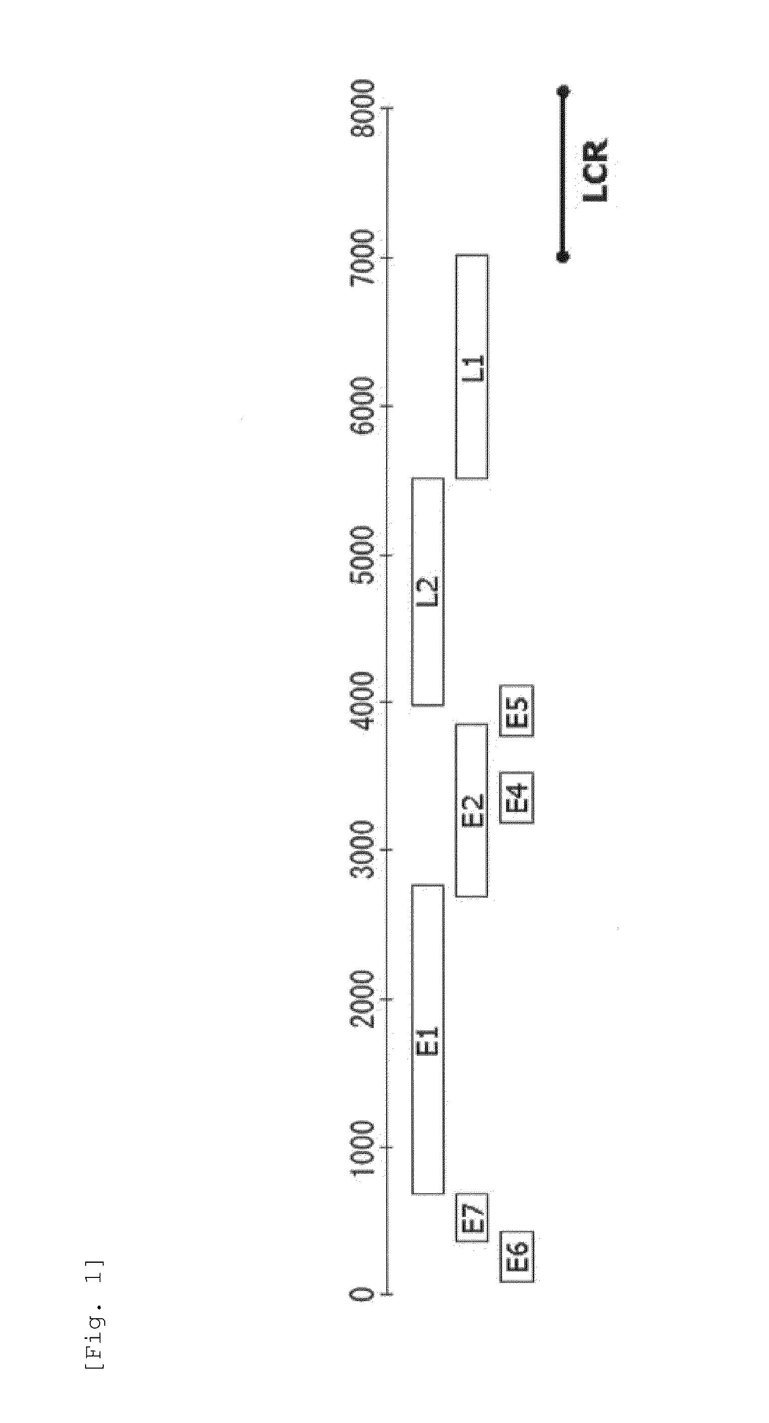
TECHNICAL FIELD[0001]The present invention relates to a method for detecting a cancer cell caused by HPV, a method for determining whether or not a tissue is at a stage of high-grade dysplasia or a more severe stage, and a primer set and a kit used therefor.BACKGROUND ART [0002]A human papillomavirus (hereinafter referred to as “HPV”) is a virus having circular double-stranded DNA as genome, which induces proliferative lesions. HPV is classified into 100 or more kinds of subtypes. Also, it is known that the subtypes of HPV share common regions, which are nonstructural protein-coding E1 region, E2 region, E4 region, E5 region, E6 region and E7 region, capsid protein-coding L1 region and L2 region, and LCR.[0003]HPV DNA has been detected in lesion sites of uterine cervical cancer and uterine cervical dysplasia, and tissues of oral cancer and pharyngeal cancer. Thus, HPV infection is regarded as one of the risk factors for uterine cervical cancer, oral cancer, and pharyngeal cancer. In...

Claims


Application Information

Patent Timeline
IPC IPC(8): C12Q1/68
CPCC12Q1/6886C12Q2600/154C12Q2600/112C12Q1/708
Inventor OKA, NORIKOKAJITA, MASAHIRO
Owner SYSMEX CORP
Learn More