
Shaped implants for tissue repair

a tissue repair and implant technology, applied in the field of tissue repair implants, can solve the problems of reduced np height, leg pain, loss of muscle control, etc., and achieve the effect of reducing the height of the np, reducing the size of the np, and reducing the degeneration or displacement of the spinal dis

Inactive Publication Date: 2011-01-06
SEMLER ERIC J +2
View PDF0 Cites 6 Cited by
  • Summary
  • Abstract
  • Description
  • Claims
  • Application Information

AI Technical Summary

Benefits of technology

[0016]Broadly speaking, the present invention involves an implant for the repair of tissue such as annulus fibrosus or meniscus. The implant has a plug section which is made of cancellous bone and is insertable in the defect. The i

Problems solved by technology

The spinal disc may degenerate or be displaced or damaged due to trauma or a disease process.
The mass of a herniated or “slipped” nucleus can compress a spinal nerve, resulting in leg pain, loss of muscle control, or even paralysis.
Subsequently, the height of the NP decreases, leading to inadequate tension on the AF.
Over time, these events may cause the AF to buckle in areas where the laminated plies are loosely bonded.
As these overlapping laminated plies of the AF begin to buckle and separate, either circumferential or radial annular tears may occur, which can result in renewed impingement by the AF on nerve structures posterior to the disc.
These events may lead to instability of the disc with increased loading on the AF.
The partially enervated AF may become painful under these conditions and cause persistent and disabling lower back pain.
Furthermore, loss of disc height resulting from loss of NP integrity may increase loading on the facet joints.
Adjacent, ancillary spinal facet joints will be forced into an overriding position, which can result in deterioration of facet cartilage and, ultimately, osteoarthritis and additional back pain.
This leads to foraminal stenosis, pinching of the traversing nerve root, and recurring radicular pain.
The most common resulting symptoms of disc degeneration are pain radiating along a compressed nerve and low back pain, both of which can be crippling for the patient.
Whenever the NP tissue is herniated or removed by surgery, the disc space will narrow and may lose much of its normal stability.
While this treatment alleviates the pain, all disc motion is lost in the fused segment.
Surgical interventions, whether discectomy or fusion of adjacent vertebrae, generally lower the functionality of the spine in some way.
Since mechanical discs are large and require a significantly invasive procedure to implant, there are substantial risks during surgery and in cases where revision is necessary.
Many prosthetic NP devices currently available do not account for this generally accepted implantation technique.
The meniscus may be injured or torn as a result of traumatic injuries such as a fall or athletic overexertion.
In addition, the meniscus begins to deteriorate with age, often developing degenerative tears.
Typically, when the meniscus is damaged, the torn piece begins to move in an abnormal fashion inside the joint.
Because the space between the bones of the joint is very small, as the abnormally mobile meniscal tissue moves, it may become caught between the bones of the joint (i.e., the femur and tibia).
When this happens, the knee becomes painful, swollen, and difficult to move.
A damaged meniscus is unable to undergo the normal healing process that occurs in other parts of the body due to the fact that, as mentioned above, the majority of the meniscus has no blood supply.
Such tears generally result in severe joint pain and locking in the short term, as well as loss of meniscal function leading to arthritis in the long term.
Currently available treatments for meniscal injuries provide little opportunity for meniscal repair or regeneration.
However, the percentage of injuries which meet the criteria to be repaired (i.e., vascularity, type of tear, stability and integrity of the meniscus, stability of the knee, and patient factors such as age and activity) in such a manner is very low (i.e., only 15% or less).
Often the meniscal tear is in an avascular region of the meniscus, and thus, will not heal even if repaired.
Moreover, some meniscal tears are frayed and cannot be sutured together.
Once the tissue is removed, however, no further treatment is conducted, and the patient is left with an abnormal meniscus.
While most patients respond well to this treatment in the short term, they often develop degenerative joint disease several years post-operatively, with the amount of tissue removed potentially playing a part in the extent and speed of degeneration.

Method used


Image

  • Shaped implants for tissue repair
  • Shaped implants for tissue repair
  • Shaped implants for tissue repair

Examples


example 1

Preparation of an Implant

[0097]Two sets of donor ilia (two ilia each from a 27 year old male and a 51 year old male with a respective yield of 12 implant prototypes and 10 implant prototypes) were selected for the preparation of three dimensional bandage-shaped implant prototypes to be utilized for mechanical testing in the context of annulus fibrosus repair.

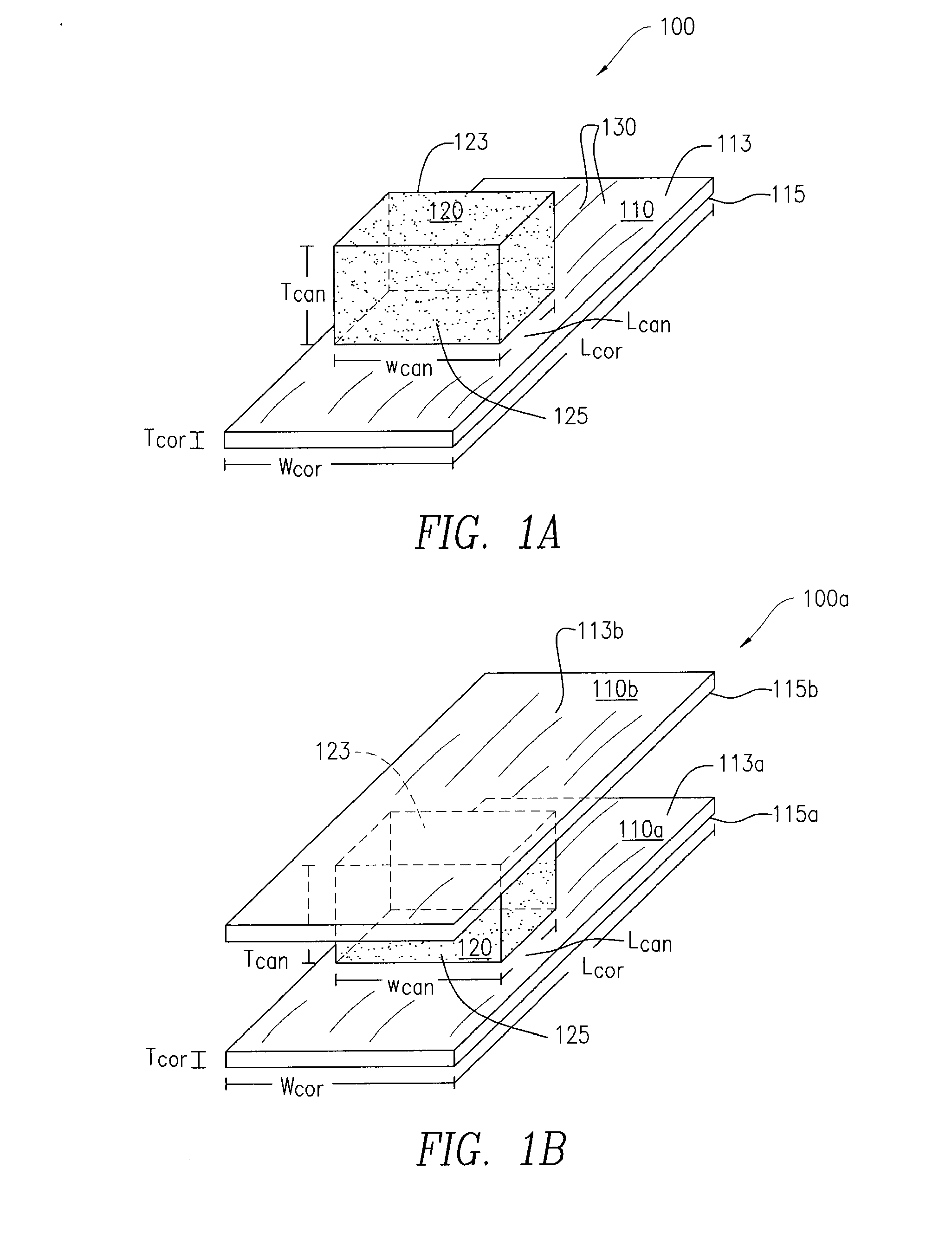
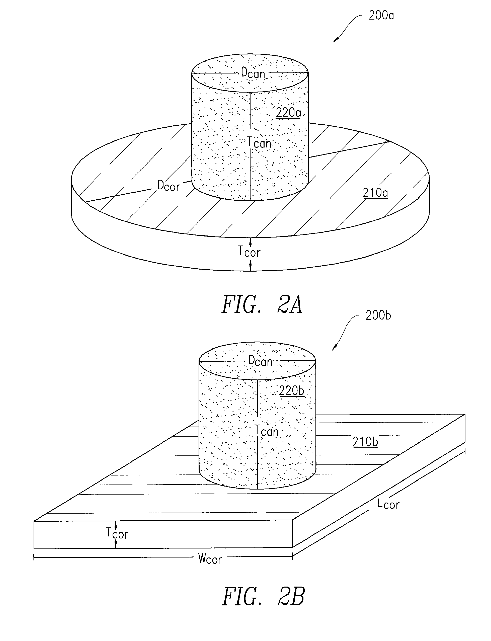
[0098]The thicker regions of donor ilia were cut into 1 cm×2 cm cross-sections (size specification shown below) using a bandsaw.

LCORTWCORTHCORTDCANCHCANCVOFFSETHOFFSET(mm)(mm)(mm)(mm)(mm)(mm)(mm)2010108561

[0099]Each of the samples contained a relatively thick cancellous layer (>5 mm) sandwiched between two thinner cortical layers. The tissue was cut so that the collagen fibers were oriented either lengthwise or widthwise and the orientation of the collagen fiber was noted. The cut tissue samples were subsequently processed and demineralized.

[0100]Next, using a scalpel, one of the thin cortical layers from each sample was strippe...


PUM


Abstract

Shaped constructs for repair of a defect in a body part or tissue of a subject are discussed herein. More specifically, implants suitable for delivery to a subject, such as a subject having a defect in a body part or body tissue, are discussed. Even more specifically, implants for intervertebral disc repair comprising corticocancellous bone, which can extend into the nucleus pulposus of an intervertebral disc and can be integrally attached to the annulus fibrosus of the disc to keep the implant in position, are described. Also, implants for meniscal repair comprising corticocancellous bone, which can extend from the outer edge of the meniscus to the inner region of the meniscus and can be integrally attached to the meniscal rim to keep the implant in position, are described. Implants for the repair of defects in bone, cartilage, and fibrocartilage are further described. Further described are methods for making such implants and for delivering the implants to a defect in a body part or body tissue of a subject.

Description

CROSS-REFERENCE TO RELATED APPLICATION[0001]The present application claims priority to U.S. Provisional Patent Application Ser. No. 61 / 186,166 filed Jun. 11, 2009, the entire disclosure of which is incorporated by reference herein in its entirety.FIELD OF THE INVENTION[0002]Shaped constructs for repair of a defect in a body part or tissue of a subject are discussed herein. More specifically, implants suitable for delivery to a subject, such as a subject having a defect in a body part or body tissue, are discussed. Even more specifically, implants for intervertebral disc repair comprising corticocancellous bone, which can extend into the nucleus pulposus of an intervertebral disc and can be integrally attached to the annulus fibrosus of the disc to keep the implant in position, are described. Also, implants for meniscal repair comprising corticocancellous bone, which can extend from the outer edge of the meniscus to the inner region of the meniscus and can be integrally attached to t...

Claims


Application Information

Patent Timeline
IPC IPC(8): A61F2/44
CPCA61F2/28A61F2310/00359A61F2/442A61F2002/2817A61F2002/30009A61F2002/30057A61F2002/30059A61F2002/30075A61F2002/30092A61F2002/30387A61F2002/30448A61F2002/30492A61F2002/30576A61F2002/30677A61F2002/4435A61F2002/4445A61F2210/0014A61F2210/0061A61F2220/0025A61F2220/005A61F2250/0028A61F2/3872
Inventor SEMLER, ERIC J.YANNARIELLO-BROWN, JUDITH I.JACOBS, MORRIS L.
Owner SEMLER ERIC J
Learn More