
Targeted Detection of Dysplasia In Barrett's Esophagus With A Novel Fluorescence-Labeled Polypeptide

a polypeptide and dysplasia technology, applied in the field of use in detecting dysplasia in barrett's esophagus, can solve the problems of ineffective detection of flat dysplasia, low detection yield, and significant limitations of approach, and achieve the effect of increasing the detectable binding of the polypeptide sequen

Inactive Publication Date: 2010-12-09
RGT UNIV OF MICHIGAN
View PDF6 Cites 7 Cited by
  • Summary
  • Abstract
  • Description
  • Claims
  • Application Information

AI Technical Summary

Benefits of technology

[0008]Described herein are compositions and methods for detecting dysplasia in Barrett's esophagus. Accordingly, in one embodiment a composition is provided consisting of or consisting essentially of a polypeptide sequence as set out in SEQ ID NO: 1 (SNFYMPL), a linker sequence and a detectable marker; the detectable marker connected to the polypeptide through the linker, the linker having a net neutral charge, and wherein the presence of the linker results in an increase in detectable binding of the polypeptide sequence to Barrett's esophageal tissue compared to the detectable binding of the polypeptide sequence to Barrett's esophageal tissue in the absence of the linker.

Problems solved by technology

However this approach has significant limitations.
This technique relies on the reflection of white light from the tissue surface to reveal structural (anatomic) changes, and is not effective for the detection of flat dysplasia, such as that which occurs in the setting of Barrett's esophagus.
Random fourquadrant biopsy is accepted as the standard of practice for screening, however, this approach is limited by a low yield for detection because dysplastic changes often occur in a spatially heterogeneous fashion [Levine et al., Gastroenterology 1993; 105:40-50].
An incorrect evaluation of biopsy specimens may result in either an unnecessary esophagectomy or in progression to frank carcinoma.
However, this method is not effective for the detection of flat dysplasia, such as that which occurs in the setting of Barrett's esophagus.
The histological evaluation of excised esophageal mucosa for the presence of dysplasia is performed in a qualitative, subjective manner, and even among expert pathologists, substantial intra- and inter-observer variability in grading dysplasia occurs, limiting patient and physician confidence in the interpretation.
Therefore, because current methods of surveillance with white light endoscopy are non-specific and are limited by sampling error, improved imaging strategies are needed to localize pre-malignant mucosa for early detection and prevention of esophagus adenocarcinoma.

Method used


Image

  • Targeted Detection of Dysplasia In Barrett's Esophagus With A Novel Fluorescence-Labeled Polypeptide
  • Targeted Detection of Dysplasia In Barrett's Esophagus With A Novel Fluorescence-Labeled Polypeptide
  • Targeted Detection of Dysplasia In Barrett's Esophagus With A Novel Fluorescence-Labeled Polypeptide

Examples


example 1

[0057]Pepide Selection. Peptide selection was performed using techniques of phage display (Ph.D.-7, New England Biolabs, Beverly, Mass.). The esophageal adenocarcinoma cell line OE33 was maintained in RPMI-1640 media supplemented with 10% FBS. The Barrett's esophagus (intestinal metaplasia) cell lines KR-42421 (Q-hTERT, non-dysplastic) was maintained in keratinocyte-serum free medium supplemented with bovine pituitary extract (BPE) and human recombinant epidermal growth factor (rEGF) (Invitrogen, Carlsbad, Calif.). All cell lines were incubated at 37° C. in 5% CO2.

[0058]Biopanning was carried out by using a subtractive whole-cell approach. Q-hTERT cells in log-phase growth were detached with cell dissociation buffer (Invitrogen, Carlsbad, Calif.) and blocked with blocking buffer (PBS with 1% bovine serum albumin) for 45 minutes on ice. Ten μl of Ph.D.-7 random phage library (1.5×10″ plaque-forming unit) was suspended in 5 ml PBS and biopanned with 1.0×10⁷ Q-hTERT cells for 30 min at...

example 2

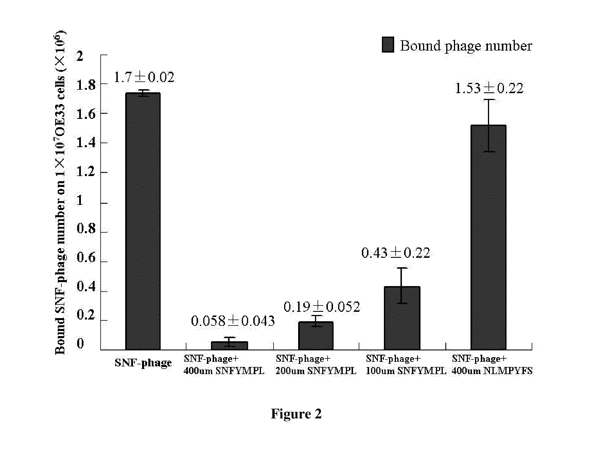
[0062]Phage Binding Assay. OE33 and Q-hTERT cells were detached and blocked as described above. The candidate SNFYMPL-phage or control phage (randomly insert) were incubated with OE33 cells (1×10⁷) or Q-hTERT cells (1×10⁷) for 30 min with gentle agitation at room temperature. After washing ten times with PBS / 0.1% (v / v) Tween-20 and one time with 0.2 M glycine, pH 2.2 / 0.1% BSA for two minutes, the bound phages were recovered and titered. Every sample was carried out in triplicate. The amount of bound phages in every sample was calculated using student's t test.

[0063]Cell ELISA for Phage Binding Assay. OE33 cells and Q-hTERT cells were grown to 100% confluence in a 96-well plate and incubated sequentially with 2×10⁷ pfu SNFYMPL-phage or control phage for 10 minutes in triplicate at room temperature, washed with PBS containing 0.1% Tween-20 six times, incubated with HRP-labeled anti-M13 antibody (Fitzgerald, Concord, Mass.), developed with TMB (Invitrogen, Carlsbad, Calif.), and absorb...

example 3

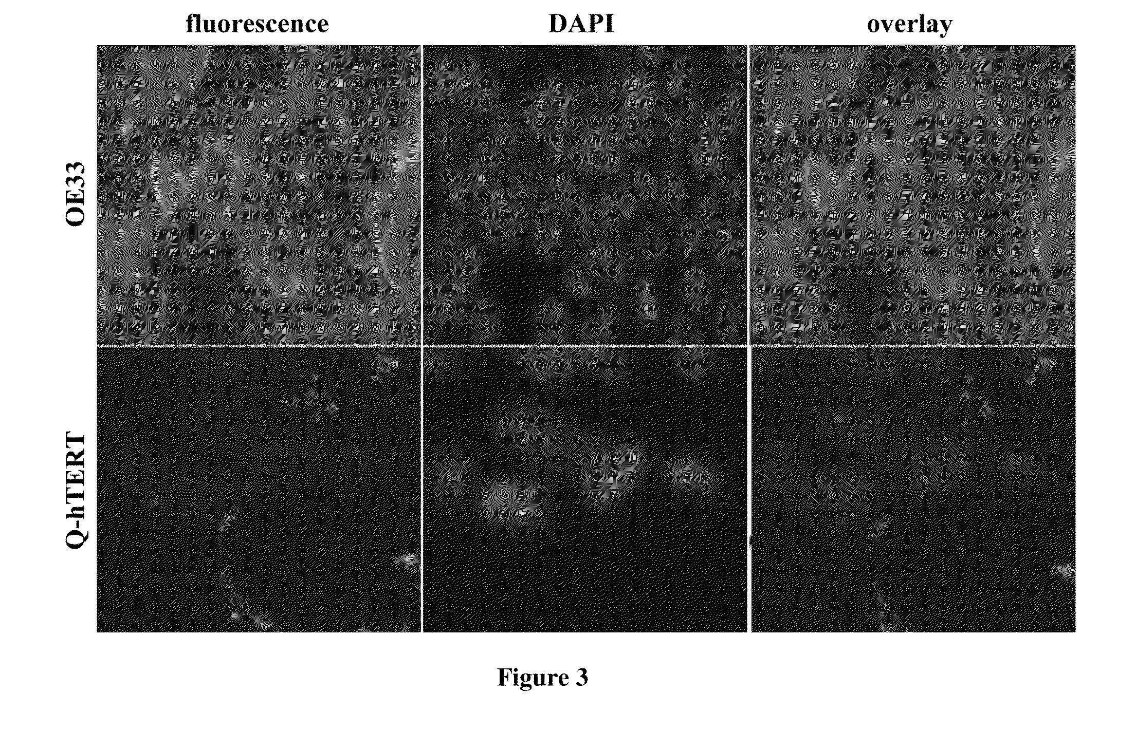
[0069]Fluoresence images for peptide binding on culture cells. The OE33 and QhTERT cells were grown in chamber slides to 80% confluence. Blocking of non-specific binding to these cells was performed by adding 200 μl of 1% BSA diluted in PBS for 30 min. The cells were then incubated with 100 μmol of the candidate FITC-labeled peptide in serum free media for 10 minutes at room temperature. The cells were washed 3 times using 200 μL PBS / 0.5% TWEEN 20 in room temperature. The cells were fixed in ice cold 4% praformaldehyde for 5 minutes. The cells were then stained with Vectashield mounting medium containing DAPI. Fluorescence images were collected with a confocal microscope (Nikon 1000) at 200×. The fluorescence intensity from the cells in 3 images was averaged to assess for peptide binding using NIH Image J software.

[0070]Under the fluoresence microscope, SNFYMPL-GGGSK-FITC was bound to the plasma membrane of >90% OE33 cells but not on Q-hTERT (normal Barrett's) cells (FIG. 3). The in...


PUM

PropertyMeasurementUnit
pHaaaaaaaaaa
pHaaaaaaaaaa
pHaaaaaaaaaa

Abstract

The present invention is directed to compositions and methods for use in detecting dysplasia in Barrett's esophagus.

Description

CROSS-REFERENCE TO RELATED APPLICATIONS[0001]This application claims the priority benefit under 35 U.S.C. §119(e) of U.S. Provisional Application No. 61 / 170,614, filed on Apr. 18, 2009, and U.S. Provisional Application No. 61 / 302,388, filed on Feb. 8, 2010, the disclosures of which are incorporated herein by reference in their entirety.STATEMENT OF GOVERNMENT INTEREST[0002]This invention was made with government support under Grant Numbers R03 CA096752, CA136429, and CA093990, awarded by the National Institutes of Health (NIH). The government has certain rights in the invention.FIELD OF THE INVENTION[0003]The present invention is directed to compositions and methods for use in detecting dysplasia in Barrett's esophagus.BACKGROUND OF THE INVENTION[0004]Adenocarcinoma of the esophagus is growing at a rate faster than any other cancer in industrialized countries. This disease is a significant cause of morbidity and mortality, and Barrett's esophagus is a known precursor condition that ...

Claims


Application Information

Patent Timeline
Patent Type & Authority Applications(United States)
IPC IPC(8): A61K49/00C07K7/06C07K7/08
CPCA61B5/1459A61K49/0043A61K49/0056G01N2800/52G01N33/57407G01N2800/06C07K7/06
Inventor WANG, THOMAS D.LI, MENG
Owner RGT UNIV OF MICHIGAN
Learn More