Magnetic Resonance Imaging For Diagnostic Mapping Of Tissues

a tissue and magnetic resonance imaging technology, applied in the field of magnetic resonance imaging, can solve the problems of ineffective halting of oa progression, pain and discomfort of subjects, and no longer serving as effective load-bearing materials, so as to improve the diagnostic accuracy, and ensure the

Active Publication Date: 2010-07-01
THE TRUSTEES OF THE UNIV OF PENNSYLVANIA
View PDF5 Cites 19 Cited by
  • Summary
  • Abstract
  • Description
  • Claims
  • Application Information

AI Technical Summary

Benefits of technology

[0017]The imaging allows for more effective diagnosis of these conditions, through improved, comprehensive, and noninvasive imaging techniques. These measurements will help monitor disease progression, evaluate potential strategies for disease management, and verify the efficacy of disease/disease modifying drugs.
[0018]In one embodiment, the use of the imaging modalities of T1ρ with sodium imaging for diagnostic imaging of pathologies such as Alzheimer's disease, osteoarthritis,...

Problems solved by technology

All of these changes in the macromolecular matrix lead to an alteration in the mechanical properties of cartilage with the result that it can no longer serve as an effective load-bearing material.
Nevertheless, while current therapies have largely been directed toward symptomatic relief of the disease, i.e., the development of drugs in animals that have shown the potential of protecting the macromolecules in cartilage from breakdown, they have proven to date to be ineffective for halting the progression of OA.
Unfortunately, arthrography is an invasive technique that causes pain and discomfort to the subjects, and is, therefore, not ideal for routine clinical use.
CT cannot provide biochemical information, but conventional MRI can assess cartilage lesions and provide morphologic information about the cartilage damage.
Nevertheless, while conventional MRI can be used to quantify structural changes in articular cartilage, it cannot quantify early-stage molecular changes.
Thus, the current lack of adequate methods for quantifying these changes has hampered research directed towards the development of potential disease modifying agents.
However, it does not yield accurate and quantifiable results on molecular changes that precede morphological changes.
However, these techniques have some practical limitations.
In dGEMRIC, long waiting period between contrast agent injec...

Method used


Image

  • Magnetic Resonance Imaging For Diagnostic Mapping Of Tissues
  • Magnetic Resonance Imaging For Diagnostic Mapping Of Tissues
  • Magnetic Resonance Imaging For Diagnostic Mapping Of Tissues

Examples


Embodiment Construction

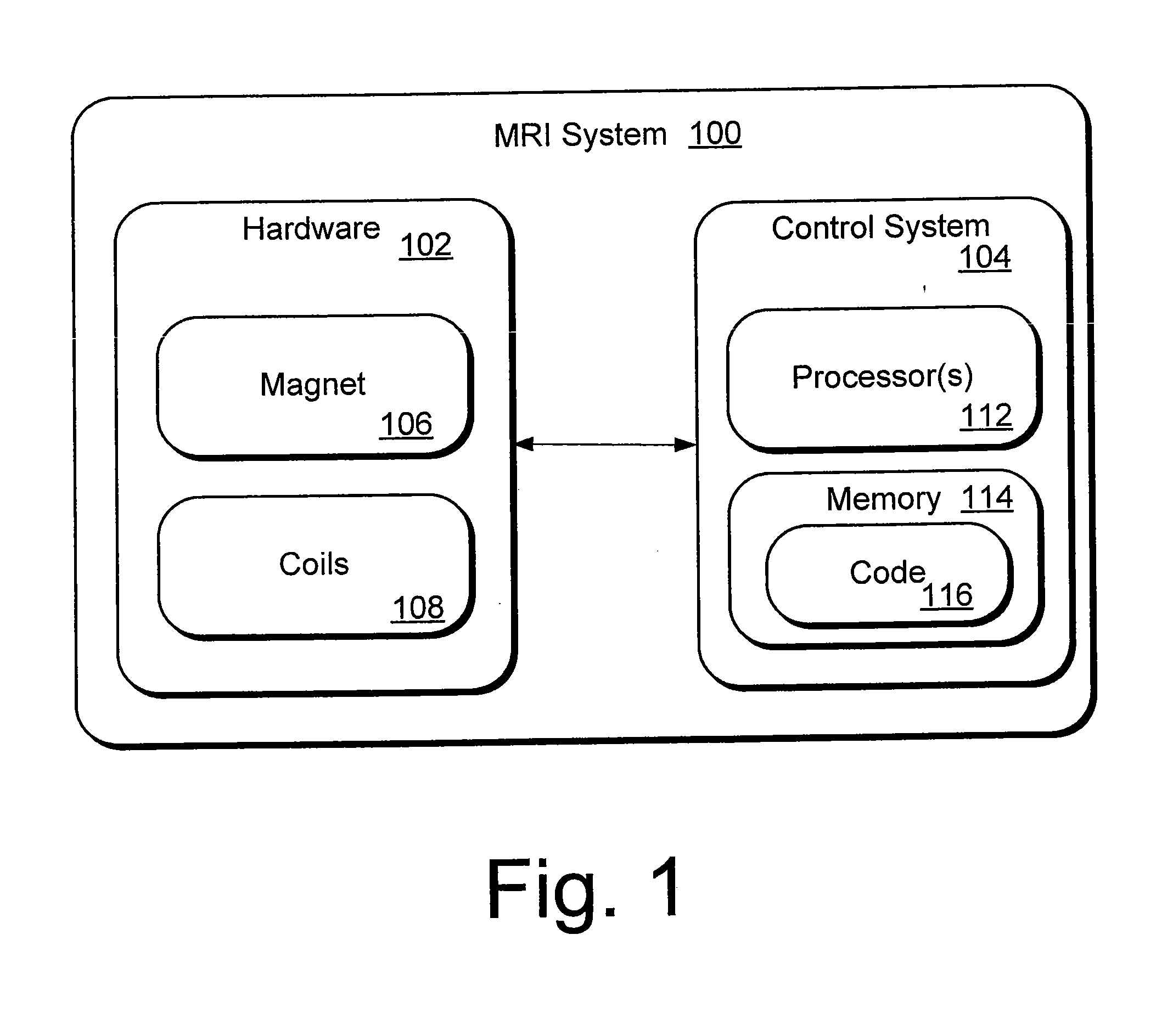
[0027]Magnetic Resonance Imaging (MRI) is an imaging technique based in part on the absorption and emission of energy in the radio frequency range. To obtain the necessary magnetic resonance (MR) images, a patient (or other target) is placed in a magnetic resonance scanner. The scanner provides a magnetic field that causes target atoms to align with the magnetic field. The scanner also includes coils that apply a transverse magnetic field. Radio-frequency (RF) pulses are emitted by the coils, causing the target atoms to absorb energy. In response to the RF pulses, photons are emitted by the target atoms and detected as signals in receiver coils.

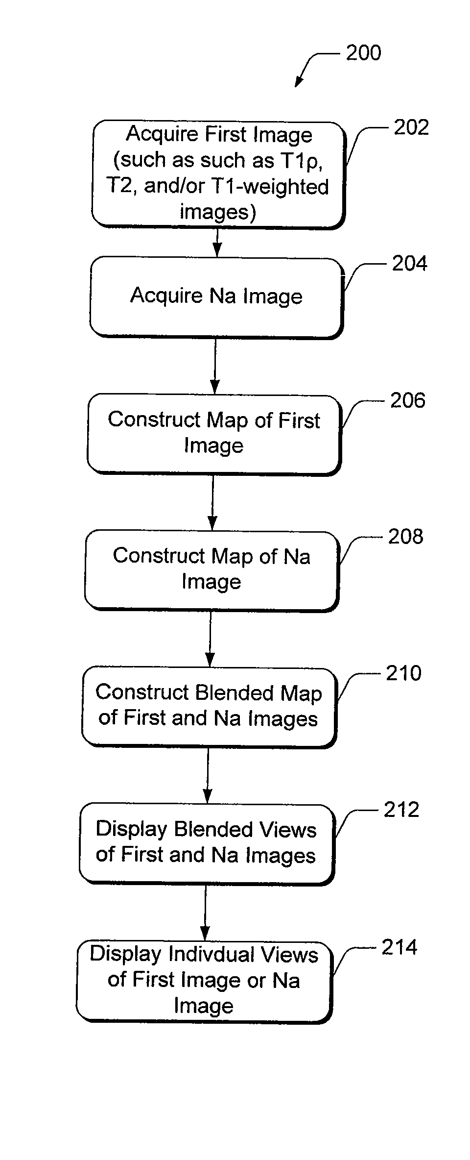
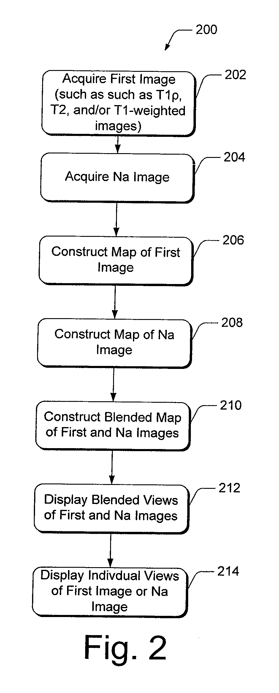
[0028]Present diagnostic strategies, such as CT, and T1- and T2-weighted magnetic resonance imaging, are only sensitive in advanced stages of the disease. Described herein is an innovative MR package that provides complete information about the biochemical and morphological state of the tissue. The MR package includes a process of pre-imaging...


PUM


Abstract

Methods of, and systems for, magnetic resonance imaging of diagnostic mapping of tissues, where sodium mapping is performed individually, as well as in combination with other images of tissue, such as T1ρ, T2, and/or T1-weighted images. In one method embodiment, a sodium image of the tissue is acquired during the same scanning session. Maps are constructed of each of the first and sodium images individually, and in combination, and further facilitate viewing in combination with each other as a single, blended image of the tissue. Maps of the images may be displayed individually or in combination with each other.

Description

CROSS-REFERENCE TO RELATED APPLICATIONS[0001]The present patent application is a continuation of International Application PCT / US2008 / 004780, filed Apr. 11, 2008 and published Oct. 23, 2008, which claims benefit of U.S. Provisional Application Ser. No. 60 / 923,215, filed Apr. 11, 2007, each of which is incorporated herein by reference.STATEMENT OF GOVERNMENT INTEREST[0002]This invention was made with government support by Grants R01AR045404 and R01AR051041 awarded by The National Institutes of Health, and performed at a NIH supported resource center (NIH RR02305). The government has certain rights in this invention.FIELD OF THE INVENTION[0003]The present invention relates to magnetic resonance imaging (MRI), and more particularly, a magnetic resonance (MR) method and system for diagnostic mapping of tissues.BACKGROUND OF THE INVENTION[0004]Osteoarthritis (OA) affects more than half of the population above the age of 65 (Felson, Epidemiol. Prev. 10:1-28 (1988); Felson, Arthritis Rheum...

Claims


Application Information

Patent Timeline
IPC IPC(8): G06K9/00
CPCA61B5/055G01R33/5608G01R33/56A61B5/4514
Inventor WITSCHEY, WALTER R.
Owner THE TRUSTEES OF THE UNIV OF PENNSYLVANIA
Why Eureka
Try Eureka