
Chromatin structure detection

a technology of chromatin structure and detection method, applied in the direction of microorganism testing/measurement, biochemistry apparatus and processes, etc., can solve the problems of limiting the access of dna to transcription factors and the transcriptional machinery, and achieve the effect of rapid generation of results

Inactive Publication Date: 2010-06-03
BIO RAD LAB INC
View PDF11 Cites 26 Cited by
  • Summary
  • Abstract
  • Description
  • Claims
  • Application Information

AI Technical Summary

Benefits of technology

[0078]One advantage of the present invention is the discovery that one can permeabilize and modify intact chromatin in cell simultaneously, e.g., by contacting cells with one buffer that includes a permeabilization agent and a DNA modifying or cleaving agent. This allows for extremely rapid generation of results. Further, this method eliminates cumbersome and potentially artifact-inducing steps such as isolation of nuclei, etc., as required in some previous methods of chromatin analysis.
[0079]The methods of the invention involve simultaneous permeabilization of a cell and contacting the cell with a DNA modifying agent under conditions such that genomic DNA in the cell has varying accessibility to the modifying agent (due to differences in chromatin structure) and then quantifying the amount of modification in a DNA region. The varying accessibility of the DNA can reflect nucleosomal structure of the genomic DNA. For example, in some embodiments, DNA regions that are more accessible to DNA modifying agents are likely in more “loose” chromatin structures.
[0080]A variety of eukaryotic cells can be used in the present invention. In some embodiments, the cells are animal cells, including but not limited to, human, or non-human, mammalian cells. Non-human mammalian cells include but are not limited to, primate cells, mouse cells, rat cells, porcine cells, and bovine cells. In some embodiments, the cells are plant cells. Cells can be, for example, cultured primary cells, immortalized culture cells or can be from a biopsy or tissue sample, optionally cultured and stimulated to divide before assayed. Cultured cells can be in suspension or adherent prior to and / or during the permeabilization and / or DNA modification steps. Cells can be from animal tissues, biopsies, etc. For example, the cells can be from a tumor biopsy.
[0081]The present methods can include correlating accessibility of a DNA region to transcription from that same region. In some embodiments, experiments are performed to determine a correlation between accessibility and gene expression and subsequently accessibility of a DNA modifying agent to a particular DNA region can be used to predict transcription from the DNA region. In some embodiments, transcription from a DNA region and accessibility of that region to DNA modifying agents are both determined. A wide variety of methods for measuring transcription are known and include but are not limited to, the use of northern blots and RT-PCR.
[0082]In some embodiments, the DNA methylation status of a region can be correlated with accessibility of a DNA region to the DNA modifying agent. In some embodiments, experiments are performed to determine a correlation between accessibility and DNA methylation in the region and subsequently accessibility of a DNA modifying agent to a particular DNA region can be used to predict DNA methylation from the DNA region. In some embodiments, methylation of a DNA region and accessibility of that region to DNA modifying agents are both determined. A wide variety of methods for measuring DNA methylation are known and include but are not limited to, the use of bisulfite (e.g., in sequencing and / or in combination with methylation-sensitive restriction enzymes (see, e.g., Eads et al., Nucleic Acids Research 28(8): E32 (2002)) and the high resolution melt assay (HRM) (see, e.g., Wodjacz et al, Nucleic Acids Research 35(6):e41 (2007)).
[0083]The invention provides for, following permeabilization / DNA modification, comparisons of quantity or other physical characteristic of a first DNA region with a second DNA region in a cell's genome. Alternatively, or in addition, one can compare quantity or other physical characteristic of the first DNA region in two different cells. For example, the two cells can represent diseased and healthy cells or tissues, different cell types, different stages of development (including but not limited to stem cells or progenitor cells), etc. Thus, by using the methods of the invention one can detect differences in chromatin structure between cells and / or determine relative chromatin structures between two or more DNA regions (e.g., genes) within one cell. In addition, one can determine the effect of a drug, chemical or environmental stimulus on the chromatin structure of a particular region in the same cells or in different cells.III. Permeabilizing and Disrupting Cells

Problems solved by technology

This tight packaging can limit the access of DNA to transcription factors and the transcriptional machinery.

Method used


Image

  • Chromatin structure detection
  • Chromatin structure detection
  • Chromatin structure detection

Examples


example 1

General Approach

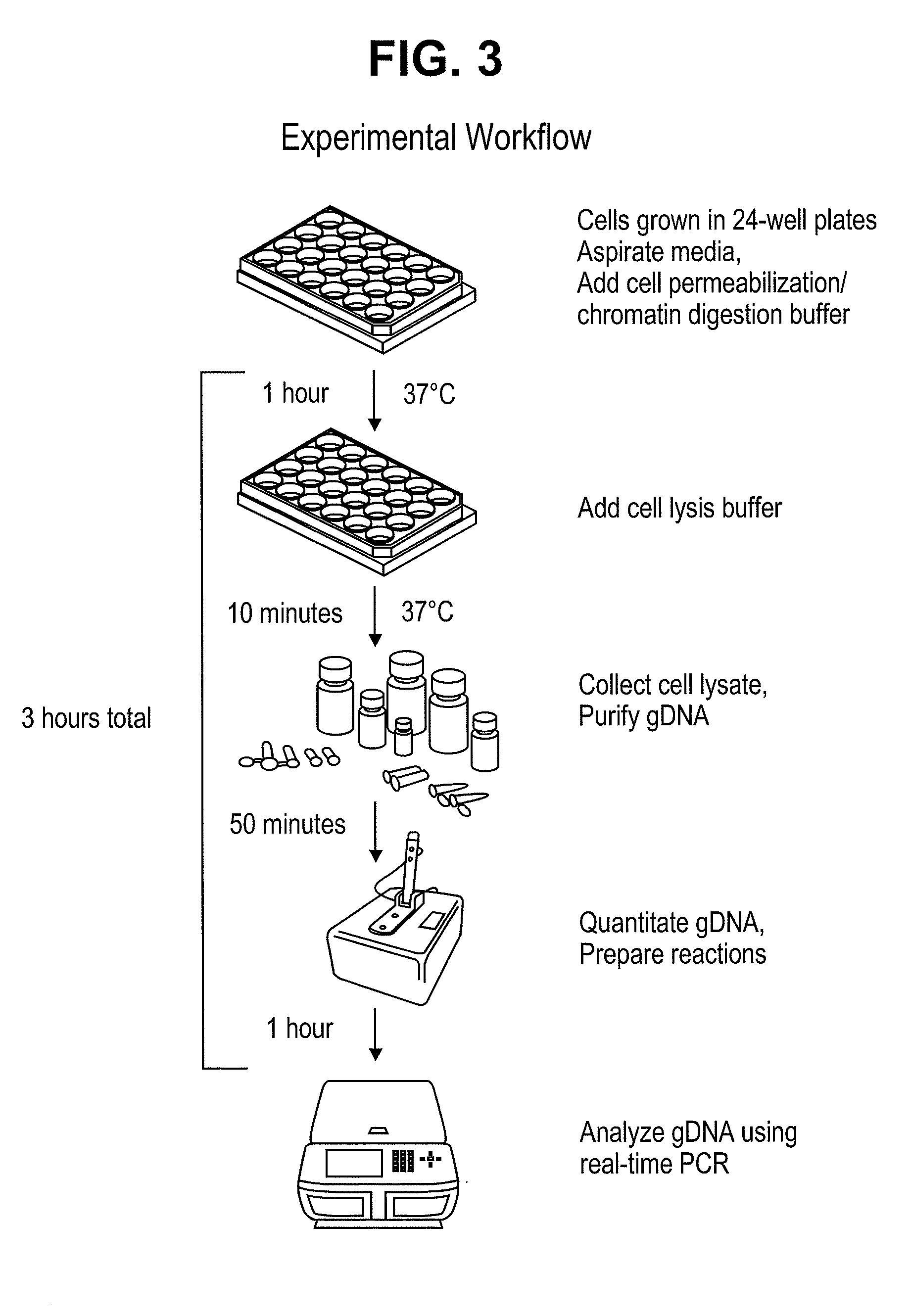
[0168]The present assay takes advantage of the difference of accessibility of a DNA modifying agent to different parts of chromatin in a cell. As illustrated in FIG. 2, DNA modifying agents can access certain portions of chromatin more readily than other parts. FIG. 3 illustrates an exemplary workflow of the assay. Adherent cells were grown in 24-well plates as starting material. In this embodiment, two wells are used for each experiment: One well is treated with permeabilization buffer and no nuclease; the other well is treated with permeabilization buffer with nuclease. The entire process, from cells to real-time PCR, takes about three hours and results are available on the day of cell harvest.

[0169]FIG. 4 illustrates a schematic for analysis of DNA isolated from cells when MnlI is used as a chromatin structure probe. The assay in this case is performed in parallel with a “no nuclease” control. Two primer sets are used for each gene. One primer set does not span an...

example 2

Data Using WI in Hela Cells

[0172]This example shows the results of data generated using an experiment approach as outlined above.

Materials and Methods

[0173]Chemicals. L-α-Lysophosphatidylcholine (lysolecithin) was purchased from Sigma-Aldrich. MnlI, DNase I, BSA and proteinase K were purchased from New England Biolabs. RNase A was purchased from Qiagen. Tissue culture plates were purchased from VWR. iQ SYBR was from Bio-Rad.

[0174]Treatment of cells. Cells grown in 24-well plates were treated when they reached 90% confluence. The culture media was aspirated and 100 ul of a permeabilization / digestion buffer was gently layered on the cells. For cells treated with MnlI the permeabilization / digestion buffer consisted of lysolecithin, NaCl, Tris-HCl, MgCl2, DTT, BSA and MnlI. For cells treated with DNase I the permeabilization / digestion buffer consisted of lysolecithin, Tris-HCl, MgCl2, CaCl2 and DNase I. The permeabilized cells were then incubated at 37° C. for 1 hour. Following incubati...

example 3

Data Using MnlI in Prostate Cancer Cells

[0186]The methods described above were also applied to different cell types to determine whether accessibility was related to expression for GSTP1. Two cell types were used: RWPE-1 cells, which are derived from non-cancerous human prostate, and in which GSTP1 is highly expressed; and LNCaP cells, which are derived from cancerous human prostate, and in which GSTP1 is silenced by epigenetic modifications.

[0187]The methods of analysis of these cells were essentially as described above. FIG. 12 shows the control reactions (i.e., for the hemoglobin gene). In both cell lines, the delta Ct in the samples with no enzyme are about the same as those with enzyme. Therefore the delta Cts were about 0, indicating that this gene was inaccessible in both cell lines.

[0188]FIG. 13 shows the results of the positive control reactions analyzing the GAPDH gene. For reactions without enzyme, both the total and intact Cts were about the same. However, for reactions ...


PUM

PropertyMeasurementUnit
pHaaaaaaaaaa
pHaaaaaaaaaa
pHaaaaaaaaaa

Abstract

The present application provides methods and compositions for determining accessibility of a DNA modifying agent in genomic DNA.

Description

CROSS-REFERENCE TO RELATED PATENT APPLICATIONS[0001]The present application claims benefit of priority to U.S. Provisional Patent Application No. 61 / 119,280, filed Dec. 2, 2008, which is incorporated by reference for all purposes.BACKGROUND OF THE INVENTION[0002]Most DNA in a cell is packaged around a group of histone proteins in a structure known as a nucleosome. This nucleosomal DNA can be further packaged into coiled structures that tightly compact the DNA. This tight packaging can limit the access of DNA to transcription factors and the transcriptional machinery. Genomic DNA packaged in this way is sometimes referred to as chromatin.[0003]Chromatin is classified into two main groups, euchromatin, where the DNA is loosely packaged, accessible and generally, but not always, transcriptionally competent, and heterochromatin, where the DNA is tightly packaged, inaccessible and generally, but not always, transcriptionally silent.[0004]What controls the transition between these two chr...

Claims


Application Information

Patent Timeline
Patent Type & Authority Applications(United States)
IPC IPC(8): C12Q1/68
CPCC12Q1/68C12Q2527/125
Inventor OKINO, STEVECHENG, MAN
Owner BIO RAD LAB INC
Learn More