
Apparatus and Method for Cardiac Tissue Modulation by Topical Application of Vacuum to Minimize Cell Death and Damage

a technology of cardiac tissue and vacuum, applied in the field of cardiac tissue modulation apparatus and vacuum topical application, can solve the problems of finite life cycle of cells and expected fatigue, and achieve the effects of minimizing the progression of pathologic processes, minimizing the disruption of physiological cardiac integrity, and minimizing the interference with cardiac blood flow

Active Publication Date: 2010-05-13
WAKE FOREST UNIV HEALTH SCI INC
View PDF99 Cites 13 Cited by
  • Summary
  • Abstract
  • Description
  • Claims
  • Application Information

AI Technical Summary

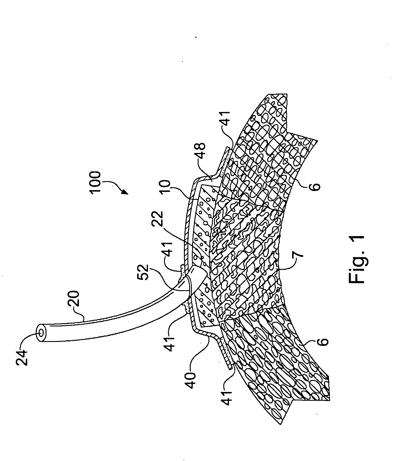
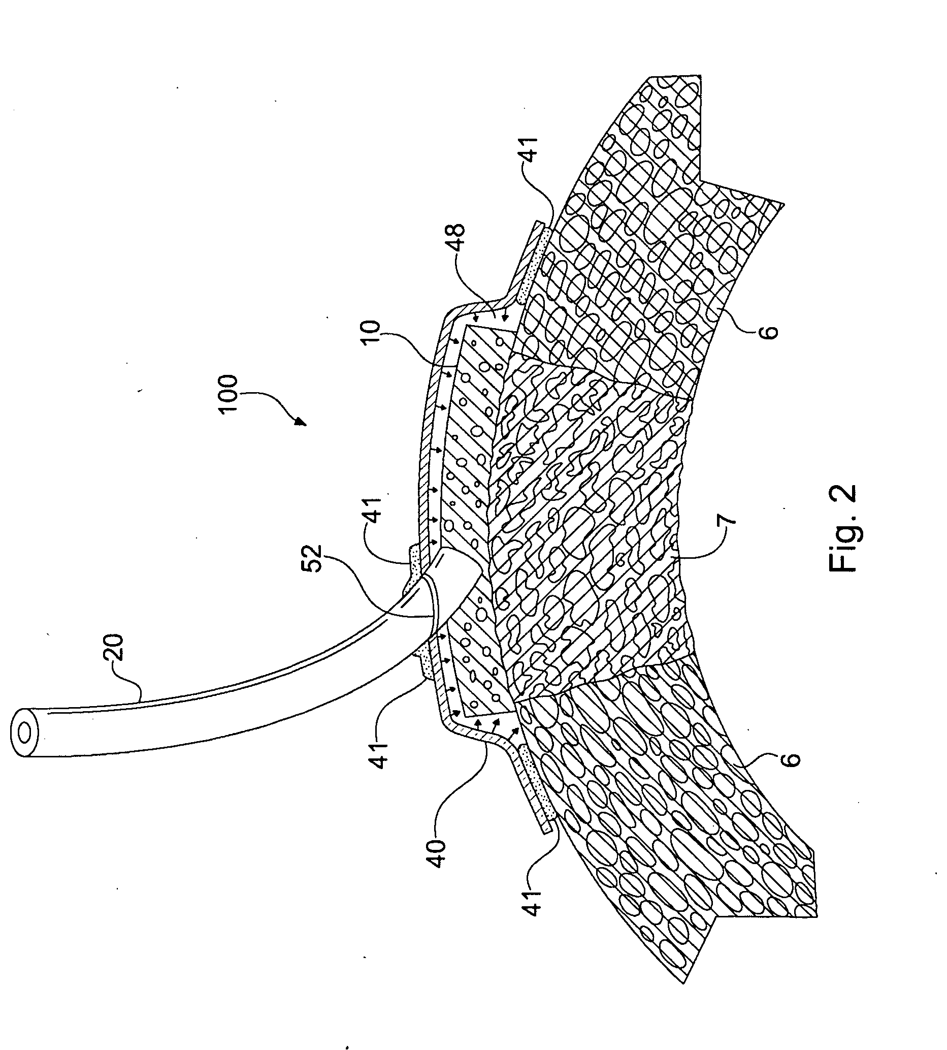
Benefits of technology

[0007]The present invention relates to devices and methods for treating damaged heart tissue, such as myocardial infarction in the ischemic or early reperfusion phase, by treatment with sub-atmospheric (or negative) pressure. Treatment with the devices and methods of the present invention may salvage cells in the zone of stasis and thereby decrease the size of the infarct. Such treatment would be especially efficacious in endstage myocardial disease where bypass or stenting would not be possible. The treatment would also be useful as an adjunct to ECMO (extracorporeal membrane oxygenation) for resting the heart, following cardiac arrest, in situations with left main artery lesions, etc.
[0010]The present invention may also provide delayed treatment of myocardial infarction where there is already a stable zone of myocardial cell death. Again through an endoscope and a small incision in the chest wall, a bio-incorporable vacuum dressing may be placed on the area that is infarcted. Again, exposure of the myocardium involved and adjacent myocardium may be required and provided with a CO2 or similar cutting device to perforate the epicardium. The vacuum dressing may be modified so that a lattice of myocardial or peripheral muscle cells may be incorporated within it. The vacuum dressing may also incorporate a small catheter with the ability to reinfuse additional myocardial cells, pleuripotent progenitor cells, or peripheral muscle cells at subsequent serial times. In areas where there is near complete cell death or there is little or no contraction of the muscle cells in the damaged cardiac tissue, new contractile cells could be seeded to replace and restore the contractile function of the damaged cardiac tissue. Initially, peripheral muscle or peripheral muscle cells grown from culture could be used. These cells have a finite life cycle and would be expected to fatigue over time. The myocardium could be biopsied at the time of the treatment of the initial treatment and myocardial cells removed and cultured to create a larger mass of viable of cells. The harvested myocardial cells could be maintained in culture and used for later periodic infusion to develop a myocardial patch that would cover the area of previous infarction. Also, progenitor cells could be harvested and immediately infused to the area of damaged cardiac tissue, or they could be grown in culture and periodically infused to the area of damaged cardiac tissue with the expectation that they would develop into cardiac myocytes. Over time the introduced cells would be induced to undergo mitosis or self-replication thus increasing the functional mass of the heart. The ability to progressively add cells that would be progressively vascularized is a major step in regenerative medicine where presently only a sheet of cells can be expected to survive.
[0012]The porous material may be sealed in situ over the damaged cardiac tissue to provide a region about the damaged cardiac tissue for maintaining sub-atmospheric pressure at the damaged cardiac tissue. The porous material may be operably connected with a vacuum source for producing sub-atmospheric pressure at the damaged cardiac tissue, and the vacuum source activated to provide sub-atmospheric pressure at the damaged cardiac tissue. The sub-atmospheric pressure may be maintained at the damaged cardiac tissue for a time sufficient to reduce edema (thus restoring contractility and compliance), decrease interstitial pressure, remove inflammatory mediators, remove inflammatory amplifiers, modulate intracellular mediators, increase reperfusion and microvascular flow, decrease microvascular plugging, and / or decrease retention of inflammatory cells within the damaged cardiac tissue. Micro and macro deformation of the cardiac tissue being treated would increase vasculoneogenesis or the formation of new blood vessels in the ischemic tissue. This would increase the survivability of the cardiocytes and ultimately improve function of the ischemic portion of the heart. In addition, macro and micro deformation of small arterioles already existing in the heart would result in their physical reorientation into the areas of ischemic tissue, thus increasing perfusion and ultimately function.
[0014]In another of its aspects the present invention provides an apparatus for treating damaged cardiac tissue. The apparatus includes a porous material for treating damaged cardiac tissue having a pore structure configured to permit gaseous communication between one or more pores of the porous material and the cardiac tissue to be treated. The porous material may include at least one of an electrospun material, a cast material, and a printed material. Alternatively or additionally, the porous material may comprise a bio-incorporable material. In such instances, it may also be beneficial for the porous material to be formulated in such a manner that the outer edges of the porous material would be resorbed or degraded more quickly than the inner portion. The rate of removal (resorption / degradation) of the porous material could be matched to the rate of formation of new tissue. One way to control the rate of degradation or resorption is by varying the number of crosslinks introduced into the porous material.
[0019]Thus, the present invention provides devices and methods for minimizing the progression of pathologic processes, minimizing the disruption of physiological cardiac integrity, and minimizing the interference with cardiac blood flow and nutrition and increasing revascularization of ischemic areas of the heart by vascular neogenesis and reorientation of existing vessels. By decreasing cardiac edema and interstitial pressure the risk of cardiac cell death and compromise may be minimized. In addition, the present invention facilitates the removal of mediators, degradation products, and toxins that enhance the inflammatory and pathophysiological response in the damaged cardiac tissue.

Problems solved by technology

Such treatment would be especially efficacious in endstage myocardial disease where bypass or stenting would not be possible.
These cells have a finite life cycle and would be expected to fatigue over time.

Method used


Image

  • Apparatus and Method for Cardiac Tissue Modulation by Topical Application of Vacuum to Minimize Cell Death and Damage
  • Apparatus and Method for Cardiac Tissue Modulation by Topical Application of Vacuum to Minimize Cell Death and Damage
  • Apparatus and Method for Cardiac Tissue Modulation by Topical Application of Vacuum to Minimize Cell Death and Damage

Examples


example 1

[0058]The porcine heart has anatomy similar to that of humans with the main vasculature consisting of the right and left coronary arteries. The left main coronary artery splits into the circumflex coronary artery and the left anterior descending (LAD) coronary artery. The LAD runs down along the anterior septum and perfuses the anterior portion of the left ventricle with diagonal branches. For these studies, a porcine model of ischemia-reperfusion was used that included the temporary ligation of 2-3 diagonal branches of the LAD in order to create an ischemic area on the anterior portion of the heart. These coronary arteries were occluded for 75 minutes and then reperfused for 3 hours to allow for ischemia / reperfusion injury to develop. The negative pressure therapy was applied only during the reperfusion phase of the experiments to simulate a clinically relevant treatment window.

[0059]To begin the study, the animals were sedated and transported to the operating room. The first 13 an...

example 2

[0067]Another experiment was conducted using 50 mm Hg vacuum for treatment for comparison to original control animals from Example 1 above. The surgical technique in this experiment was similar to that used for those of Example 1. These animals were sedated and prepped for surgery. The heart was exposed through a midline sternotomy. Branches of the left anterior descending artery were ligated for 75 minutes. A polyvinyl alcohol vacuum dressing was placed over the ischemic area and an AlloDerm® cover was placed over the vacuum dressing and sealed into place with a combination of sutures and fibrin glue. Negative pressure of 50 mm Hg was applied for 3 hours. At the end of this time the heart was stained for area of risk, removed and then counter stained for area of necrosis. The infarct size results for these five, 50 mm Hg negative pressure therapy animals were significantly smaller (P<0.001) than for the control animals. The infarct size for the 50 mm Hg treated animals was smaller ...

example 3

[0071]A subsequent study was performed to examine resorbable vacuum dressings and overlay covers. One animal was sedated, prepared for surgery as described, and the heart exposed through a mid-line sternotomy. Branches of the LAD were ligated for 90 minutes. The dressing was prepared by freeze drying. A solution of chitosan (1.33% weight / volume in 2% acetic acid, 20 ml total volume) was poured into an appropriately sized mold. The solution was frozen for 2 hours at −70° C., then transferred to the lyophylizer for 24 hours. The dressing was cross-linked by 2.5% glutaraldehyde vapor for 12 hours to provide a porous material. The overlay cover was an electrospun mixture of Type I collagen and poly 1,8-octanediol citrate (POC) (80%:20% weight / weight). The solution concentration was 15% dissolved in hexafluoro-20proponal (HFIP) with a total volume of 9.5 ml. The solution was ejected from a syringe through an 18 gauge needle at a flow rate of 3 ml / hour. The voltage was 25 KV with a workin...


PUM


Abstract

A method and apparatus are provided for treating cardiac tissue to modulate ischemic heart tissue with topical sub-atmospheric pressure to minimize cell death and damage.

Description

RELATED APPLICATIONS[0001]The present application claims the benefit of priority of U.S. Provisional Application 61 / 088,558, filed on Aug. 13, 2008 and U.S. Provisional Application No. 61 / 081,997, filed on Jul. 18, 2008, the entire contents of which are incorporated herein by reference.FIELD OF THE INVENTION[0002]The present invention relates generally to a method and apparatus for treating cardiac tissue, and more particularly, but not exclusively, to modulating ischemic and reperfused heart tissue with topical sub-atmospheric pressure to minimize cell death and damage.BACKGROUND OF THE INVENTION[0003]Myocardial ischemia occurs when a portion of the heart does not receive sufficient oxygen and energy substrates to meet its demand. This usually occurs because of a blockage in the artery due to either atherosclerotic plaque or thrombus formation. In a myocardial infarction there is an area of injury where the cells, because of lack of blood flow, will die immediately. There is a laye...

Claims


Application Information

Patent Timeline
Patent Type & Authority Applications(United States)
IPC IPC(8): A61H7/00
CPCA61B17/00234A61B2019/464A61B2017/306A61B2017/00243A61M1/916A61M1/96A61M1/915A61M2210/125A61M2210/1082A61B90/06A61B2090/064A61F2013/0028A61M1/75A61M1/74A61M1/917A61M1/984A61F13/05A61B17/00A61M1/00A61L17/10A61L17/08
Inventor ARGENTA, LOUIS C.CARROLL, DAVID L.JORDAN, JAMES E.LEVI, NICOLE H.LIU, JIEMORYKWAS, MICHAEL J.WAGNER, WILLIAM D.
Owner WAKE FOREST UNIV HEALTH SCI INC
Learn More