P2x7 receptor targeted therapy
a p2x7 receptor and targeted therapy technology, applied in the field of cancer treatment, can solve the problems of treatment failure, persistent development of chemoresistance, and failure of clinical evaluation approach,
- Summary
- Abstract
- Description
- Claims
- Application Information
AI Technical Summary
Benefits of technology
Problems solved by technology
Method used
Image
Examples
example 1
[0288]Chemotherapy Treatment in Myeloma RPMI-8226 and Neuroblastoma Kelly Cell Lines.
[0289]Materials and Methods
[0290]4,000 cells were seeded in a 96 well plate and allowed to adhere over-night. Cells were then treated with increasing amount of chemotherapy or vehicle control for 72 hours to induce varying amount of cell killing. Cell viability was then measured using CellTiter-Blue Cell Viability Assay form Promega following manufacturer's instruction.
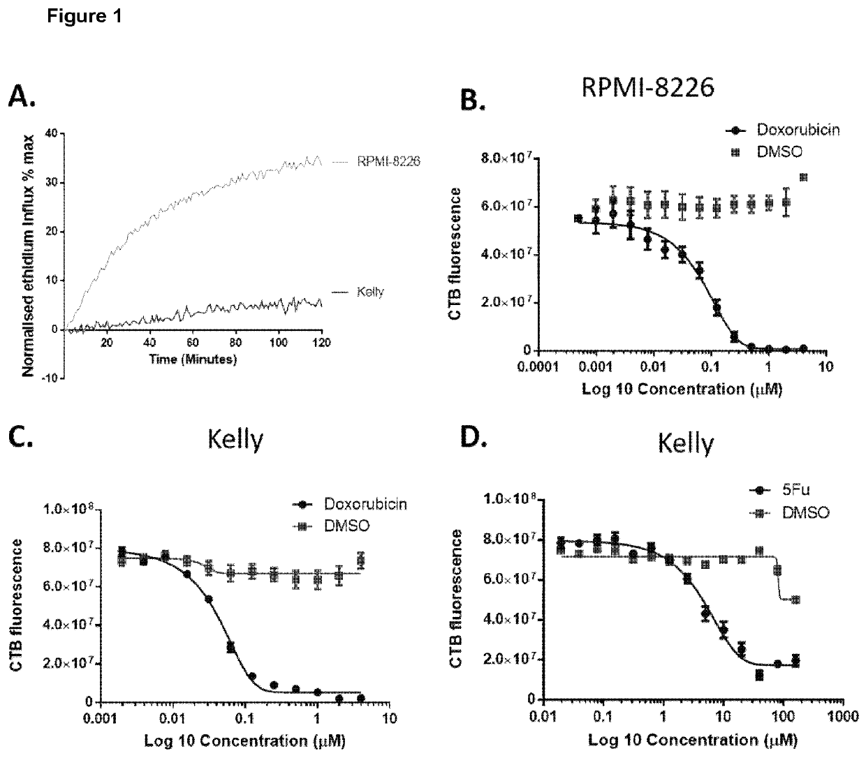
[0291]Normalised ethidium influx in response to 0.5 mM BzATP stimulation in the myeloma RPMI-8226 and neuroblastoma Kelly cell lines. Mean of three independent experiments is shown.
[0292]Results
[0293]FIG. 1A shows that the myeloma cell line, RPMI-8226, is able to open the P2X7 pore in response to 0.5 mM BzATP whilst the neuroblastoma cell line Kelly is not. RPMI-8226 and Kelly cell lines were selected as representative models of blood born cancer (myeloma) and solid tumour (Neuroblastoma). FIGS. 1B, C and D shows the effect of increas...
example 2
[0295]Chemotherapy Treatment in Functional Myeloma RPMI-8226 and Non-Functional Neuroblastoma Kelly Cell Lines and Induction of nfP2X7 as Detected by BPM09.
[0296]Materials and Methods
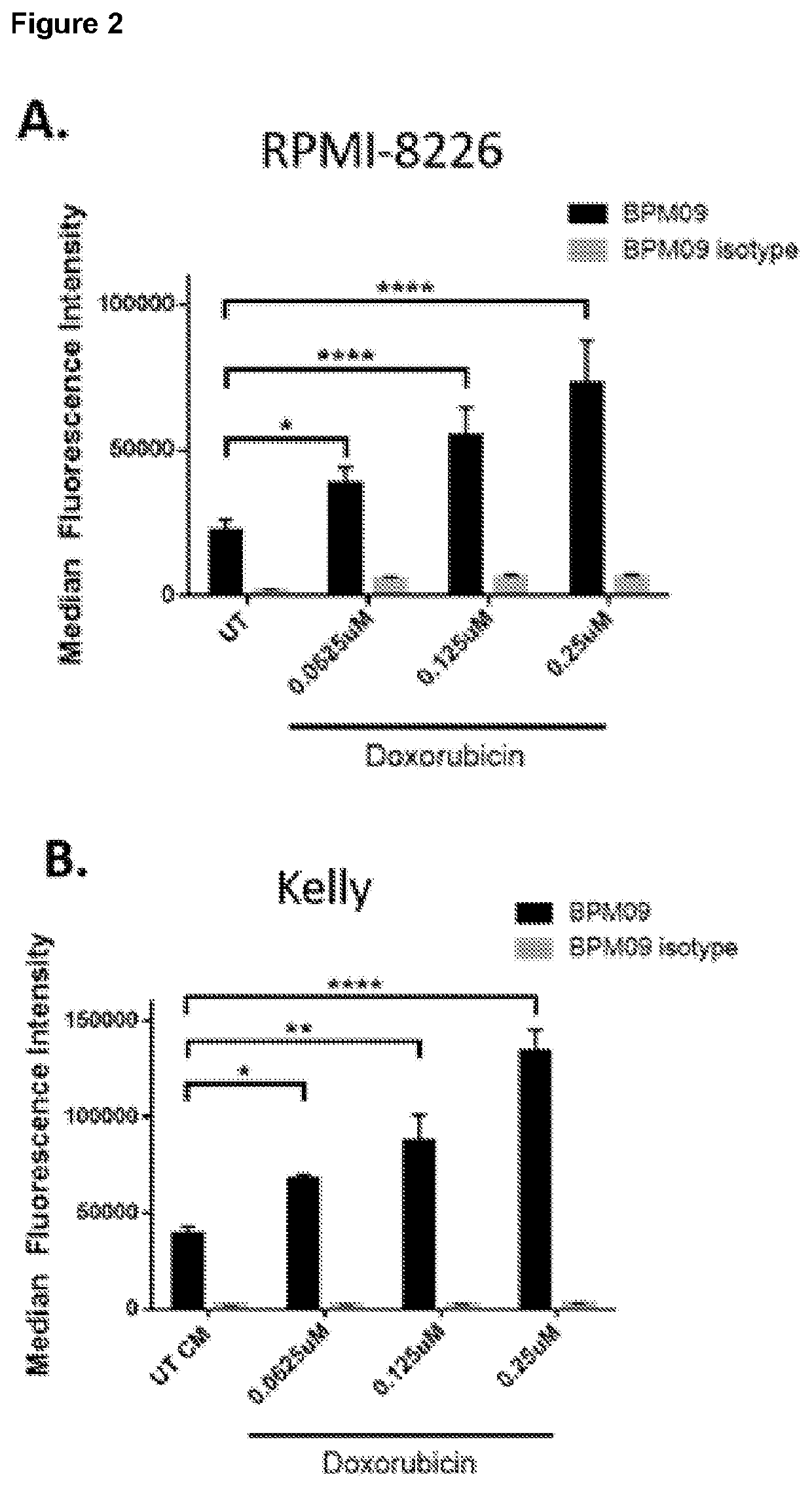
[0297]500,000 cells were seeded in a 6 well plate and allowed to adhere over-night. Cells were then treated with increasing amount of chemotherapy or vehicle control for 72 hours to induce varying amount of cell killing. The remaining live cells were dissociated in PBS based enzyme-free dissociation buffer, washed and re-suspended in staining buffer (PBS, 2% FCS). Cells were then stained for 1 h with primary antibody raised against non-functional P2X7 (here 2-2-1hFc), washed 3 times in staining buffer prior to a 1 h incubation with fluorescently coupled secondary antibody and 7AAD. Fluorescence staining on live cells was acquired using a BD Accuri flow cytometer and analysed with FlowJo-flow cytometry analysis software. Median fluorescence intensity of the live cell population was analysed using 7AAD li...
example 3
[0300]Cell Lines with Acquired Resistance to Chemotherapy have Increased Expression of nfP2X7
[0301]Materials and Methods
[0302]A2780 parental cells, A2780 cells with acquired resistance to doxorubicin and A2780 cells with acquired resistance to cisplatin were cultured were obtained from ECACC repository (References: Proc Amer Assoc Cancer Res 1984; 25:336; Semin Oncol 1984; 11:285; Cancer Res 1987; 47:414; Cancer Res 1988; 48:5713).
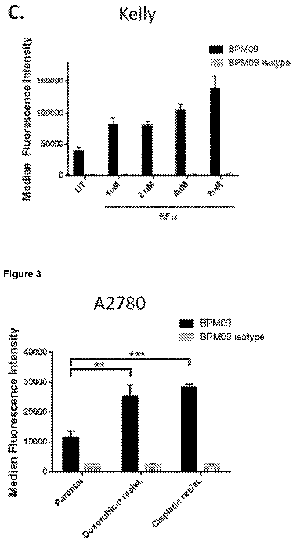
[0303]500,000 A2780, A2780 with doxorubicin resistance and A2780 with cisplatin resistance cells were seeded in a 6 well plate and allowed to adhere over-night. Cells were dissociated in PBS based enzyme-free dissociation buffer, washed and re-suspended in staining buffer (PBS, 2% FCS). Cells were then stained for 1 h with primary antibody raised against non-functional P2X7 (here BPM09), washed 3 times in staining buffer prior to a 1 h incubation with fluorescently coupled secondary antibody and 7AAD. Fluorescence staining on live cells was acquired using ...
PUM
| Property | Measurement | Unit |
|---|---|---|
| dissociation constant | aaaaa | aaaaa |
| dissociation constant | aaaaa | aaaaa |
| dissociation constant | aaaaa | aaaaa |
Abstract
Description
Claims
Application Information
 - R&D
- Intellectual Property
- Life Sciences
- Materials
- Tech Scout
- Unparalleled Data Quality
- Higher Quality Content
- 60% Fewer Hallucinations
Browse by: Latest US Patents, China's latest patents, Technical Efficacy Thesaurus, Application Domain, Technology Topic, Popular Technical Reports.
© 2025 PatSnap. All rights reserved.Legal|Privacy policy|Modern Slavery Act Transparency Statement|Sitemap|About US| Contact US: help@patsnap.com


