
Radiological image capturing apparatus and radiological image capturing system

a radiological image and imaging apparatus technology, applied in the direction of instruments, patient positioning for diagnostics, applications, etc., can solve the problems of difficult periodic mri photographing operation, difficult to determine whether or not rheumatic disease has actually developed, and difficult to perform mri photographing operation in the framework of regular physical examination, etc., to achieve good x ray image

Active Publication Date: 2010-05-13
KONICA MINOLTA MEDICAL & GRAPHICS INC
View PDF9 Cites 38 Cited by
  • Summary
  • Abstract
  • Description
  • Claims
  • Application Information

AI Technical Summary

Benefits of technology

[0019]To overcome the abovementioned drawbacks in conventional radiological image capturing apparatuses and systems, it is one of objects of the present invention to provide a radiological image capturing apparatus, which makes it possible to obtain a good X ray image in which contrast of the peripheral portions (edge portions), such as a cartilage tissue of human body, etc., are emphasized by employing the Talbot interferometer method and the Talbot-Lau interferometer method, and to provide a radiological image capturing system in which the above-captured X ray image is processed.

Problems solved by technology

However, the above-mentioned early symptoms of the rheumatic disease have been hardly detected by observing the X-ray photographic image, which has been widely accepted as a simple and convenient inspection method, and accordingly, it has been difficult for a doctor or the like to determine whether or not the rheumatic disease has actually developed.
However, since the patient has been heavily burdened with the MRI photographing operation from the viewpoint of the cost and time required for making a diagnosis, it has been difficult to perform the MRI photographing operation in the framework of the regular physical examination.
Therefore, there has been such a problem that it has been difficult to periodically perform the MRI photographing operation so as to observe (inspect) the changes of the joint portions, such as fingers, etc., over time, as a longitudinal diagnosis.
Further, since a huge image-capturing installation is necessary for conducting the image-capturing operation that employs the radiant beam, and, sometimes, several tens of minutes are required for completing the image-capturing operation, it has been virtually impossible for a general-purpose medical facility to employ the radiant beam for conducting the image-capturing operation.
However, since the radiant beam X ray source, which requires a special facility, is employed in the Talbot interferometer method set forth in Patent Document 2, there has been a problem that it is virtually impossible for general-purpose medical facilities, widely exiting in the society, to employ the Talbot interferometer method.
However, since the excessively low energy X-rays tend to be absorbed into the human body, and accordingly, since an amount of the X rays arriving at a detector is relatively small, it is necessary to increase the dose of radiation exposure in order to acquire an appropriate S / N (Signal to Noise) ratio at the detector, resulting in an increase of the X-ray exposure.
Further, the increase of the X-ray exposure will cause an extension of the image capturing time interval.
However, it is difficult to make the movement of the human body, serving as the subject, freeze for a long time during the image capturing time interval.
On the other hand, if the energy of the X rays to be irradiated onto the human body is excessively high, it has been acquired such a knowledge that an image contrast being sufficient for depicting bone tissues and soft tissues that constitute the human body cannot be obtained.
Accordingly, there has been a problem that an X ray image, which is sufficiently usable for making a diagnosis on the human body, serving as the subject, cannot be obtained, unless the contrast in the X ray image can be obtained.
However, the structure and configuration of the concerned apparatus become more complicated than ever, and, further, since various kinds of conditions, such as positional relationships between the multi-slit element and the other elements, etc., are added as new limitations for configuring the apparatus, the structural conditions for the concerned apparatus become still more stricter than ever, and it is required for the concerned apparatus to fulfill such the extremely strict conditions.
Whereas, since the X ray emitting source is converted to the multi (plural) radiant sources by employing the multi-slit element, the Talbot-Lau interferometer method is inferior to the Talbot interferometer method in sharpness of the reproduced X ray image to some extent.

Method used


Image

  • Radiological image capturing apparatus and radiological image capturing system
  • Radiological image capturing apparatus and radiological image capturing system
  • Radiological image capturing apparatus and radiological image capturing system

Examples


Embodiment Construction

[0036]Referring to the drawings, the radiological image capturing apparatus and the radiological image capturing system, both embodied in the present invention, will be detailed in the following. However, the scope of the present invention is not limited to the examples indicated in the drawings.

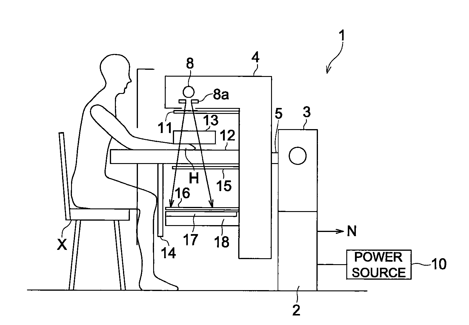
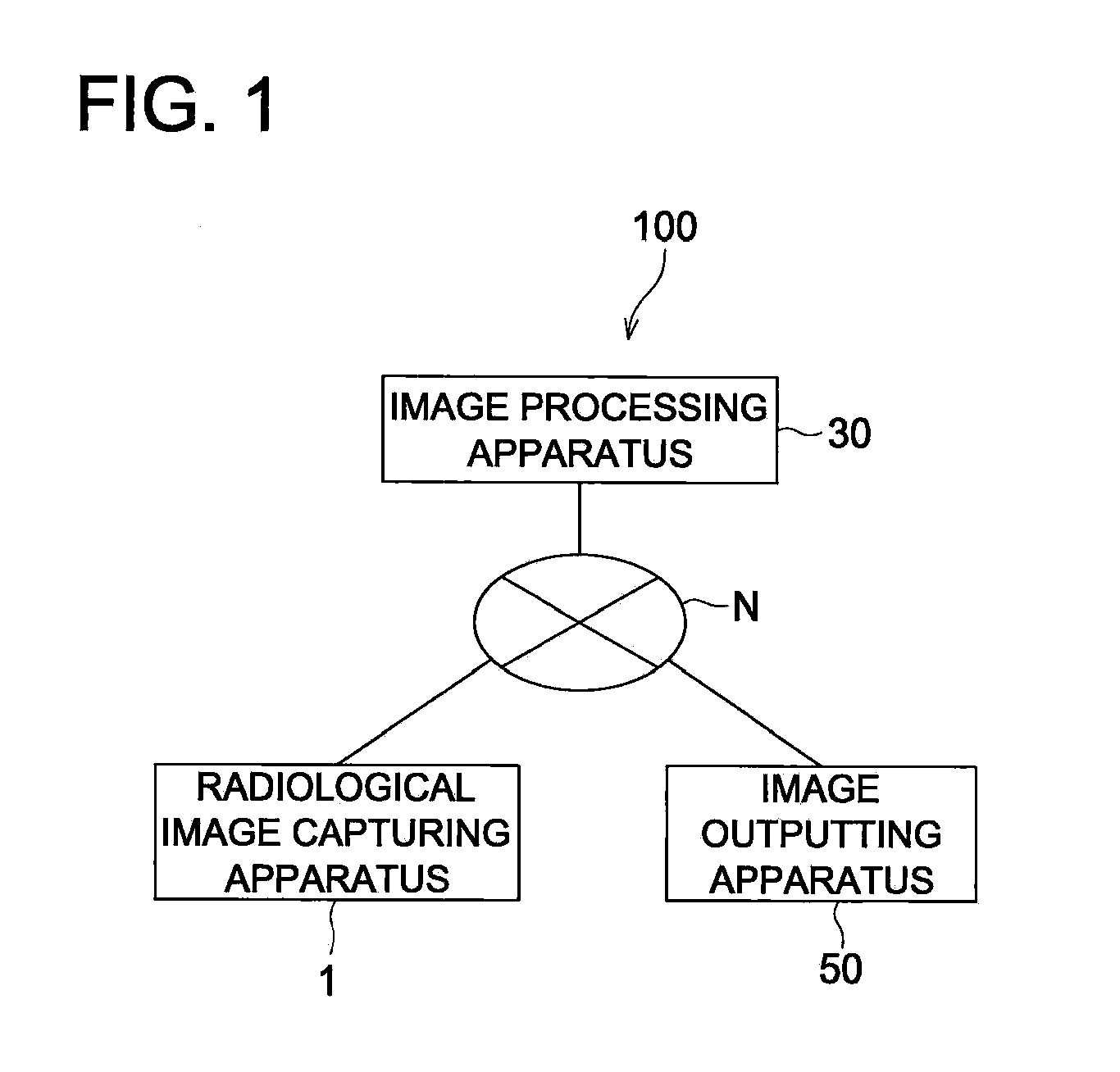
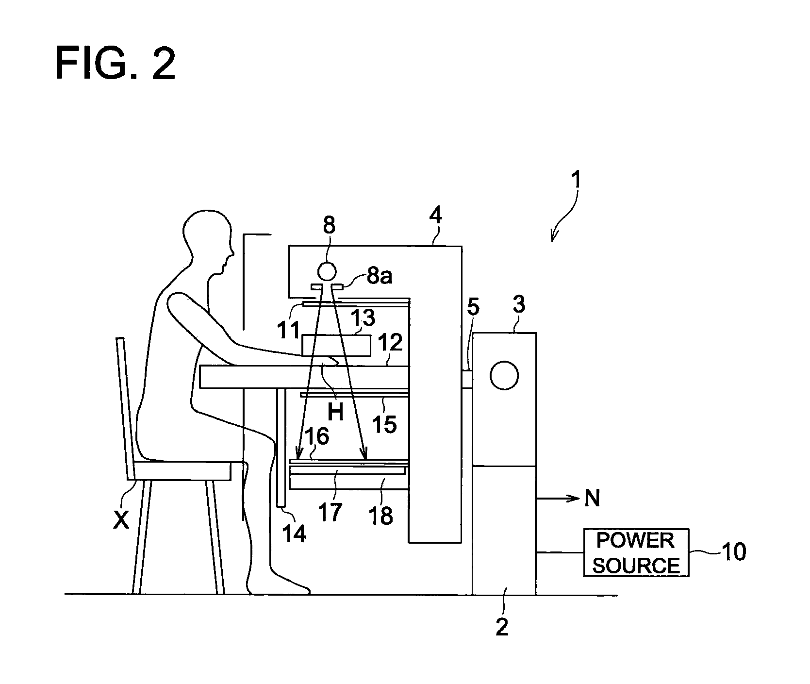
[0037]In the present embodiment, a radiological image capturing system 100 is constituted by: a radiological image capturing apparatus 1 that irradiates X rays, serving as radial rays, onto a subject so as to generate radiological image data of the subject; an image processing apparatus 30 that applies various kinds of image processing to the radiological image data generated by the radiological image capturing apparatus 1; and an image outputting apparatus 50 that outputs a radiological image, etc., onto a display screen or a film, based on processed image data generated by applying the various kinds of image processing to the radiological image data in the image processing apparatus 30. Ea...


PUM


Abstract

There is described a radiological image capturing apparatus, which makes it possible to obtain a good X ray image in which contrast of the peripheral portions are emphasized by employing the Talbot interferometer method and the Talbot-Lau interferometer method. The apparatus is provided with an X-ray tube, a multi-slit member, a first diffraction grating, a second diffraction grating and an X-ray detector. The second diffraction grating contacts the X-ray detector. A distance L between the multi-slit element and the first diffraction grating is set to be not less than 0.5 m, a distance Z1 between the first diffraction grating and the second diffraction grating is set to be not less than 0.05 m, and a slit interval distance d0 of the multi-slit element is set to be not less than 2 μm. With the settings, the abovementioned good X-ray image can be obtained by using the Talbot-Lau interferometer system.

Description

FIELD OF THE INVENTION[0001]The present invention relates to a radiological image capturing apparatus and a radiological image capturing system, and specifically relates to such a radiological image capturing apparatus that employs a Talbot-Lau interferometer method and such a radiological image capturing system that applies various kinds of processing to an image captured by the radiological image capturing apparatus.TECHNICAL BACKGROUND[0002]In recent years, the morbidity rate of the rheumatic disease in Japan has reached to 1% of the national population, and accordingly, the rheumatic disease has been regarded as a kind of a folk disease at present. An abrasion loss at a cartilage portion (destruction of cartilage) and / or subtle changes of a bone shape and a bone trabeculae are observed as its early symptoms, and then, at the time when the symptoms have worsened, considerable changes of shape of the bone sections can be observed significantly.[0003]Accordingly, by observing the s...

Claims


Application Information

Patent Timeline
IPC IPC(8): G01N23/201A61B6/00
CPCA61B6/4488A61B6/484A61B6/505A61B6/4291A61B6/48A61B6/5282A61B6/586G21K2201/067G01N23/20075G21K2207/005A61B6/4007G21K1/06A61B6/04A61B6/5258
Inventor OHARA, HIROMU
Owner KONICA MINOLTA MEDICAL & GRAPHICS INC
Learn More