Prognostic Methods in Colorectal Cancer
a colorectal cancer and colorectal cancer technology, applied in the field of colorectal cancer prognosis, can solve the problems of increasing the number of mutations in tumour cells, standard treatment is far from ideal for these patients, and accumulation and nuclear translocation of -catenin
- Summary
- Abstract
- Description
- Claims
- Application Information
AI Technical Summary
Benefits of technology
Problems solved by technology
Method used
Image
Examples
example 1
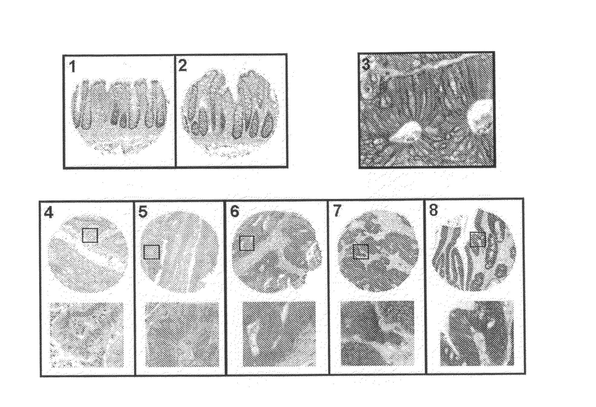
Determination of the EphB4 Levels in Colorectal Tumours by Immunohistochemistry
[0039]For these experiments a tissue microarray of paired tumour and normal colorectal samples of 137 patients was used. Each tissue was present in triplicate. The samples were collected in several medical institutes from the south of Finland, alter informed consent of all the patients. The expression levels of EPHB4 were determined by immunohistochemical staining with an antibody directed against the C-terminal region of human EPHB4 (Clone 3D7G8; Zymed Laboratories, San Francisco, Calif., USA). The specificity of this antibody has been demonstrated previously in formalin fixed in paraffin embedded tissues (Berclaz et al., (2003) Activation of the receptor protein tyrosine kinase EphB4 in endometrial hyperplasia and endometrial carcinoma. Ann Oncol, 14, 220-6). For the immunohistochemical staining, the commercial PowerVision Poly-HRP IHC (ImmunoVision Technologies, Brisbane, Calif., USA) kit was used acco...
example 2
Determination of the EPHB4 Levels in Colorectal Tumours by Image Analysis after Immunohistological Staining
[0040]The same tissue microarray with paired samples employed in example 1; stained as described in this example, was analyzed by image analysis. All sections without irregularities (tissues en bad state) were quantified. In total, 86 tissue samples from patients with colorectal cancer were analyzed. Each of the samples was present in triplicate, and sections corresponding to tumour tissue were analyzed.
[0041]The images corresponding to each of the sections present in the microarray were acquired with an optical microscope using the AnalySIS, Soft Imaging System GMBH software. The image analysis was performed with the same software.
[0042]The original image was treated using the following filters: a DCE (Differential Contrast Enhancemente) contrast filter. The parameter for bandwidth was fixed at 60; enhancement at 40; Edge Enhance Filter, particle size was fixed at 3 pixels and...
example 3
Determination of the Levels of EphB4 in Colorectal Cancer Cell Lines Using Western Blot
[0046]100 microgram fractions of protein extracts from the colorectal cancer cell lines were separated in a 7% SDS-polyacrylamide gel. The proteins were transferred to a nitrocellulose membrane and stained with an anti-EPHB4 antibody (dilution 1 / 200; Clone 3D7G8; Zymed Laboratories, San Francisco, Calif.) as described previously (Arango et al., (2003) c-Myc overexpression sensitizes colon cancer cells to camptothecin-induced apoptosis. British Journal of Cancer, 89, 1757-65). Afterwards, the membrane was stained with an anti-actine antibody (clone AC74, 1 / 1000; Sigma) to ensure equal loading in each lane (Arango et al., (2003) c-Myc overexpression sensitizes colon cancer cells to camptothecin-induced apoptosis. British Journal of Cancer, 89, 1757-65).
PUM
| Property | Measurement | Unit |
|---|---|---|
| mean survival time | aaaaa | aaaaa |
| mean survival time | aaaaa | aaaaa |
| gel electrophoresis | aaaaa | aaaaa |
Abstract
Description
Claims
Application Information
 - R&D
- Intellectual Property
- Life Sciences
- Materials
- Tech Scout
- Unparalleled Data Quality
- Higher Quality Content
- 60% Fewer Hallucinations
Browse by: Latest US Patents, China's latest patents, Technical Efficacy Thesaurus, Application Domain, Technology Topic, Popular Technical Reports.
© 2025 PatSnap. All rights reserved.Legal|Privacy policy|Modern Slavery Act Transparency Statement|Sitemap|About US| Contact US: help@patsnap.com


