
Low-cost device for c-scan photoacoustic imaging

a low-cost, photoacoustic imaging technology, applied in the field of imaging, can solve the problems of insufficient reliability of trus, inability to use only as a biopsy template, and inability to demonstrate the efficacy of color and power doppler ultrasound, etc., to enhance the probability of early detection, small size, and low cost

Active Publication Date: 2010-01-21
ROCHESTER INSTITUTE OF TECHNOLOGY
View PDF43 Cites 65 Cited by
  • Summary
  • Abstract
  • Description
  • Claims
  • Application Information

AI Technical Summary

Benefits of technology

[0010]It is a further object of the invention to provide a low-cost imaging technology that can take C-scan images of the prostate gland in real time based on the photoacoustic phenomenon.
[0017]The imaging system can be lightweight, small in size, and therefore portable. The entire lens and sensor assembly can fit in a capsule no larger than 1.5×1×1 inches that can be used transrectally. A low power laser source and a computer monitor is all that will be required externally.
[0020]CSPIP may also prove to be unique and better than Doppler ultrasound in detecting angiogenesis, because Doppler flow has poor detection sensitivity for slow flow velocities. In CSPIP, contrast in the image depends on the blood concentration in cancer tissue and not on the blood flow velocity; therefore, it is expected to have better contrast and improved delineation of prostate cancer.

Problems solved by technology

As men age, the risk of developing prostate cancer increases.
TRUS however, is not reliable enough to be used solely as a template for biopsy.
Efficacy of color and power Doppler ultrasound has not been demonstrated, probably due to limited resolution and small flow velocities.
These modalities are helpful, but have drawbacks and limitations.
MRI is expensive and not mobile.
Nuclear scintillation is expensive, provides low resolution planar images, and there are problems with radiotracer excretion through the kidneys.
Both these modalities are not available for general use.
Ultrasound is not reliable enough to use solely as a template for diagnosing prostate cancer.
First, in many cases prostate cancer appears as an isoechoic lesion (similar gray scale value as surrounding tissue) causing high miss rate (FIG. 10).
Secondly, when it is visible (hyper or hypoechoic), it is not possible to say with certainty if it is cancer or benign because many other noncancer conditions such as prostate atrophy, inflammation of the prostate gland, and benign tumors may also look similar in appearance on ultrasound examination.
Biopsies are uncomfortable and bleeding may result as a complication.
Because of poor lesion detection, even the current prostate biopsy techniques miss approximately 30% of prostate cancer.
Utility of color flow and power Doppler in conjunction with gray scale ultrasound has been explored, but not successfully.
U.S. Patent Application Publication No. U.S. 2009 / 0024038 A1 to Arnold teaches an acoustic imaging probe incorporating photoacoustic excitation, but does not overcome all of the above-noted deficiencies of the prior art.

Method used


Image

  • Low-cost device for c-scan photoacoustic imaging
  • Low-cost device for c-scan photoacoustic imaging
  • Low-cost device for c-scan photoacoustic imaging

Examples


first embodiment

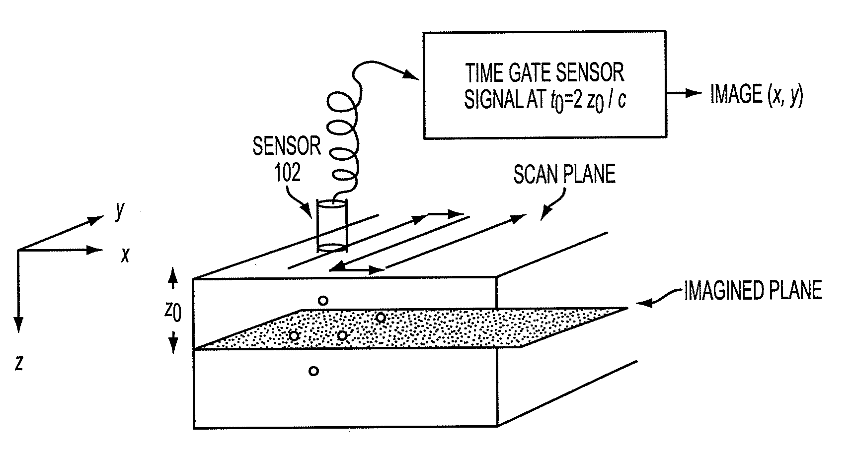
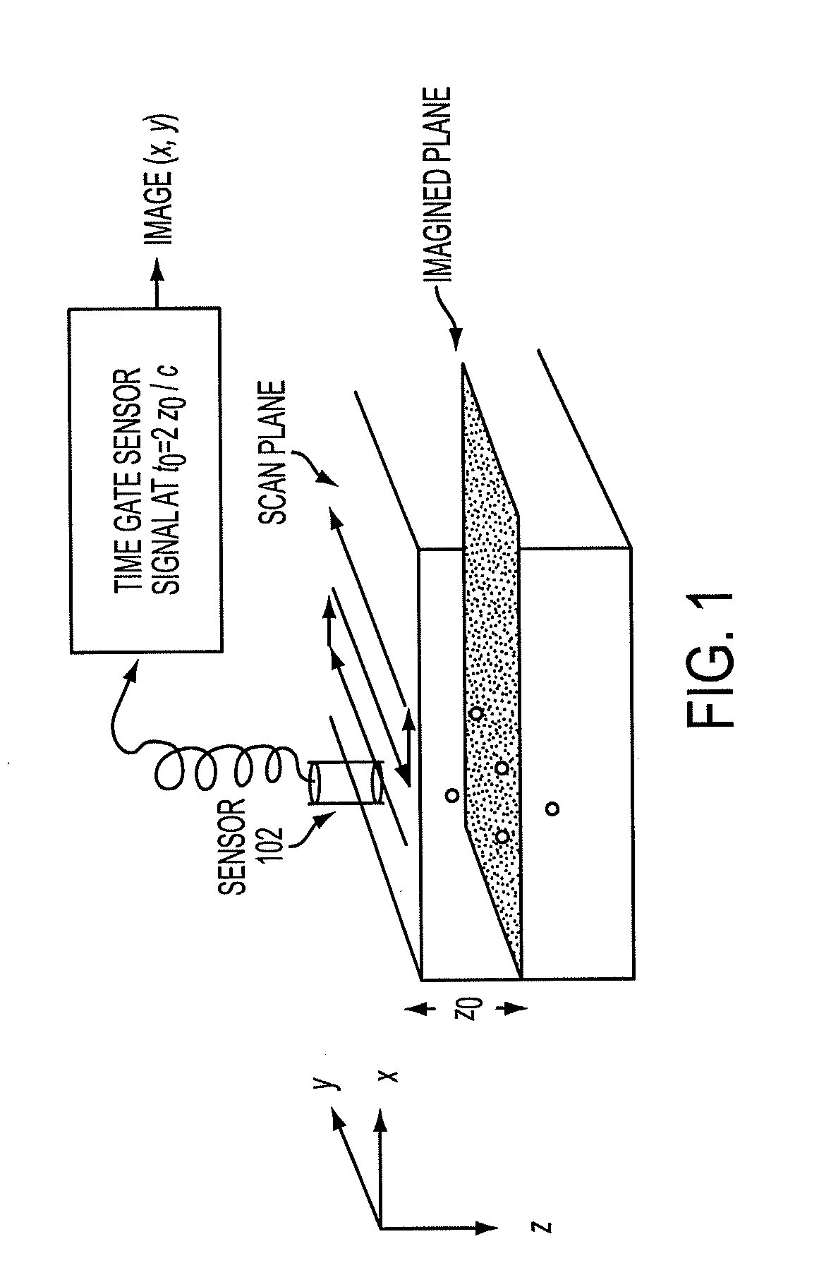
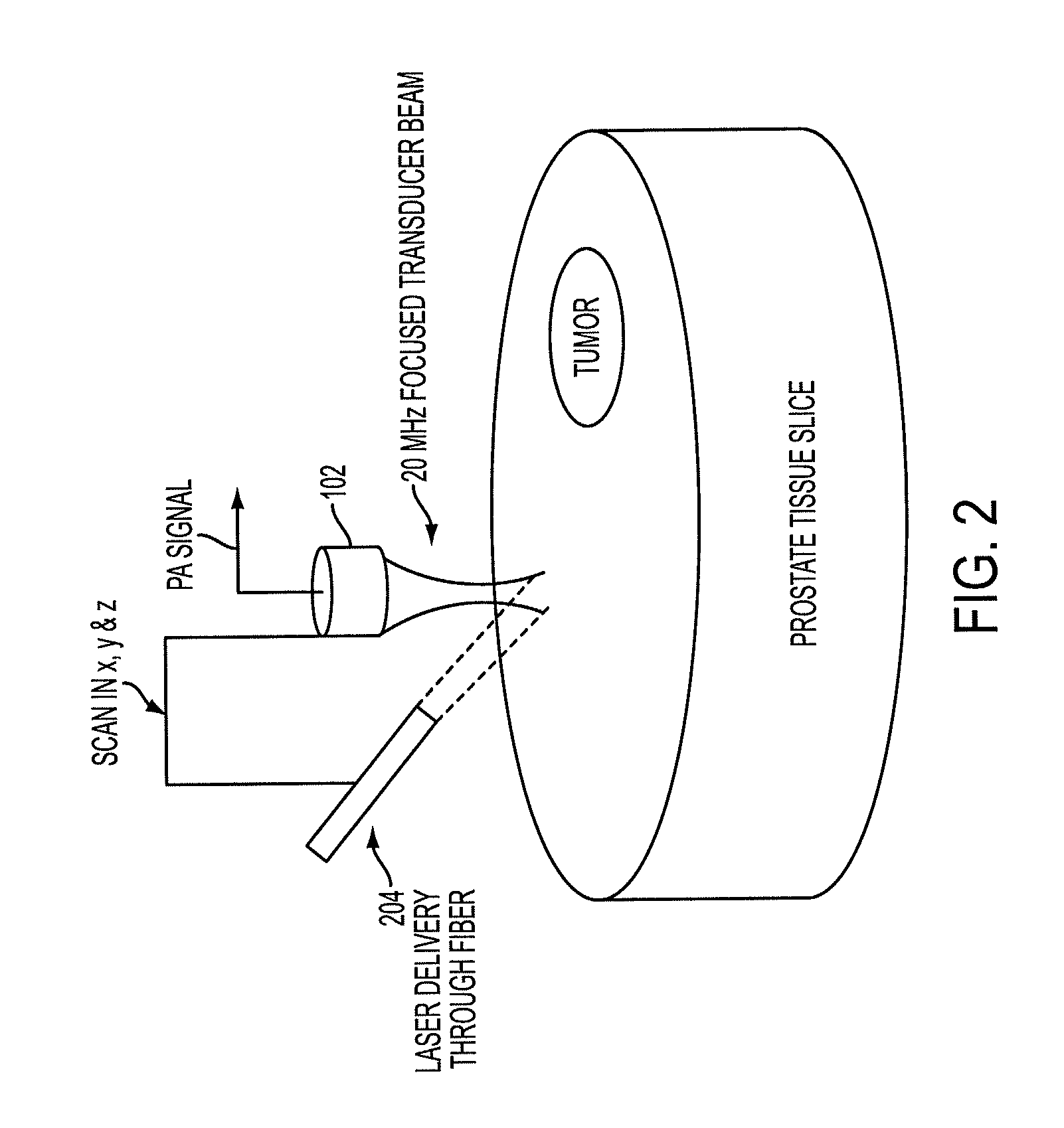
[0047]Tests of the concept will be described. FIG. 5A shows the setup. The image plane was scanned with a 1.5 mm diameter hydrophone. C-scan image plane was 10×25 pixels. The object was 0.2 mm lead pencil illuminated by a laser beam of 2 mm diameter. The laser generated pulses of wavelength 1064 nm with 10 ns pulse width. The point object was located at an object distance (OD) from the lens. The lens had a focal length of 27.5 mm. Hydrophone was located in the image plane at an image distance (ID) from the lens. An acoustic reflector was used to simulate first embodiment of prostate probe.

[0048]FIG. 5B shows a single PA signal and several time gates chosen for C-Scan. C-Scan planar slices at different time gate centers are also depicted in the figure. FIG. 4 shows the C-Scan image of the point source with fixed OD=55 mm and different ID. This demonstrates that the sharp image is located at an ID of 55 mm corresponding to an OD of 55 mm for a lens of focal length 27.5 mm. FIG. 5C dep...

embodiment 800

[0088]A second variation of the third preferred embodiment, shown in FIG. 8E as 800″, is used for breast ultrasound. It can be constructed like the third preferred embodiment 800, except for the omission of the elements concerned only with the photoacoustic effect and for any differences described herein.

[0089]The device 800″ will facilitate the definitive detection of breast cancer and generates C-scan multiplanar images of the breast. An input ultrasound source plane wave generator) is used for ultrasound pulse echo C-scan imaging. Focusing of each plane happens in real time with the help of the acoustic lens. The lens will be large enough for focusing the ultrasound signals reflected from the entire breast volume to propagate towards the receiver. The breast is compresses during the examination and the C-scan images over the entire volume of the breast are produced by the device. The probe including the acoustic window, acoustic lens and the 2d sensor is brought in contact with t...


PUM


Abstract

The prostate gland or other region of interest is stimulated with laser light, resulting in ultrasound waves (photoacoustic effect) which are focused by an acoustic lens and captured by a specific 1- or 2D sensor array and subsequently displayed as a C-scan on a computer screen. The amplitude of the ultrasound waves generated by laser stimulation is proportional to the optical absorption of the tissue element at that spatial location. Variability in tissue absorption results in C-scan image contrast. The photoacoustic imaging is combined with an ultrasound C-scan image produced with a plane ultrasound wave applied to the region of interest.

Description

REFERENCE TO RELATED APPLICATION[0001]The present application claims the benefit of U.S. Provisional Patent Application No. 61 / 081,946, filed Jul. 18, 2008, whose disclosure is hereby incorporated by reference in its entirety into the present disclosure.FIELD OF THE INVENTION[0002]The present invention is directed to imaging, e.g., of prostate tissue for detection of cancer and more particularly to photoacoustic imaging and ultrasound imaging to produce C-scans.DESCRIPTION OF RELATED ART[0003]Prostate cancer is the most prevalent newly diagnosed malignancy in men, second only to lung cancer in causing cancer-related deaths. Adenocarcinoma of the prostate is the most common malignancy in the Western world. There were a projected 218,890 new cases of prostate cancer diagnosed in the United States in 2007, with an estimated 27,050 deaths. As men age, the risk of developing prostate cancer increases. Prostate cancer has been found incidentally in approximately 30% of autopsy specimens o...

Claims


Application Information

Patent Timeline
Patent Type & Authority Applications(United States)
IPC IPC(8): A61B8/00
CPCA61B5/0059A61B5/0095A61B5/415A61B5/418A61B5/4381A61B8/4416A61B5/4337A61B5/4571A61B5/0091G01S7/52061A61B5/4312
Inventor DOGRA, VIKRAM S.RAO, NAVALGUND A.H.K.
Owner ROCHESTER INSTITUTE OF TECHNOLOGY
Learn More