
Novel oligonucleotide compositions and probe sequences useful for detection and analysis of micrornas and their target mrnas

a technology of oligonucleotide compositions and probe sequences, applied in the field of detection and analysis of target nucleotide sequences, can solve the problems of low throughput, poor sensitivity, inability to achieve, etc., and achieve the effect of high usefulness

Inactive Publication Date: 2009-11-19
EXIQON AS
View PDF6 Cites 15 Cited by
  • Summary
  • Abstract
  • Description
  • Claims
  • Application Information

AI Technical Summary

Benefits of technology

"The patent text describes a method for detecting and analyzing non-coding RNA molecules in complex nucleic acid samples. The method uses oligonucleotide probes that are designed to have improved sensitivity and specificity for detecting specific RNA sequences. The probes are substituted with high-affinity nucleobase analogues to increase their ability to detect target RNA molecules. The method can be used to design detection probes for specific RNA sequences and can also be used in computer systems for automated design of optimal detection probes. Overall, the method provides a better way to analyze the complex RNA transcriptomes of higher eukaryotic organisms."

Problems solved by technology

The disadvantage of all the gel-based assays (Northern blotting, primer extension, RNase protection assays etc.) as tools for monitoring miRNA expression includes low throughput and poor sensitivity.
Consequently, a large amount of total RNA per sample is required for Northern analysis of miRNAs, which is not feasible when the cell or tissue source is limited.
The drawback of all DNA-based oligonucleotide arrays regardless of the capture probe length is the requirement of high concentrations of labelled input target RNA for efficient hybridization and signal generation, low sensitivity for rare and low-abundant miRNAs, and the necessity for post-array validation using more sensitive assays such as real-time quantitative PCR, which is not currently feasible.
In addition, at least in some array platforms discrimination of highly homologous miRNA differing by just one or two nucleotides could not be achieved, thus presenting problems in data interpretation, although the 60-mer microarray by Barad et al.
This method is useful to clone miRNAs, but highly impractical for routine miRNA expression profiling, since it involves gel isolation of small RNAs and ligation to linker oligonucleotides.
Although apparently sensitive and specific for the mature miRNA, the drawback of the Invader quantitation assay is the number of oligonucleotide probes and individual reaction steps needed for the complete assay, which increases the risk of cross-contamination between different assays and samples, especially when high-throughput analyses are desired.
The disadvantage of this method is that it only allows quantification of the precursor miRNAs, which does not necessarily reflect the expression levels of mature miRNAs.
However, these techniques lack the resolution for addressing the spatial and temporal expression patterns of mature miRNAs.
Due to the small size of mature miRNAs, detection of them by standard RNA in situ hybridization has proven difficult to adapt in both plants and vertebrates, even though in situ hybridization has recently been reported in A. thaliana and maize using RNA probes corresponding to the stem-loop precursor miRNAs (Chen et al.
Although sensitive, this approach is time-consuming since it requires generation of the expression constructs and transgenes.
The large number of miRNAs along with their small size makes it difficult to create loss-of-function mutants for functional genomics analyses.
Another potential problem is that many miRNA genes are present in several copies per genome occurring in different loci, which makes it even more difficult to obtain mutant phenotypes.
Thus, the success rate for using DNA antisense oligonucleotides to inhibit miRNA function would most likely be too low to allow functional analyses of miRNAs on a larger, genomic scale.
A drawback of this method is the need of high 2′-O-methyl oligonucleotide concentrations (100 micromolar) in transfection and injection experiments, which may be toxic to the animal.
In conclusion, the biggest challenge in detection, quantitation and functional analysis of the mature miRNAs as well as siRNAs using currently available methods is their small size of the of 18-25 nt and often low level of expression.
Without the ability to detect and quantify the splice variants present in one tissue, the transcript content or the protein content cannot be described accurately.
At present, there is little understanding of the rates at which alternative splicing patterns or RNA editing change, and the factors influencing these rates.
However, clinical information can be incomplete or misleading.
These difficulties may result in diagnostic confusion, with the need for mandatory second opinions in all surgical pathology cases (Tomaszewski and LiVolsi 1999, Cancer 86: 2198-2200).
Molecular diagnostics offer the promise of precise, objective, and systematic human cancer classification, but these tests are not widely applied because characteristic molecular markers for most solid tumors have yet to be identified.
However, studies are still limited and have utilized different array platforms making it difficult to compare the different datasets (Golub et al.
In addition, comprehensive gene expression databases have to be developed, and there are no established analytical methods yet capable of solving complex, multiclass, gene expression-based classification problems.
Another problem for cancer diagnostics is the identification of tumor origin for metastatic carcinomas.
The lack of unique microscopic appearance of the different types of adenocarcinomas challenges morphological diagnosis of adenocarcinomas of unknown origin.
The application of tumor-specific serum markers in identifying cancer type could be feasible, but such markers are not available at present (Milovic et al.
98: 15149-15154), but the lack of a standard for array data collection and analysis make them difficult to use in a clinical setting.
The drawback of this method is, however, its low throughput, making it inappropriate for routine clinical applications.
2002, Hum. Mol. Genet. 11: 199-206), the measurement of such a large number of randomly selected genes by PCR is clinically impractical.
The biggest challenge, on the other hand, in detection and quantitation of the mature miRNAs using currently available methods is the small size of 18-25 nt and sometimes low level of expression.

Method used


Image

  • Novel oligonucleotide compositions and probe sequences useful for detection and analysis of micrornas and their target mrnas
  • Novel oligonucleotide compositions and probe sequences useful for detection and analysis of micrornas and their target mrnas
  • Novel oligonucleotide compositions and probe sequences useful for detection and analysis of micrornas and their target mrnas

Examples


example 1

Synthesis, Deprotection and Purification of LNA-Substituted Oligonucleotide Probes

[0174]The LNA-substituted probes of Example 2 to 11 were prepared on an automated DNA synthesizer (Expedite 8909 DNA synthesizer, PerSeptive Biosystems, 0.2 μmol scale) using the phosphoramidite approach (Beaucage and Caruthers, Tetrahedron Lett. 22: 1859-1862, 1981) with 2-cyanoethyl protected LNA and DNA phosphoramidites, (Sinha, et al., Tetrahedron Lett. 24: 5843-5846, 1983). CPG solid supports derivatised with a suitable quencher and 5′-fluorescein phosphoramidite (GLEN Research, Sterling, Va., USA). The synthesis cycle was modified for LNA phosphoramidites (250 s coupling time) compared to DNA phosphoramidites. 1H-tetrazole or 4,5-dicyanoimidazole (Proligo, Hamburg, Germany) was used as activator in the coupling step.

[0175]The probes were deprotected using 32% aqueous ammonia (1 h at room temperature, then 2 hours at 60° C.) and purified by HPLC (Shimadzu-SpectraChrom series; Xterra™ RP18 column, ...

example 2

List of LNA-Substituted Detection Probes for Detection of Fully Conserved Vertebrate microRNAs in All Vertebrates

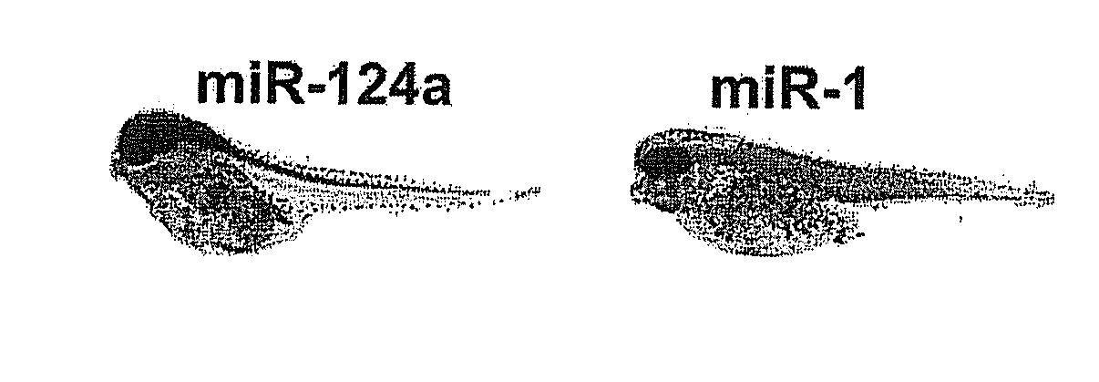
[0176]LNA nucleotides are depicted by capital letters, DNA nucleotides by lowercase letters, mC denotes LNA methyl-cytosine. The detection probes can be used to detect and analyze conserved vertebrate miRNAs by RNA in situ hybridization, Northern blot analysis and by silencing using the probes as miRNA inhibitors. The LNA-modified probes can be conjugated with a variety of haptens or fluorochromes for miRNA in situ hybridization using standard methods. 5′-end labeling using T4 polynucleotide kinase and gamma-32P-ATP can be carried out by standard methods for Northern blot analysis. In addition, the LNA-modified probe sequences can be used as capture sequences for expression profiling by LNA oligonucleotide microarrays. Covalent attachment to the solid surfaces of the capture probes can be accomplished by incorporating a NH2—C6— or a NH2—C6-hexaethylene glycol monomer or d...

example 3

List of LNA-Substituted Detection Probes for Detection of Fully Conserved Vertebrate microRNAs in All Vertebrates

[0177]LNA nucleotides are depicted by capital letters, DNA nucleotides by lowercase letters, mC denotes LNA methyl-cytosine. The detection probes can be used to detect and analyze conserved vertebrate miRNAs by RNA in situ hybridization, Northern blot analysis and by silencing using the probes as miRNA inhibitors. The LNA-modified probes can be conjugated with a variety of haptens or fluorochromes for miRNA in situ hybridization using standard methods. 5′-end labeling using T4 polynucleotide kinase and gamma-32P-ATP can be carried out by standard methods for Northern blot analysis. In addition, the LNA-modified probe sequences can be used as capture sequences for expression profiling by LNA oligonucleotide microarrays. Covalent attachment to the solid surfaces of the capture probes can be accomplished by incorporating a NH2—C6— or a NH2—C6-hexaethylene glycol monomer or d...


PUM

PropertyMeasurementUnit
affinityaaaaaaaaaa
lengthaaaaaaaaaa

Abstract

The invention relates to ribonucleic acids and oligonucleotide probes useful for detection and analysis of microRNAs and their target mRNAs, as well as small interfering RNAs (siRNAs).

Description

CROSS-REFERENCE TO RELATED APPLICATIONS[0001]This application is a continuation of U.S. application Ser. No. 11 / 324,177, filed Dec. 29, 2005, which claims benefit of the filing dates of U.S. provisional patent application No. 60 / 640,098, filed Dec. 29, 2004, and of Danish patent application numbers PA 2004 02018, PA 2005 00638, PA 2005 00637, and PA 2005 01351, filed Dec. 29, 2004, Apr. 29, 2005, Apr. 29, 2005, and Sep. 27, 2005, respectively, each of which is hereby incorporated by reference.[0002]The present invention relates to ribonucleic acids and oligonucleotide probes useful for detection and analysis of microRNAs and their target mRNAs, as well as small interfering RNAs (siRNAs). The invention furthermore relates to oligonucleotide probes for detection and analysis of other non-coding RNAs, as well as mRNAs, mRNA splice variants, allelic variants of single transcripts, mutations, deletions, or duplications of particular exons in transcripts, e.g. alterations associated with ...

Claims


Application Information

Patent Timeline
Patent Type & Authority Applications(United States)
IPC IPC(8): A61K38/16C07H21/00A61P43/00C12N15/11G16B25/20
CPCC12N15/111C12Q2600/178C12N2320/11C12Q1/6813C12Q1/6827C12Q1/6832C12Q1/6841C12Q1/6883G06F19/20G06F19/22C12N2310/141C12Q2600/158C12Q2527/107C12Q2525/207C12Q2525/101G16B25/00G16B30/00A61P43/00G16B25/20
Inventor KAUPPINEN, SAKARIPLASTERK, RONALDWIENHOLDS, ERNOKLOOSTERMAN, WIGARD
Owner EXIQON AS
Learn More