
Tissue Conditioning Protocols

a tissue and conditioning technology, applied in the field of tissue conditioning protocols, can solve the problems of increasing the analysis cost and exposure risk associated with each tissue sample tested, affecting the use of diagnostics, and rapidly deteriorating tissue samples, so as to prevent tissue exposure, and reduce the risk of ihc and ish integration

Inactive Publication Date: 2009-07-02
VENTANA MEDICAL SYST INC
View PDF10 Cites 21 Cited by
  • Summary
  • Abstract
  • Description
  • Claims
  • Application Information

AI Technical Summary

Benefits of technology

[0021]Some embodiments use high boiling point, low vapor pressure tissue conditioning fluids. The fluids or mixtures exhibit minimal fluid loss or loss in fluid volume during the tissue conditioning reaction.

Problems solved by technology

Without preservation, tissue samples rapidly deteriorate.
This deterioration quickly compromises their use in diagnostics.
These organic solvents are very volatile causing problems that require special processing (e.g., traditionally de-waxing is performed in ventilated hoods) or special waste disposal.
The use of these organic solvents increases the analysis cost and exposure risk associated with each tissue sample tested and has serious environmental effects.
All such prior art schemes entail system design complexities or limitations.
For example, 120 degrees C. processing requires pressure vessel containment limiting easy integration with downstream IHC processing in a single platform.
Microwave processing and sonication entail instrumentation complexities of their own, significantly challenging downstream IHC and ISH integration.
Generally, higher temperatures cause larger evaporation losses, which challenge fluid retention schemes.
Operating too close to the boiling point of the retrieval solution also causes “fluidic instabilities”.
First, solvent evaporation causes the solution to concentrate and the tissue potentially to dry.
Second, dissolved gases come out of solution as temperature rises for many liquid systems.
Entrained gas bubbles can prevent exposure of the tissue to solution leading to insufficient treatment and inconsistent staining.
Third, the solution may locally boil at hot spots.
Boiling and entrained or nucleated gas bubbles in or around tissue causes morphological damage.
While such a process serves to achieve retrieval in only a few minutes, substantial time is still consumed with sample loading, apparatus heating, apparatus cooling, and sample unloading.
Also, high pressure processing can be dangerous if high pressure steam inadvertently escapes.
Furthermore, incorporating a pressure vessel into an automated integrated system, which otherwise provides reliable, cheap, simple, and small-footprint processing, is not practical.
The issues and difficulties are similar to those of the high-pressure steam.

Method used


Image

  • Tissue Conditioning Protocols
  • Tissue Conditioning Protocols
  • Tissue Conditioning Protocols

Examples


example 1

Ki67 on Tonsil on DISCOVERY® Autostainer Using Automated Antigen Retrieval

[0077]Tissue Block A contained a piece of paraffin-embedded, neutral-buffered, formalin fixed human tonsil. The block was micro-sectioned in approximately 4-micron thick sections, one section mounted per slide for ˜200 slides provided for testing. Tissue cross section diameter was approximately 1.0 cm. Slides had been stored for a minimum of ˜1 month and so were effectively dried and adhered to the glass. Slides were de-waxed off-line in xylenes and graded alcohols and thoroughly rinsed with de-ionized water. Antigen Ki-67 was selected for testing retrieval characteristics because it is known to be masked by formalin fixation. Hematoxylin counterstain was selected to improve visualization of tissue morphology. The following reagents were all obtained from Ventana Medical Systems, Inc., Tucson, Ariz.: Antibody CONFIRM™ anti-Ki67 (K-2 clone) catalogue #790-29 10; DAB MAP™ Kit cat #760-124; Universal Secondary An...

example 2

Time Dependence of Antigen Retrieval

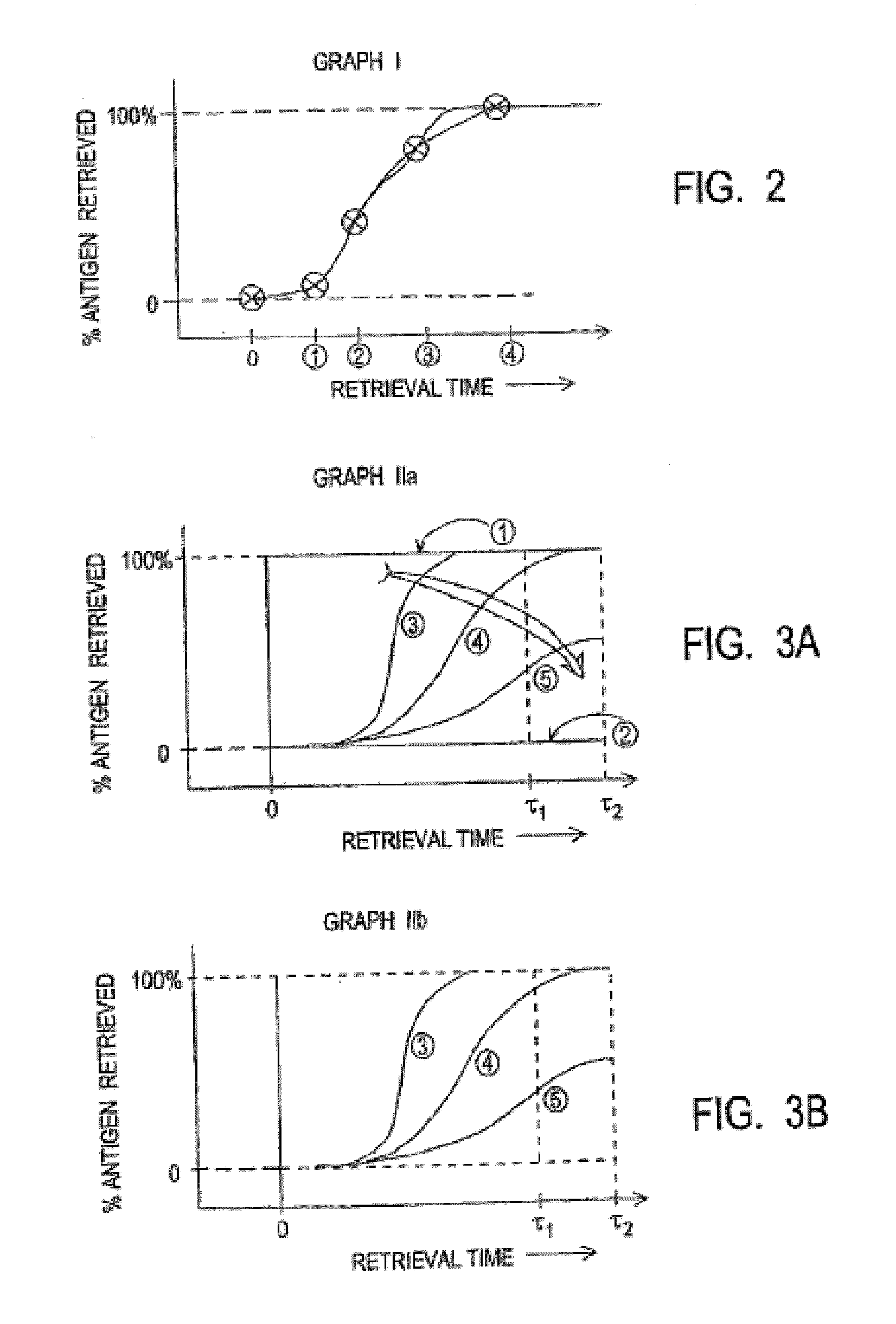
[0080]Tissue Block B contained a piece of paraffin-embedded, neutral-buffered, formalin-fixed human tonsil. Four slides each with a single tissue section were run at various conditions of antigen retrieval processing with nominal set point reaction temperature of 100 C: “Short”, “Mild”, “Standard”, and “Extended” protocols. All four protocols began with the same heat ramp processing taking ˜18 minutes. Short tissue conditioning total time was 24 minutes; Mild was 42 minutes; Standard was 72 minutes; and Extended was 102 minutes. Each condition therefore progressively exposed the tissue sample to greater antigen retrieval reaction times. Table 1, below illustrates the effect of retrieval time on observable stain intensity. Greater exposure time during antigen retrieval processes increases the degree of antigen retrieval as measured by observable detection, illustrated in Graph I as shown in FIG. 2.

TABLE 1Exposure ConditionStain intensityShort (24 m...

example 3

Temperature Dependence of Antigen Retrieval

[0082]Tissue Blocks B and C each contained a piece of paraffin-embedded, neutral-buffered, formalin fixed human tonsil. One slide each was stained using standard tissue conditioning and an additional slide was stained using the same protocol except that the tissue conditioning temperature was changed to 95 degrees C. and 90 degrees C. Results are reported in Table 2, below.

TABLE 2TemperatureStain Intensity -Stain Intensity -deg C.Tissue CTissue B100darkmedium95darklight90mediumfaint

[0083]Tissue B required greater retrieval in order to recover an equivalent amount of antigen signal compared to Tissue C for each of the retrieval processes listed. Tissue morphology was good in all cases.

[0084]Three various retrieval processes are illustrated in Table 2, above differentiated by process temperature with various staining intensity results. Higher temperature provided greater antigen retrieval. Graph IIb (FIG. 3b) illustrates this temperature effe...


PUM


Abstract

Solutions exhibiting little or no evaporative loss at elevated temperatures, i.e., in excess of 100° C., are employed in place of conventional aqueous-based antigen retrieval solutions.

Description

RELATED APPLICATION DATA[0001]This claims the benefit of U.S. utility patent application Ser. No. 11 / 720,705, filed on 14 Dec. 2005, which application claims the benefit of U.S. provisional application No. 60 / 637,245, filed on 17 Dec. 2004; all of these applications are hereby incorporated by reference in their entirety.FIELD[0002]The present invention relates to the processing of tissue samples, and more particularly to methods, materials, and apparatus for processing of preserved tissue samples. The invention description particularly references processing of embedded biological tissue samples for staining and will be in connection with such utility, although other utilities are contemplated.BACKGROUNDSummary of the Related Art[0003]The diagnosis of disease based on the interpretation of tissue or cell samples taken from a diseased organism has expanded dramatically over the past few years.[0004]In addition to traditional histological staining techniques and immunohistochemical ass...

Claims


Application Information

Patent Timeline
IPC IPC(8): G01N1/28
CPCG01N1/30G01N1/36G01N1/312
Inventor REESER, RYANKRAM, BRIAN H.RIZZO, VINCENT R.CHAFIN, DAVIDKOSMEDER, JEROME W.BIENIARZ, CHRISTOPHER
Owner VENTANA MEDICAL SYST INC
Learn More