
Methods for detecting colon carcinoma

a colon carcinoma and cancer technology, applied in the field of cancer diagnosis, can solve the problems of increasing national expenditures by $0, affecting the probability of cancer cells, and being inferior to other strategies such as fecal occult blood testing, fobt, colonoscopy, etc., and achieves the effect of relatively low probability of cancer cells

Inactive Publication Date: 2009-07-02
PEVSNER PAUL H
View PDF5 Cites 5 Cited by
  • Summary
  • Abstract
  • Description
  • Claims
  • Application Information

AI Technical Summary

Benefits of technology

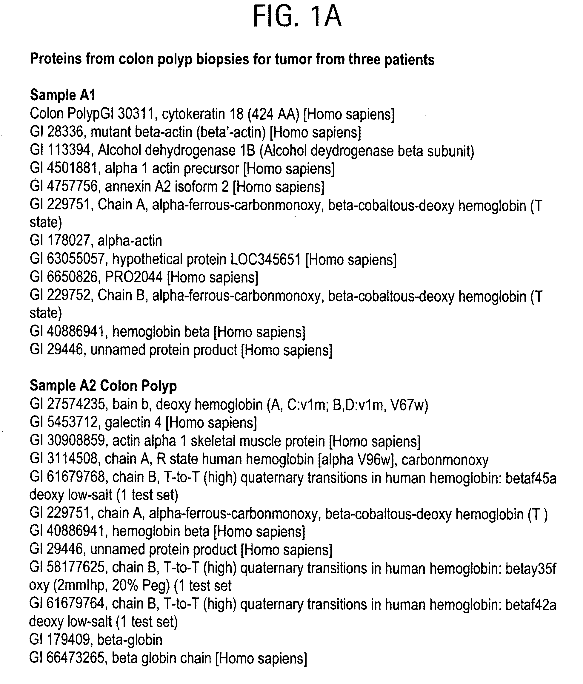
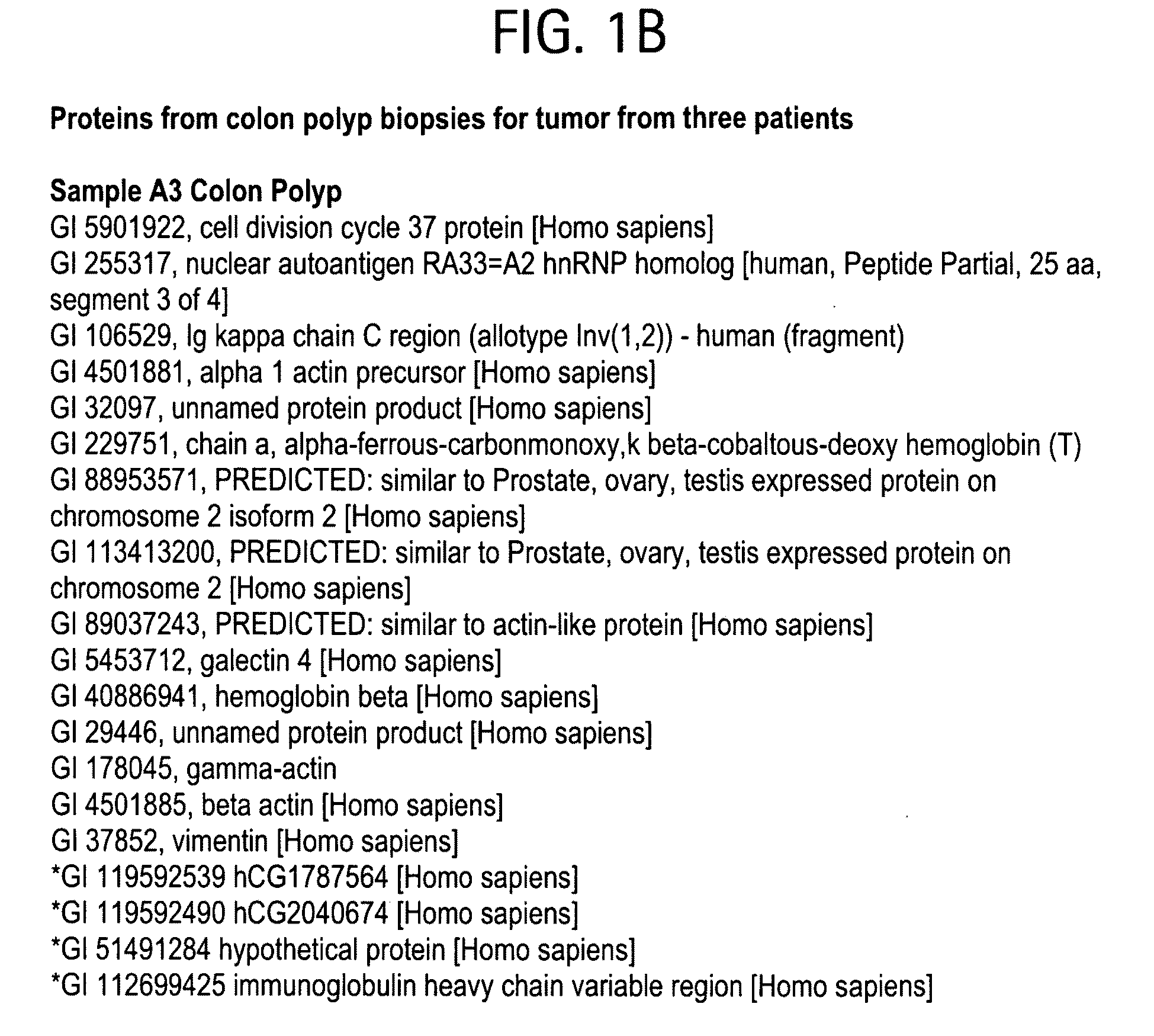
[0020]The methods may optionally include quantifying one or more of the carcinoma associated markers such as those presented in FIG. 1. The methods may further include comparing the amount of one or more carcinoma associated markers in the sample with either (i) the amount determined for normal samples know...

Problems solved by technology

In an extensive study, Song et al. demonstrated that fecal DNA testing every 5 years appeared both diagnostically effective and cost-effective compared with no screening, but inferior to other strategies such as fecal occult blood testing, FOBT, and colonoscopy.
The conclusion was that despite savings in care, widespread screening is unlikely to be cost saving and may increase national expenditures by $0.8-$2.8 billion per year with conventional tests.
Fourth, in some cases, mutant tumor suppressor genes appear to exert a phenotypic effect even when present in the heterozygous state; thus, some tumor suppressor genes may not be “recessive” at the cellular level.
Furthermore, bcl-2 expression was frequently abnormal in nondysplastic epithelium surrounding dysplastic lesions, suggesting that altered expression occurred before the development of morphological dysplasia.
Furthermore, he proposed that cells with mutations and resistance to apoptosis in “normal” appearing mucosa represented a field defect that could potentially become carcinoma.
There was also a distinctive alteration in the distribution of proliferating cells within the colonic crypts, which was particularly marked in the left colon, and this change implied an additional risk for mutational events because highly vulnerable nondifferentiated cells at the mouth of the crypts were more exposed to intraluminal factors.
The majority of these ACF's never reach macroscopic dimensions and many even regress.
This leads to “field defects” in the mucosa of patients with colon carcinoma.
Although ACF and microadenomas are expected as biomarkers of colon cancer risk when an entire animal colon is examined with methylene blue staining, such lesions are not easily detected in biopsy-sized tissue samples taken from human colons.
The complexity of genetic analysis of CRC was further discussed in the report in which the authors stated that “these genetic analyses can only identify candidate genes that may play a role in cancer and do not definitively implicate any gene in the neoplastic process”.
Such alterations result in microsatellite instability, and synchronous and metachronous lesions which develop into cancers.
2006; 15:1778-1784) Therefore, histopathology alone may significantly affect therapy by underestimating the extent of metaplastic or malignant disease.

Method used


Image

  • Methods for detecting colon carcinoma
  • Methods for detecting colon carcinoma
  • Methods for detecting colon carcinoma

Examples


example 1

[0074]Specimens from individuals with colon adenocarcinoma were compared with normal appearing colonic tissue from average risk individuals who completed a screening colonoscopy. During endoscopy using standard biopsy forceps, eight samples each measuring 2 mm×3 mm were obtained from each patient. Samples were immersed immediately in a solution of dimethyl sulfoxide (DMSO) 2%, glycerol 20%, and ethyl alcohol 78% and stored at 4° C. This mixture both fixes without cross-linking the proteins and cryoprotects the tissue. (Terracio et al., J Histochem Cytochem. 1981; 29:1021-1028; Rosene et al., J Histochem Cytochem. 1986; 34:1301-1315) Cryosections were obtained for IMS, hematoxylin and eosin histopathological staining, and protein extraction. A pathologist blinded to the clinical data, reviewed each specimen. Proteins were extracted from the colon tissue with organic solvent and high pressure using ProteoSolve and the Barocycler respectively (Pressure BioSciences, West Bridgewater, Ma...

example 2

[0077]Biopsy tissue was cryo protected by immediate immersion in a mixture of dimethyl sulfoxide, DMSO 2% / glycerol 20% / ethanol 78%. Cryo sections, 1μ thick, could be obtained without freezer artifact that would otherwise destroy the tissue architecture. Contiguous 1μ sections were obtained for histology, IMS, and protein extraction with high pressure (Barocycler, Pressure BioSciences). The complete protocols are detailed in the references, herein incorporated by reference. (Pevsner et al. “Microtubule Associated Proteins (MAP) and Motor Molecules: Direct Tissue MALDI Identification and Imaging.” 2007, British Mass Spectrometry Society, Edinburgh, Scotland ; Pevsner et al., “Colon Cancer: Protein Biomarkers in Tissue and Body” 2007; Pevsner et al. British Mass Spectrometry Society, Edinburgh, Scotland, “Colorectal Carcinoma—Field Defects in Satellite Tissue” 2007; Vecchione et al., “Prophylactic Estrogen (Estradiol) Therapy” 2007, British Mass Spectrometry Society, Robinson College, ...


PUM


Abstract

The present invention provides methods for diagnosing carcinoma, providing a prognosis for a carcinoma or assessing the likelihood that a tissue may become cancerous by identifying the presence or absence of or determining the amount of one or more carcinoma associated markers. Further, the present invention provides methods for determining whether a tissue should be surgically resected and for determining the territorial extent of resection. The carcinoma markers may be those provided in FIG. 1. The present invention also provides a diagnostic kit for diagnosing carcinoma, providing a prognosis for a carcinoma or assessing the likelihood that a tissue may become cancerous by identifying the presence of or determining the amount of one or more carcinoma associated markers. The methods and kits are especially useful regarding colon, rectal or colorectal carcinoma.

Description

CROSS REFERENCE TO RELATED APPLICATIONS[0001]The present application claims the benefit of U.S. provisional patent application 60 / 007,225, filed on Dec. 11, 2007, the disclosure of which is herein incorporated by reference.FIELD OF THE INVENTION[0002]This invention relates to methods of diagnosing carcinoma, providing a prognosis for a carcinoma or assessing the likelihood that a tissue may become cancerous.BACKGROUND OF THE INVENTION[0003]Colorectal cancer (CRC) is the second-leading cause of death from cancer in the USA, with more than 155,000 predicted new cases per year. (Vogelaar et al., Cancer 2006; 107:1624-1633) The projected number of colorectal cancer deaths for 2008 is 53,000. The standard diagnostic paradigm is based on histopathology of either biopsy or surgical specimens.[0004]In an extensive study, Song et al. demonstrated that fecal DNA testing every 5 years appeared both diagnostically effective and cost-effective compared with no screening, but inferior to other st...

Claims


Application Information

Patent Timeline
IPC IPC(8): G01N33/574G01N33/68G01N33/72C12Q1/32G01N33/566
CPCG01N33/57419G01N33/574
Inventor PEVSNER, PAUL H.
Owner PEVSNER PAUL H
Learn More