
Method and compositions for polymer nanocarriers containing therapeutic molecules

a technology of polymer nanocarriers and therapeutic molecules, which is applied in the direction of biocide, peptide/protein ingredients, microcapsules, etc., can solve the problems of limited utility of potent but labile therapeutic proteins, affinity for desired target sites, and comparatively little success

Active Publication Date: 2009-04-30
THE TRUSTEES OF THE UNIV OF PENNSYLVANIA
View PDF9 Cites 9 Cited by
  • Summary
  • Abstract
  • Description
  • Claims
  • Application Information

AI Technical Summary

Benefits of technology

[0011]In still another aspect, a pharmaceutical composition contains the filamentous polymeric nanocarrier-encapsulated protein particles in which the encapsulated protein is a therapeutically useful protein with retained biological activity. In one such embodiment, such a protein is a therapeutically useful enzyme. In yet another embodiment, the substrate for the encapsulated enzyme is freely diffusible through the polymer material of the nanoparticles, which permits prolonged enzymatic activity by the polymeric nanocarrier-encapsulated protein particles protecting protein from proteolysis.

Problems solved by technology

Rapid clearance from the circulation, inactivation by proteases and inhibitors, and a lack of affinity for the desired target sites of action limit the utility of potent but labile therapeutic proteins (Muzykantov, V. R., J Control Release 2001, 71, (1), 1-21).
Comparatively little success has been achieved, however, in nanocarrier mediated delivery of therapeutic proteins, which is especially challenging because protein's biological activity requires maintaining it's native folded state.
Loading therapeutic proteins into biodegradable polymer nanocarriers (PNC) can be complicated by protein unfolding and inactivation.
Loss of enzymatic activity due to protein unfolding in harsh conditions of PNC formulation has represented a major barrier to the use of biodegradable co-polymers for delivery of therapeutic enzymes.
However, encapsulation of large therapeutic proteins, especially enzymes, without loss of their biological activity into these polymer nanoparticles formed by self-assembly mechanism has not been reported, because conditions providing their formulation via this mechanism are not compatible with retaining enzymatic activity.
Despite the specificity of therapeutic enzymes, medical utility is often limited by inadequate delivery and insufficient stability in the body.
However, catalase and other antioxidant enzymes (e.g., superoxide dismutase) have no practical medical utility due to inadequate delivery to therapeutic sites, especially the endothelial cells lining the luminal surface of blood vessels.

Method used


Image

  • Method and compositions for polymer nanocarriers containing therapeutic molecules
  • Method and compositions for polymer nanocarriers containing therapeutic molecules
  • Method and compositions for polymer nanocarriers containing therapeutic molecules

Examples


example 1

Synthesis of Diblock Copolymers

[0082]DL-lactide was re-crystallized twice in anhydrous ether, before mixing with methoxy poly(ethylene glycol) (mPEG) MW 5,000 (Polysciences, Warrington, Pa.) in stoichiometric ratios to achieve desired molecular weights. Reactants were heated to 140° C. under nitrogen while stirring for 2 hours to remove trace water from samples. The temperature was reduced to 120° C. and stannous octoate (1 wt %) was added to catalyze the ring opening polymerization (ROP) of lactide with mPEG as the initiator. The polymerization was allowed to continue for 6 hours. The diblock copolymer was then dissolved in dichloromethane (DCM) and twice precipitated in cold diethyl ether. Residual solvent was then removed by first drying via rotary evaporation (SAFETY VAP® 205 system, Buchi, Switzerland), followed by lyophilization (RCT 60, Jouan, Winchester, Va.).

[0083]Number average molecular weights ( Mn) of bulk copolymers were determined using proton nuclear magnetic resonan...

example 2

Nanoparticle Formation

[0087]A freeze-thaw double emulsion solvent evaporation technique was used as previously described in Dziubla et al 2005 cited above and US Patent Application Publication No. 2006 / 0127386. Briefly, mPEG-PLA diblock copolymer is dissolved in DCM at 25 mg / ml. A 1 mg / ml bovine liver catalase (242 kDa) (Calbiochem, EMD Biosciences, San Diego, Calif.) solution and a polyvinyl alcohol (PVA) surfactant solution (2 wt %, 87-89% hydrolyzed, Mw=13,000-23,000) in 20 mM PBS are prepared. The primary emulsion consisted of the organic phase (1 ml polymer-DCM mixture) and the aqueous phase (100 μl catalase solution) homogenized at 15 krpm for 1 minute in a dry ice-acetone bath with a 7 mm-blade homogenizer (KINEMETICA POLYTRON 3100 instrument with a PDTA3007 / 2 generator, Brinkmann Instruments, Westbury, N.Y.). The primary emulsion was then added to 5 ml of the PVA surfactant solution and homogenized at 15 krpm for 1 minute. The resultant mixture was added to 10 ml of PVA solu...

example 3

Enzyme Loading Determination

[0088]Protein loading was determined via radioisotope labeling and enzymatic activity. Loading via radiolabeling was determined as described before, by formulating PNC with 125I-labeled catalase following the directions of the above-referenced in Dziubla et al 2005 cited above. Catalase was radiolabeled with Na125I (Perkin Elmer, Boston, Mass.) via the Iodogen method (Pierce Biotech., Rockford, Ill.). Unbound 125I was removed from catalase using BIOSPIN 6 columns in accordance with the manufacturer's instructions (Bio-Rad labs, Hercules, Calif.). Total solution 125I-catalase content was measured before centrifugation, and then radioactivity of the 125I-catalase / PNC-composed pellet after centrifugation was measured. A WIZARD 1470 gamma counter (Wallac, Oy, Turku, Finland) was used for radiotracing.

[0089]To determine loading via enzymatic activity, a catalase activity assay (Shuvaev, V. V. et al, Methods Mol Biol 2004, 283, 3-1) was used, both for the total...


PUM


Abstract

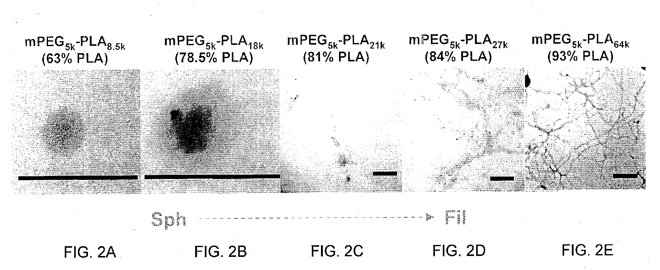
A method of controlling a physical characteristic of polymeric nanocarrier-encapsulated protein particles includes altering or selecting a weight percentage of a hydrophobic polymer block in a total amphiphilic diblock copolymer of a primary emulsion of a double emulsion, freeze-thaw technique. The primary emulsion is formed using a freeze-thaw cycle of the amphiphilic diblock copolymer and a protein having a molecular weight of up to or equal to 300,000 Da. Selection of the hydrophobic polymer block percentage alters one or more characteristics of the resulting nanoparticles, such as shape. Thus, as one aspect, a method of producing filamentous polymeric nanocarrier-encapsulated protein (i.e., active enzyme) particles involves forming a primary emulsion using a freeze-thaw cycle of (i) an amphiphilic diblock copolymer, which has a molecular weight of about 10,000 to about 100,000 Da and comprises a conjugate of the hydrophobic polymer block and a hydrophilic polymer block, wherein the amphiphilic diblock copolymer comprises greater than 81% to about 95% by weight of the hydrophobic polymer block; and a protein having a molecular weight of up to or equal to about 300,000 Da. Various compositions comprising such filamentous-shaped nanocarrier particles, and methods of use for diagnosis and therapy are disclosed.

Description

STATEMENT REGARDING FEDERALLY SPONSORED RESEARCH OR DEVELOPMENT[0001]The present invention was supported by Grant Nos. HL007954, HL073940-01-A1, PO1-HL079063 from the National Institutes of Health. The government has an interest in the invention.BACKGROUND OF THE INVENTION[0002]Rapid clearance from the circulation, inactivation by proteases and inhibitors, and a lack of affinity for the desired target sites of action limit the utility of potent but labile therapeutic proteins (Muzykantov, V. R., J Control Release 2001, 71, (1), 1-21). Diverse drug delivery systems (e.g., natural lipoproteins, liposomes and polymer nanocarriers) are being widely designed in order to maximize drug efficacy and minimize side effects (Langer, R., Nature 1998, 392, (6679 Suppl), 5-10). For example, polyethylene glycol (PEG), a hydrophilic polymer that enhances aqueous solubility, masks drugs and carriers from host defense systems and prolongs circulation in the bloodstream (“stealth” technology) (Moghimi...

Claims


Application Information

Patent Timeline
Patent Type & Authority Applications(United States)
IPC IPC(8): A61K9/16A61K9/50A61K38/00A61K38/54
CPCA61K9/5153
Inventor SIMONE, ERICMUZYKANTOV, VLADIMIR R.DZIUBLA, THOMAS D.
Owner THE TRUSTEES OF THE UNIV OF PENNSYLVANIA
Learn More