
Apparatus for Thin-Layer Cell Smear Preparation and In-situ Hybridization

a thin-layer cell smear and in-situ hybridization technology, applied in the field of in-situ hybridization of thin-layer cell smear preparation, can solve the problems of poor sampling technique, high false negative result, and difficult sampling, and achieve the effect of simplifying the preparation and operation of thin-layer cell smear

Active Publication Date: 2009-04-23
KING CAR FOOD IND
View PDF3 Cites 14 Cited by
  • Summary
  • Abstract
  • Description
  • Claims
  • Application Information

AI Technical Summary

Benefits of technology

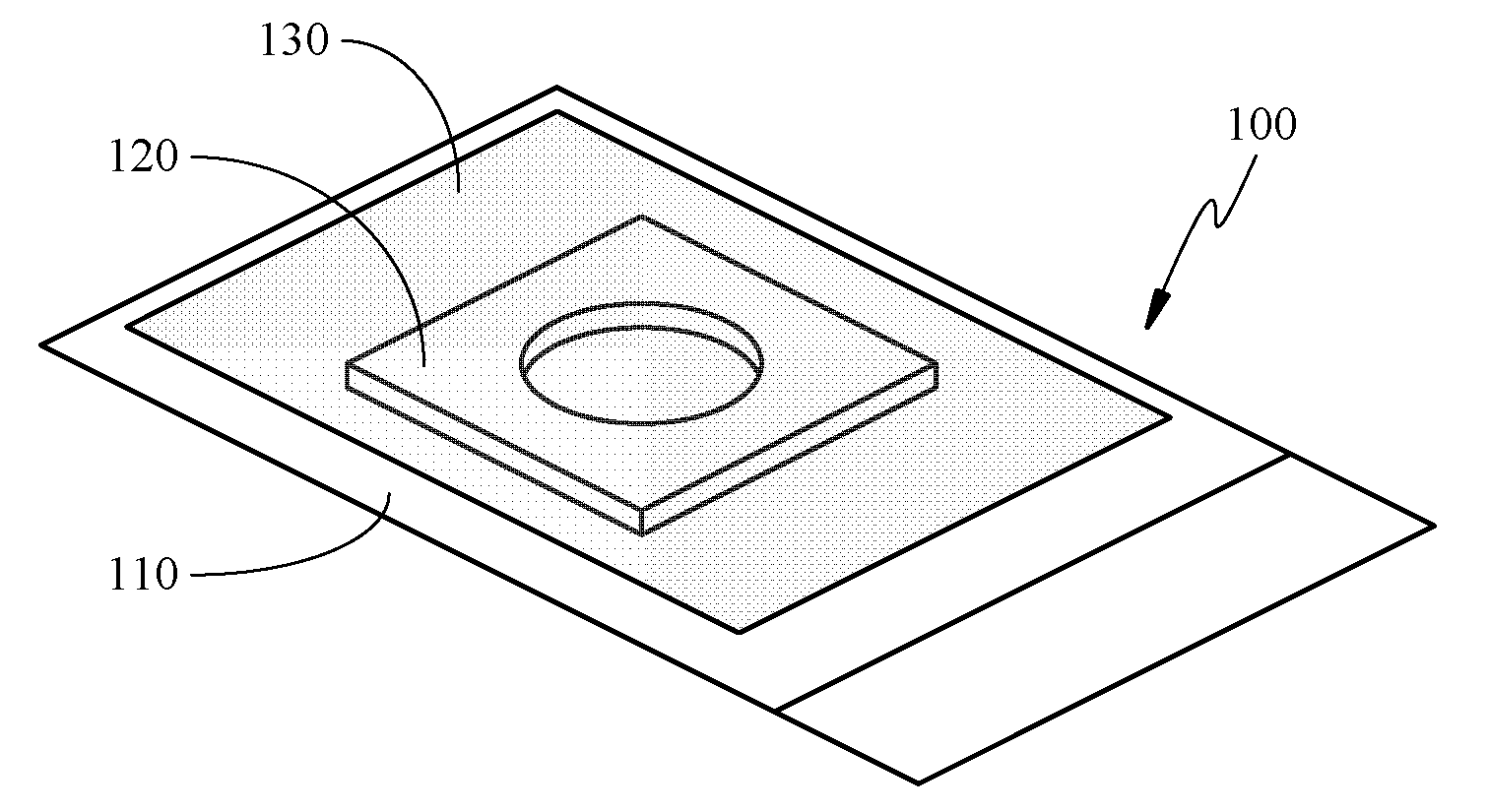
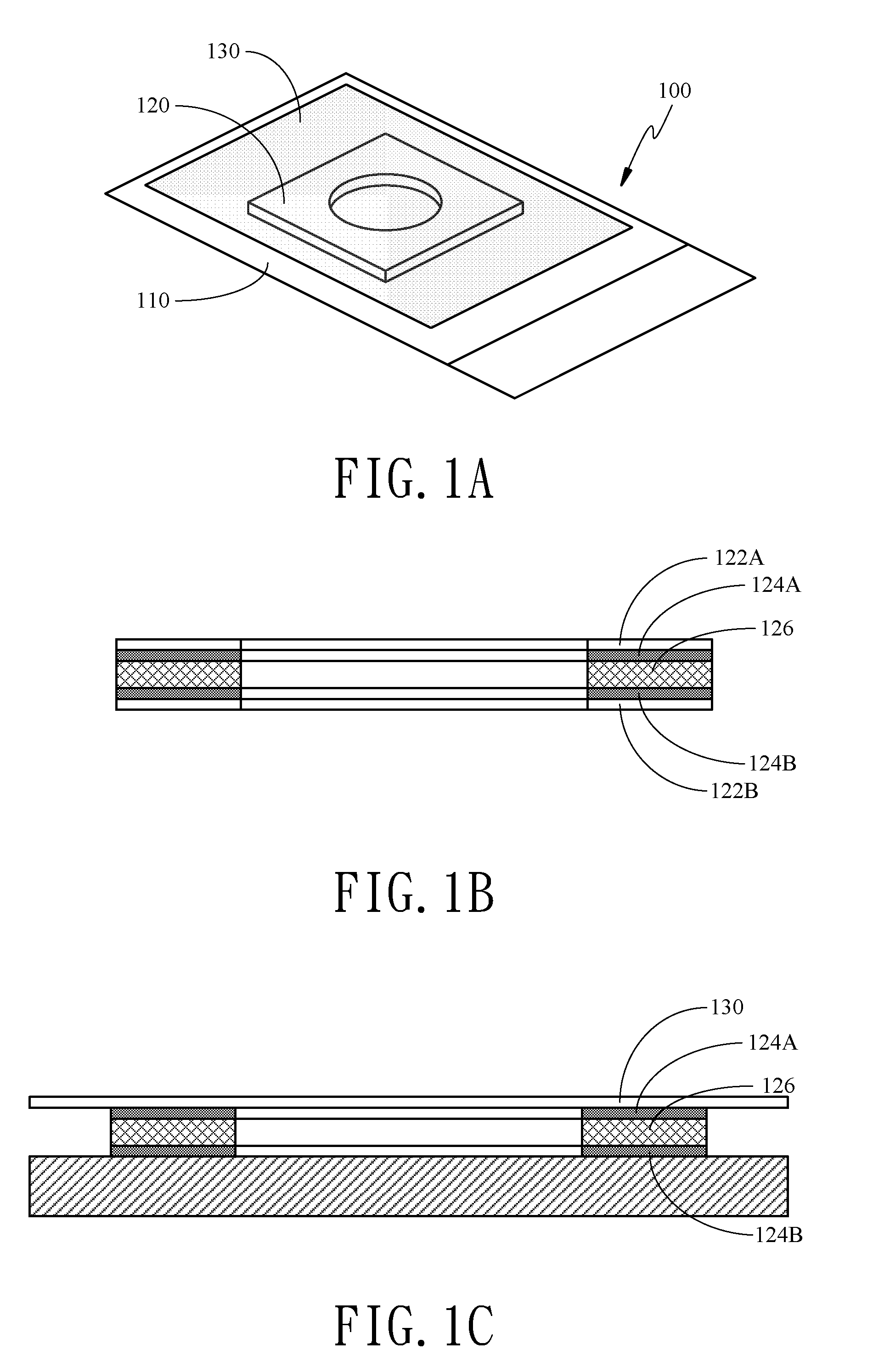
[0013]One object of the present invention is to provide an apparatus for thin-layer cell smear preparation and in-situ hybridization to simplify the thin-layer cell smear preparation and operations for the in-situ hybridization experiment by integrating two functions in one apparatus.

Problems solved by technology

The controversial point in medicine is that the false negative result is as high as 10˜40%.
Some is caused by poor sampling technique while some is caused by the examinee, such as cervical atresia, cervical atrophy without hormone replacement therapy after menopause, and serious anteflexion or retroflexion of uterus, to make sampling difficult.
On the other hand, if the specimen cells on the smear are too thick, that is, many layers of cells are overlapped, the diagnosis becomes difficult.
In addition, the common false negative result is caused by misdiagnosis.
Since typically there are two or three hundred thousand cells on a smear, it is difficult to make the diagnosis in a few minutes.
These operations are tedious and the labor and material costs are high.

Method used


Image

  • Apparatus for Thin-Layer Cell Smear Preparation and In-situ Hybridization
  • Apparatus for Thin-Layer Cell Smear Preparation and In-situ Hybridization
  • Apparatus for Thin-Layer Cell Smear Preparation and In-situ Hybridization

Examples


example 1

[0032]1. A slide (glass or plastic slide) is coated with a cross-linking agent, in order to have cells be adhered thereon. The slide is immersed in the poly-L-lysine or silane solution for 10 minutes and then taken out to dry at room temperature.

[0033]2. Specimen cells in the preservation solution or fixation solution is then centrifugal settling. After collected, the cells are then suspended in a buffer solution to prepare a cell suspension.

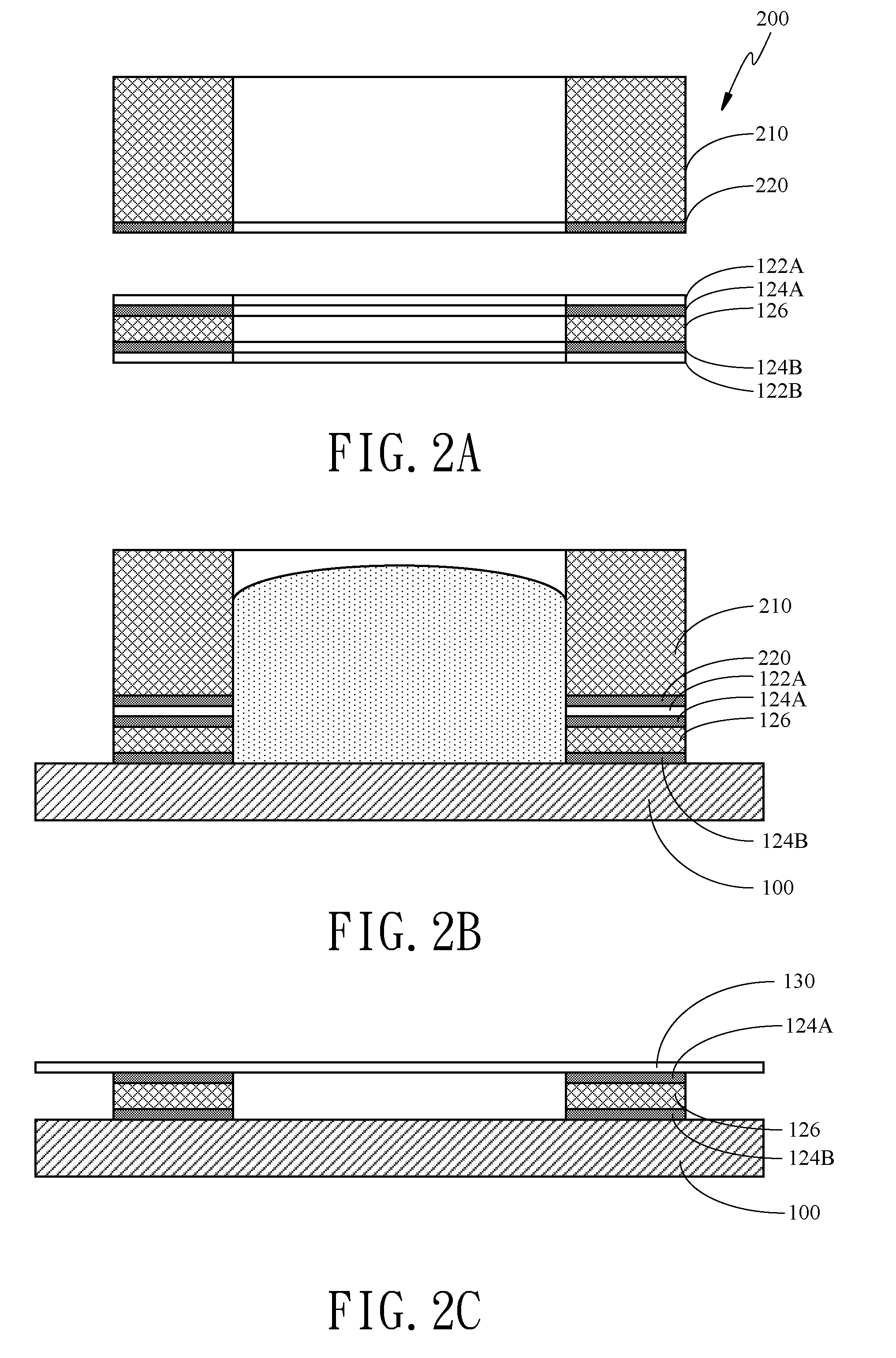
[0034]3. The release paper of the positioning device is removed and the positioning device is adhered to the slide. The cell suspension is added and set for 15 minutes to have the cells naturally precipitate and be adsorbed on the slide. Then, the cell suspension is poured out and the buffer solution is used to rinse once. It is stood to dry at room temperature.

[0035]A hybridization buffer solution comprising a fluorescence labeled nucleic acid probe is added. After the release paper is removed, it is sealed by the sealing device. At 37° C., the...

example 2

[0036]1. A slide (glass or plastic slide) is coated with a cross-linking agent, in order to have cells be adhered thereon. The slide is immersed in the poly-L-lysine or silane solution for 10 minutes and then taken out to dry at room temperature. The slide is assembled with the positioning device.

[0037]2. Specimen cells in the preservation solution or fixation solution is then centrifugal settling. After collected, the cells are then suspended in a buffer solution to prepare a cell suspension.

[0038]3. The release paper of the positioning device having the thickening device is removed and the positioning device is adhered to the slide. The cell suspension is added and set for 15 minutes to have the cells naturally precipitate and be adsorbed on the slide. Then, the cell suspension is poured out and the buffer solution is used to rinse once. It is stood to dry at room temperature.

[0039]4. The thickening device and the release paper of the positioning device are removed. A hybridizatio...


PUM

PropertyMeasurementUnit
thicknessaaaaaaaaaa
temperatureaaaaaaaaaa
adhesiveaaaaaaaaaa

Abstract

The present invention discloses an apparatus for thin-layer cell smear preparation and in-situ hybridization, comprising at least one positioning device and a sealing device. The positioning device comprises at least one first opening. When the positioning device is set on a carrier device, the wall of the first opening and the carrier device form a cavity. The cavity is used to accommodate a cell suspension. The sealing device is provided on the positioning device for sealing the cavity to form an enclosed space.

Description

BACKGROUND OF THE INVENTION[0001]1. Field of the Invention[0002]The present invention is generally related to an apparatus for thin-layer cell smear preparation and in-situ hybridization, and more particularly to an apparatus for cell smear preparation and in-situ hybridization to prepare a thin-layer cell smear and carry out in-situ hybridization.[0003]2. Description of the Prior Art[0004]At present, a cervical smear is a major method for cervical cancer prophylaxis. The conventional cervical smear examination uses a cytology spatula or brush to collect cervical cells and coats on a slide for pathological diagnosis in a laboratory. Although the accuracy is not high enough, almost all of the gynecologist-obstetricians agree that the cervical smear examination should be done every year and no problem for three consecutive years can be generally considered as safe from the cervical cancer.[0005]The cervical smear examination has been practiced for years. The controversial point in med...

Claims


Application Information

Patent Timeline
Patent Type & Authority Applications(United States)
IPC IPC(8): C12M1/00
CPCA61B10/0045A61B2010/0074A61B10/0096Y10S435/803Y10S435/805
Inventor LEE, BOR-HENGYAN, CHIU-CHOWANG, YING-CHIHLIN, CHING-YU
Owner KING CAR FOOD IND
Learn More