
Systems and methods for improving image-guided tissue ablation

Active Publication Date: 2008-08-28
NUVUE THERAPEUTICS
View PDF12 Cites 54 Cited by
  • Summary
  • Abstract
  • Description
  • Claims
  • Application Information

AI Technical Summary

Benefits of technology

[0023]It is an object of the invention to teach novel materials and procedures for enhanced safety and efficacy of focal cryoablation of unwanted tumor tissue while preserving the body's immune system.
[0024]It is a further object of the invention to teach a method that decreases the operative time for cryoablation of a tumor target, and protects surrounding tissues from harmful cooling.
[0026]It is an even further object of the invention to teach a method that uses repeated challenges or treatments of the target tissue or tumor with the imageable agent(s) or microcapsule carriers of those agents to facilitate and complete the initial combined treatment.
[0027]It is yet another object of the invention to teach using, during cryoablation of a tumor, a vibrating delivery device that allows for continuous, real time visualization through Ultrasound imaging assuring the precise delivery of any needles, probes, and catheters used for the precise intratumor deposition of unique thermo sensitive, biodegradable, and imageable carriers, such as microcapsules, containing imaging contrast agent(s) and combinations of tumor inhibiting drugs. A synergistic kill effect on tumor cell results from the degradation of the thermosensitive carriers during transient controlled freezing / cooling of the target margins and after the tumor has returned to its pre-cooled normal temperature.

Problems solved by technology

Despite a strict adherence to these standards, which are time consuming, certain tumors like prostate or metastatic liver cancer show a 20 to 40% post procedure recurrence.
Systemic chemotherapy has long been used to enhance the kill effect of cryosurgery on experimental and human solid tumors, but results have been inconsistent.
A number of papers have shown that in vitro moderate freezing temperatures combined with low dose chemotherapy increased the rate of cell death for prostate and colo-rectal cancer cells.
However, these findings weren't transferred to in vivo experiments.
The issue with systemic chemotherapy is that the side effects cannot be prevented, tumor exposure to therapeutic doses is intermittent, and tumor penetration is unpredictable.
Another drawback is that tumor cells still need to be frozen.
Therefore, there is a risk of damage to neighboring normal tissue by excessive freezing.
Moreover, the cytotoxic drug penetration into tumor may be difficult and imprecise upon initiation of cryo-induced microvascular impairments particularly if a precise timing between the drug administration and the cryo-application hasn't been respected.
Since these carriers cannot be imaged, there is no method to determine, in real time, the optimum delivery, in terms of spatial distribution and dose.
However, the prescribed mode of application doesn't call for a specific geometric deposition of the cream, i.e. preferentially at lesion margins, or timing between cryoablation and chemoablation, and therefore isn't optimized to increase the cryo-kill at warmer temperatures and spare the neighboring normal skin.
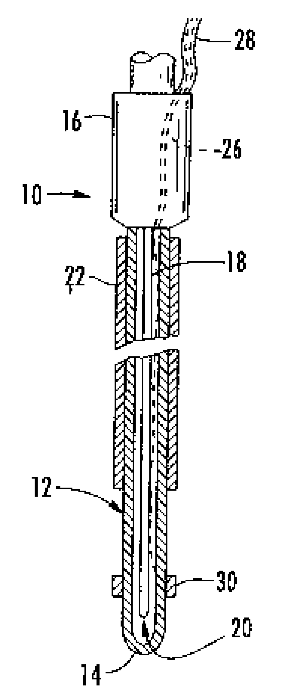
However, embolization techniques aren't easy, require specific and costly technologies and highly specialized departments, and the drug distribution isn't necessarily homogeneous.
A major drawback of the sustained-release drug carriers—delivery carriers (microspheres, liposomes, microcapsules, gel-foam particles, etc.) is that they aren't visible continuously, in real time using most of the available clinical imaging systems, i.e. ultrasound imaging, C-T radiography or fluoroscopy.
As a consequence, the physician doesn't know whether the desired target site of deposition has been reached, nor whether the drug carriers are correctly distributed throughout the tumor or target tissues.
However since the carrier and the contrast agent diffusion / distributions in tissues are different, the imaging of the contrast in the mixture doesn't give precise clue of the carrier location beyond a few minutes period.
Another drawback is that a pinpoint placement of the depots into tumor requires a Perfect visibility of the delivery device throughout the procedure until the delivery tip reaches the targeted tumor region, particularly for deep-seated lesions.
Although a number of techniques are described to increase the echogenicity of delivery needles or catheters during various procedures, their characteristics aren't helpful for visualization in deep-seated lesions, where their effectiveness would be most desirable.

Method used


Image

  • Systems and methods for improving image-guided tissue ablation
  • Systems and methods for improving image-guided tissue ablation
  • Systems and methods for improving image-guided tissue ablation

Examples


Embodiment Construction

[0034]This invention incorporates and improves on the subject matter of several patents: e.g., U.S. Pat. No. 6,255,018 for monitoring cryosurgery; U.S. Pat. No. 5,425,370 that oscillates the delivery device(s) at its resonant frequency; and U.S. Pat. No. 5,827,531 that discloses the unique microcapsules. All of these patents are incorporated herein by reference. The patent material is summarized below for a clear understanding of the objects and advantages of the present invention.

[0035]The computer-aided monitoring method disclosed in U.S. Pat. No. 6,235,018 predicts, in real-time, the extent of the ice ball kill zone, and, alone, or in conjunction with conventional imaging techniques, such as, Ultrasound, “US”, Computerized Tomography, “CT”, Magnetic Resonance, “MR” allows a precise location of the target regions for complementary treatment with unique imageable drug(s) carriers.

[0036]The imageable drug(s) carriers of this invention are unique microcapsules. The new microcapsules ...


PUM

PropertyMeasurementUnit
Temperatureaaaaaaaaaa

Abstract

The cryosurgical kit and method of use relates to the combined use of biodegradable, imageable, drug(s) carriers, vectors or microcapsules deposited into a target tissue region inside a body and the controlled cooling of the target tissue region. A minimally invasive method of treatment that applies a cooling device to a selected tissue region, uses a vibrating resonant frequency delivery device to inject drug(s) carriers (microcapsules) containing therapeutic and contrast agents into tissue region(s) submitted to cooling temperatures, successive real-time imaging of the microcapsule deposition and later detection of the progressive degradation of microcapsules with ultrasound imaging which provides estimation of sustained release rates and for planning re-dosing regimens. The thermosensitive microcapsules preferably contain ethiodol, a contrast agent for Ultrasound and / or X-Ray imaging, and cytotoxic drug(s).

Description

RELATED APPLICATIONS[0001]Priority is claimed under 35 USC 120 to Apr. 16, 2004, the filing date of U.S. Provisional Application 60 / 562,759.FIELD OF THE INVENTION[0002]This invention relates to the field of cryosurgery and the treatment of tumors.BACKGROUND OF THE INVENTION[0003]Modern approaches to minimally invasive ablative treatment of solid tumors involve the use of miniature instruments, and combined treatments like thermal ablation and systemic chemotherapy.[0004]Cryosurgery is now recognized as an efficient, thermo-ablative, minimally invasive, method for a large number of solid tumors like prostate, lung, liver, kidney, to cite only a few. Cryosurgery affects tumor tissue viability in three different ways with immediate and delayed alterations: the freezing of tumor cells, and vasculature results in tumor kill through direct cell alterations, and indirect vascular occlusion.[0005]Recently another mechanism of cell kill consecutive to cryothermal changes is apoptosis, a prog...

Claims


Application Information

Patent Timeline
IPC IPC(8): A61B8/14A61B18/02A61B17/20A61K31/513A61N7/00
CPCA61B18/02A61B2018/0212A61N7/00A61K31/513A61B2019/5276A61B2090/378A61P35/00
Inventor LEPIVERT, PATRICKKOLASINSKI, ROGER STEVEN
Owner NUVUE THERAPEUTICS
Learn More