
Constrained Hiv V3 Loop Peptides as Novel Immunogens and Receptor Antagonists

a hiv v3 loop, novel technology, applied in the direction of peptides, viruses, cyclic peptide ingredients, etc., can solve the problem that the peptides do not permit the determination of the global conformation of the v3 loop, and achieve the effect of broad neutralizing activity and potent

Inactive Publication Date: 2008-08-28
NEW YORK UNIV +1
View PDF2 Cites 8 Cited by
  • Summary
  • Abstract
  • Description
  • Claims
  • Application Information

AI Technical Summary

Benefits of technology

[0014]It is now appreciated that, though the sequence in V3 is variable, the V3 loop is characterized by a constant size of 30-35 amino acids, a conserved type II β-turn at its tip, a disulfide bond at its base and a net positive charge (Kwong et al., 2000, supra). These structural constraints on the V3 loop appear to be imposed by the requirement for V3 / chemokine receptor interaction (Hill, C M et al., 1997. J Virol 71:6296; Trkola et al., supra). This suggested to the present inventors that V3 must have conserved conformational aspects despite the sequence variation. This is borne out by reports that conserved elements in the V3 crown and stem are mandatory elements for coreceptor interaction (Wang, W K et al. (1999) Proc Natl Acad Sci USA 96:4558-62; Suphaphiphat, P et al., 2003, J Virol 77:3832; Cormier, E G et al., 2002, J Virol 76:8953). Cast in this new light, the present inventors predicted that antibodies to constrained V3 conformational epitopes would have potent and broad neutralizing activity.
[0032]In another embodiment, the constrained peptides can be used as antagonists that inhibit interactions between HIV virions and co-receptors on target lymphocytes (generally R5 receptors) or target cells of the monocyte / macrophage or other myeloid lineage (generally X4 receptors). Such inhibition can suppress viral infectivity and intercellular viral spread by reducing the ability of virions to bind productively to target cells.
[0035]Definition of the peptide conformations that are “adapted” to fit the antigen binding pocket of broadly reactive neutralizing antibody are based on NMR structures for each peptide, which appear as X, Y, and Z orthogonal coordinates in Tables 3-6. The NMR constraints and structural statistics for the refined peptide structures are shown in Tables 1 and 2. Similar information derived from X-ray crystallographic studies are also presented briefly in Example VII. The X ray diffraction results confirm the structural parameters first obtained by NMR analysis. The inventors' NMR analysis has identified two subtypes of V3 β hairpin structures (termed R5A and R5B) that differ in the C-terminus of the P strand (residue positions approximately 324-327 of the gp120 sequence). X-ray analysis has the added advantage of providing information that better defines the fine structure of the antibody cleft and the residues therein that contact the amino acids of the peptide / mimetic.

Problems solved by technology

However, the short epitopes recognized by these antibodies which had been induced using synthetic peptides did not permit the determination of the global conformation of the V3MN loop.

Method used


Image

  • Constrained Hiv V3 Loop Peptides as Novel Immunogens and Receptor Antagonists
  • Constrained Hiv V3 Loop Peptides as Novel Immunogens and Receptor Antagonists
  • Constrained Hiv V3 Loop Peptides as Novel Immunogens and Receptor Antagonists

Examples


example i

Experimental Procedures and Materials

[0196]Note on letter / number codes: The MN or IIIB superscript preceding the single letter amino acid code indicates the HIVMN or HIVIIIB strain origin of the sequence; the number following the amino acid code represents the position of the residue in the full length gp120MN or gp120IIIB sequence. The number is sometimes followed by the position of the hydrogen (H) involved in the hydrogen bonding—i.e., an amino hydrogen (HN) or a hydrogen atom bonded to the α carbon (Hα)

Sample Preparation

[0197]The V3MN peptide, 308-332gp120MN (YNKRKRIHI--GPGRAFYTTKNIIG; SEQ ID NO:13) linked to a fusion protein was expressed in E. coli, cleaved and purified as previously described by M. Sharon et al. (2002) Protein Expr. Purif. 24:374-383.). Note that the sequential numbering system in V3MN is interrupted due to a rare two residue insertion in HIV-1IIIB and therefore residues 317 and 318 are not present in V3MN. The 447Fv was expressed in BL21(DE3)pLysS strain.

[01...

example ii

Mapping the V3MN Epitope

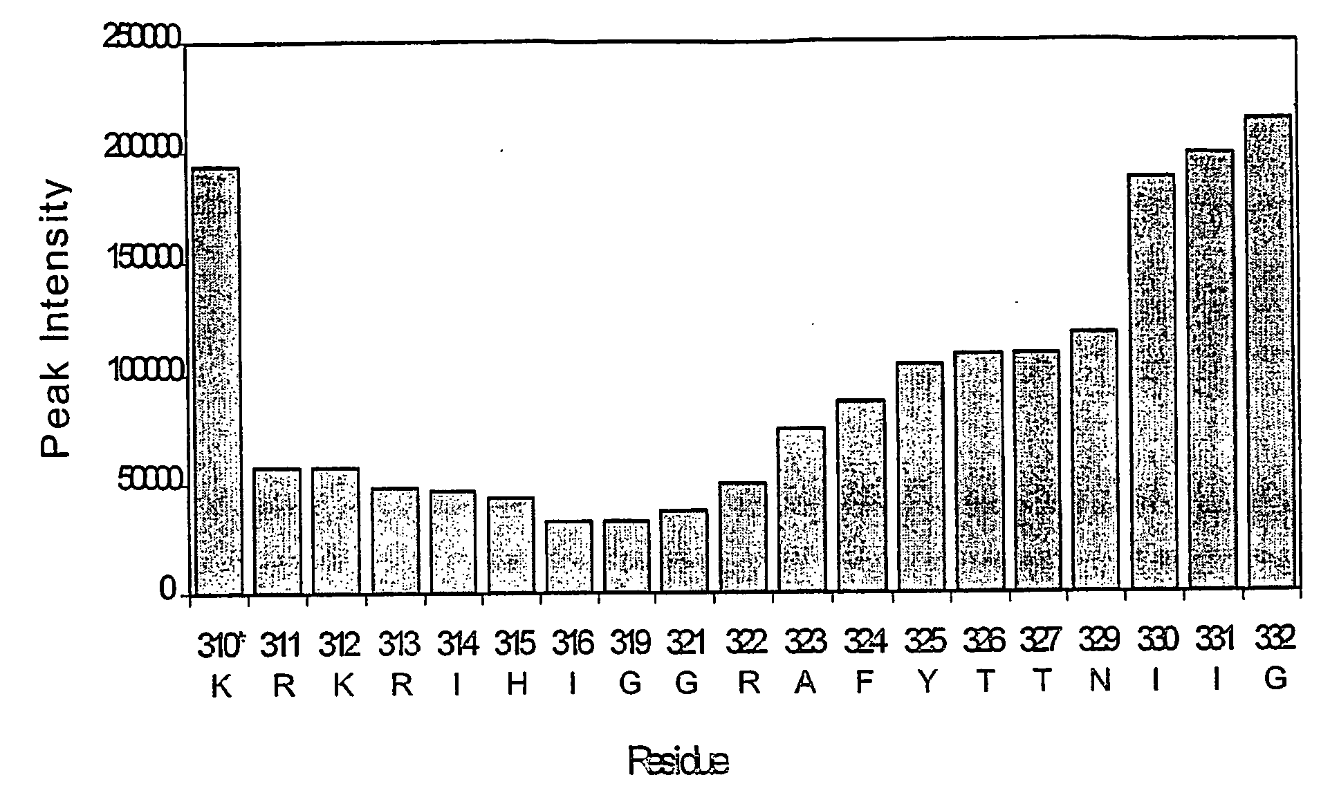
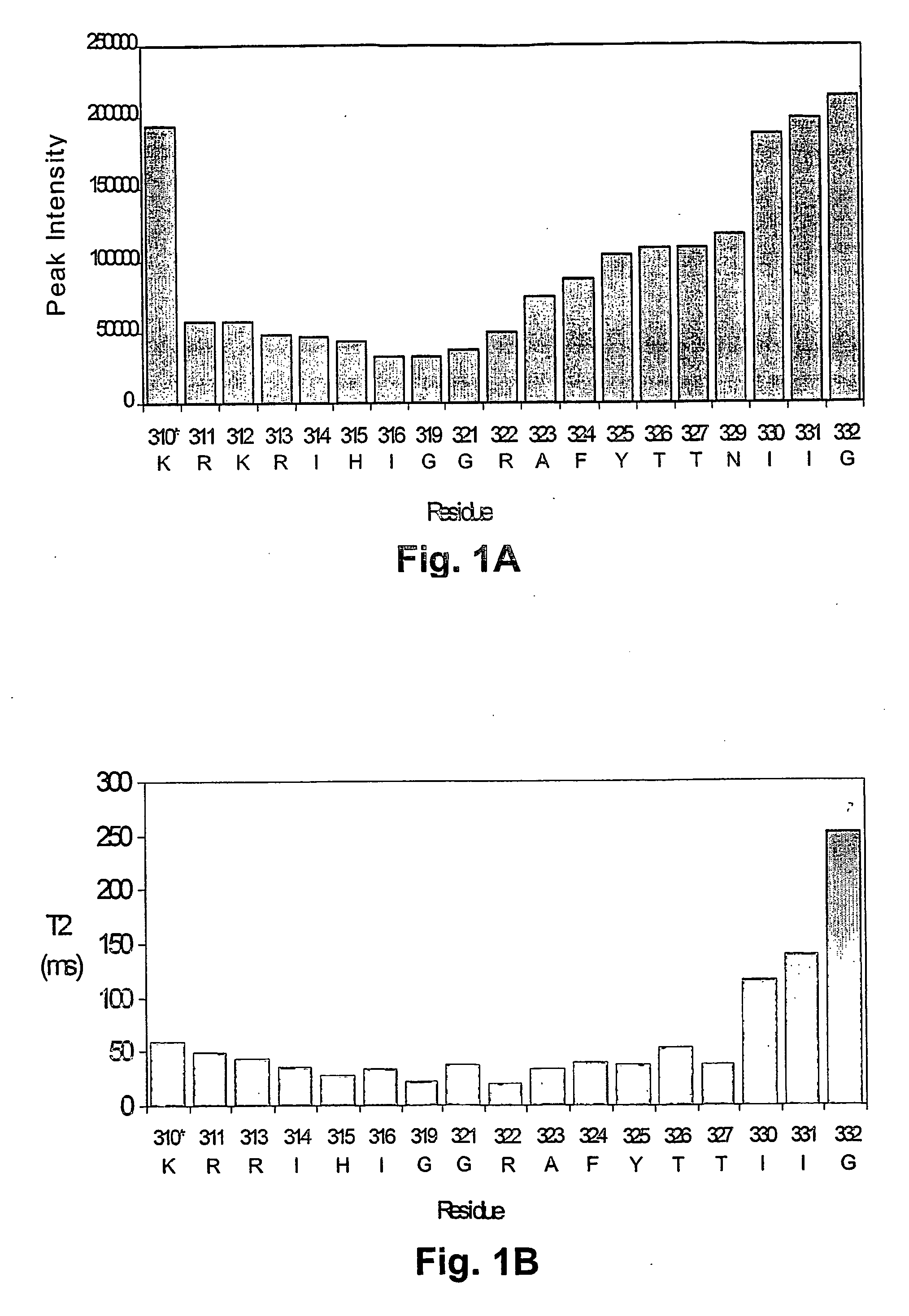
[0211]NMR dynamic filtering was used to map the epitope within the V3 peptide recognized by the 447Fv. Peptide protons that do not interact with the Fv retain considerable mobility in comparison to peptide protons which do interact. As a result of the long mixing period used in the HOHAHA and ROESY spectra, the cross peaks of peptide protons interacting with the Fv as well as of most Fv protons vanish while the cross peaks of residues in the flexible parts of the peptide that do not interact with the Fv continue to be observed. These include seven residues of the C-terminal region (MNT326-MNG332) and two of the N-terminal segment (MNN309, MNR311). The proton chemical shifts of these residues were practically identical to those observed for the free peptide, confirming that they do not interact, or have only very minor interactions with the antibody. The HOHAHA cross-peaks of MNK312-MNR322 were undetectable in the spectra, implying strong interactions with 447...

example iii

Solution Structure of the Antibody-Bound V3MN Peptide

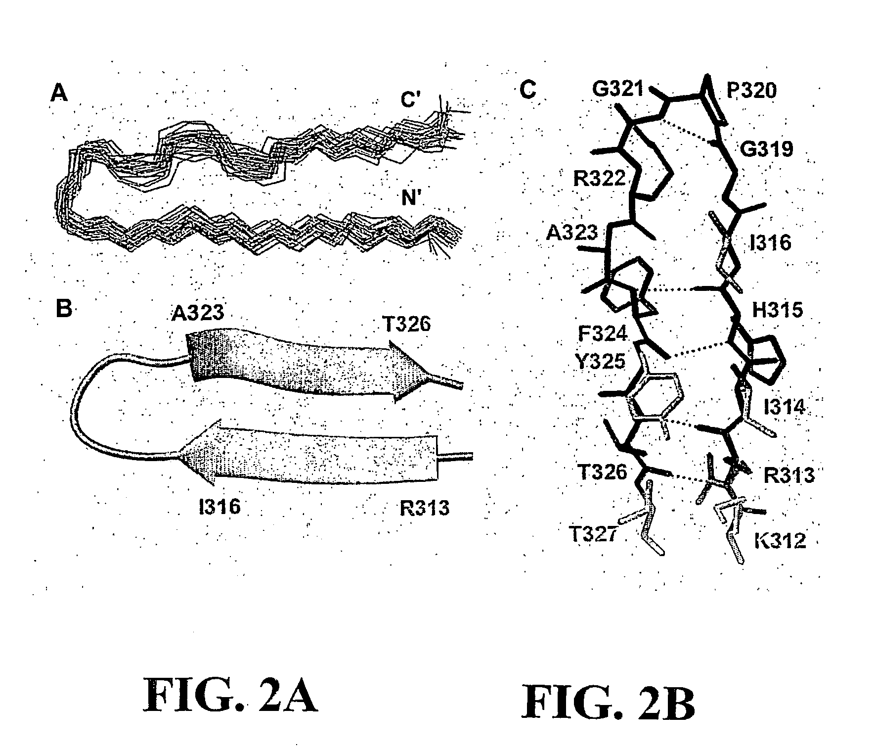
[0213]The structure of the bound V3MN epitope was determined using 305 NMR-derived distance (90 long and medium range), 10 dihedral angle and 2 hydrogen bonds constraints. The superposition of the 29 lowest energy structures that satisfied the experimental restraints with no NOE violations larger than 0.5 Å and no torsion angle violations exceeding 5° is shown in FIG. 2A. The overall structure of the epitope (312-327gp120) is well defined with root-mean-square deviations (rmsd) values of 0.37 Å and 1.17 Å for the backbone and heavy atoms, respectively. The structural statistics and rmsd are presented in Table 1. A Ramachandran plot (not shown) of the mean structure of the complex suggests that the φ and ψ angles of the structure predominantly occupy allowed regions except for MNG319 and MNG321.

[0214]The average NMR coordinates for the V3 MN peptide as bound by and constrained by the 447Fv antibody fragment are shown in Table 3. Th...


PUM

PropertyMeasurementUnit
Molar densityaaaaaaaaaa

Abstract

The present invention provides constrained peptides and other organic molecules, that mimic the three dimensional characteristics of the HIV-1 V3 loop peptide when bound by a highly potent human neutralizing monoclonal antibody specific for a V3 conformational epitope, which structure is determined by NMR. Methods for screening for, and designing such molecules are disclosed. These molecules are useful as immunogens for inducing broadly-neutralizing antibodies against HIV-1 as well as antagonists for inhibiting the binding of HIV-1 to the relevant co-receptors, and may therefore be used in method of preventing or treating HIV-1 infection and disease.

Description

BACKGROUND OF THE INVENTION[0001]1. Field of the Invention[0002]The present invention in the fields of structural chemistry, immunology and medicine relates to novel molecules including constrained peptides and other organic molecules, that mimic the three dimensional (3D) characteristics of the HIV-1 V3 loop peptide when bound by a highly potent human neutralizing monoclonal antibody (mAb) specific for a V3 conformational epitope. These novel molecules are useful as immunogens for inducing neutralizing antibodies to HIV-1 as well as antagonists for inhibiting the binding of HIV-1 to the relevant co-receptors.[0003]2. Description of the Background Art[0004]The binding of the human immunodeficiency-virus type-1 (HIV-1) to its target cells is mediated primarily by the envelope glycoprotein (gp120) of the virus. Binding of gp120 to CD4, a molecule found on the surface of both T cells and macrophages triggers conformational changes in gp120 that expose a binding site for either the CCR5...

Claims


Application Information

Patent Timeline
IPC IPC(8): A61K39/00C07K2/00C07K16/46A61P31/18G06F17/50C12N5/06G01N33/68C07K14/16G01N33/569
CPCA61K39/00C07K14/005C12N2740/16122G01N2500/02G01N2333/16G01N2333/715G01N33/56988A61P31/18
Inventor ANGLISTER, JACOBSHARON, MICHALSCHAPIRA, MATTHIEUZOLLA-PAZNER, SUSANROSEN, OSNAT
Owner NEW YORK UNIV
Learn More