
Implant designs and methods of improving cartilage repair

a cartilage repair and implant technology, applied in the field of in situ cartilage repair implants, can solve the problems of inability to improve cartilage repair, degenerate, bone spur formation and decrease motion, lack of healthy cartilage (fibrocartilage), etc., to prevent the influx of synovial fluid

Inactive Publication Date: 2008-05-29
WARSAW ORTHOPEDIC INC
View PDF43 Cites 47 Cited by
  • Summary
  • Abstract
  • Description
  • Claims
  • Application Information

AI Technical Summary

Benefits of technology

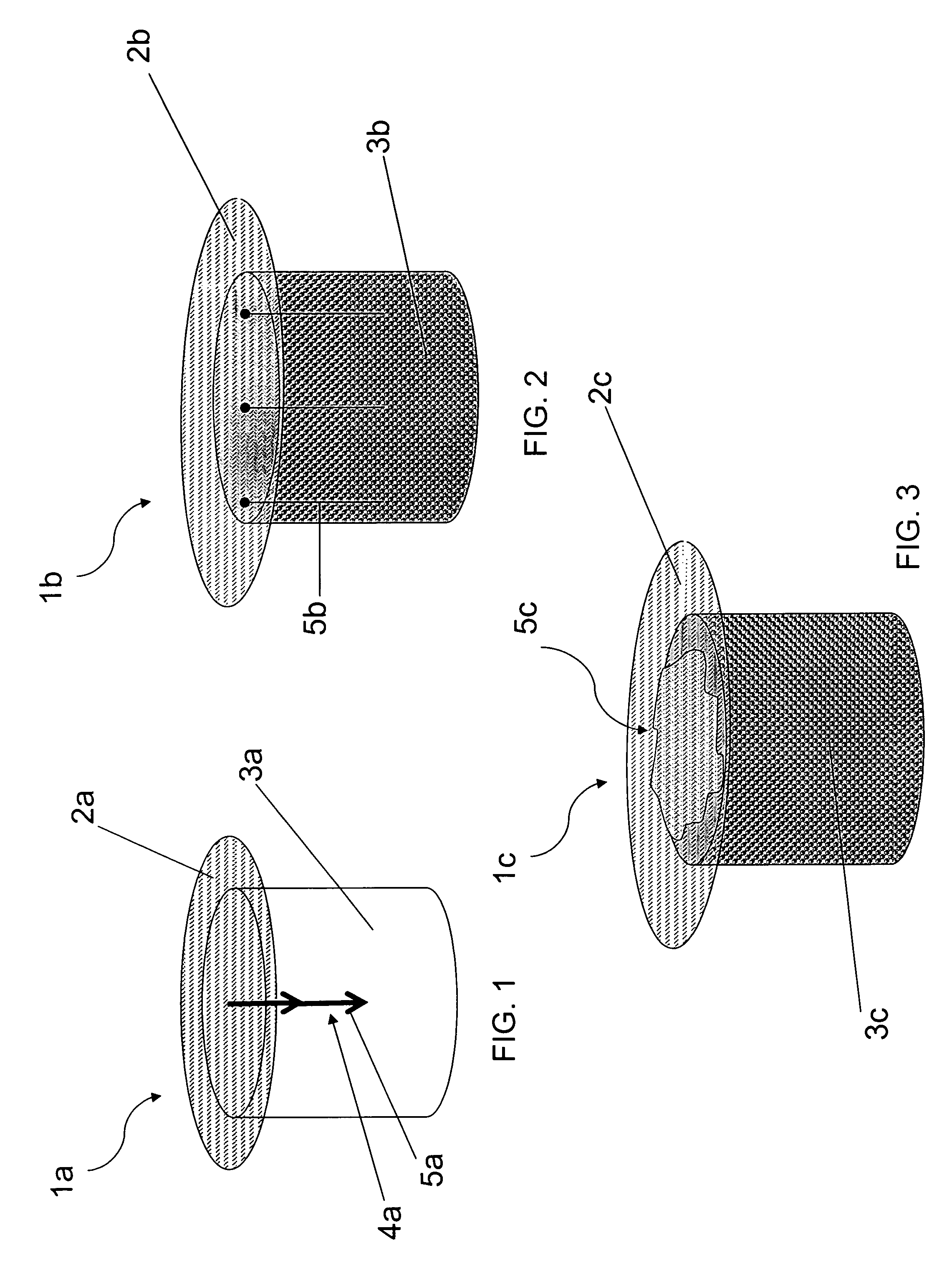
[0022]The present invention overcomes the drawbacks of prior art by providing a novel cartilage implant that comprises a cover, a cartilage repair scaffold, a means for axially fixing the cover to a scaffold end surface, and optionally a gasket. The implant may prevent the influx of synovial fluid and inflammatory cytokines from the synovial cavity into a surgically prepared defect meant for cartilage repair.
[0034]In various embodiments, cover prevents the influx of synovial fluid and inflammatory cytokines into a surgically prepared defect that extends from the surfaces of hyaline cartilage into sub-chondral bone.

Problems solved by technology

Degenerative diseases wear away hyaline cartilage covering the end of bones, causing inflammation-related pain, swelling, bone spur formation and decreased motion.
Typically, when hyaline cartilage heals, it lacks the structural and physical properties of healthy cartilage (fibrocartilage) and will degenerate over time.
However, mosaicplasty can be compromised if the donor cartilage is diseased, if there is damage to the collagen-forming chondrocytes, or if there is a wearing of the graft over time.
A drawback to this procedure is that there must be a fresh donor and the tissue must be stored at low temperatures and used within a month to ensure a greater than 50% cell viability.
These methods provide temporary relief of pain but do little for the formation of new cartilage tissue.

Method used


Image

  • Implant designs and methods of improving cartilage repair
  • Implant designs and methods of improving cartilage repair
  • Implant designs and methods of improving cartilage repair

Examples


example 1

Cartilage Repair Implant

[0122]Having generally described the implant, the following specific example is offered for purposes of illustration and only for illustration. No intention to limit the invention should be inferred. An implant for cartilage repair in keeping with the present disclosure may be prepared as follows:

[0123]The implant is manufactured by dissolving PLGA polymer in a solvent and adding 50% by wt. biphasic calcium phosphate particles (100-250 microns in diameter). This mixture is poured into large flat trays 20 mm in depth. These trays are placed into ovens to drive off the solvent creating a highly porous structure.

[0124]From these large porous PLGA / BCP sheets, 4-15 mm diameter plugs are cored and then cut to a desired 10-15 mm lengths. Similarly, porous collagen sheets 2-3 mm thick are made by pouring collagen slurry into trays and freeze drying under vacuum conditions. 4-15 mm diameter plugs are cut from the large sheet. Separately, 100-500 micron thick impermeab...


PUM

PropertyMeasurementUnit
diameteraaaaaaaaaa
depthaaaaaaaaaa
lengthsaaaaaaaaaa

Abstract

The invention herein generally refers to an in situ cartilage repair implant. The implant promotes cartilage repair by providing a sealed barrier that prevents the flow of synovial fluid and inflammatory cytokines into a surgically prepared hole that accommodates the implant. Optionally, additives are associated with the implant to promote cartilage repair.

Description

FIELD OF INVENTION[0001]An in situ cartilage repair implant is disclosed. More specifically, a device is disclosed that comprises a scaffold crowned with a protective cover. The implant promotes cartilage repair by providing a sealed barrier that prevents the flow of synovial fluid and inflammatory cytokines located in the synovial cavity into a surgically prepared defect that accommodates the implant. Optionally, additives are associated with the implant to induce cartilage repair.BACKGROUND OF THE INVENTION[0002]Hyaline cartilage is connective tissue found in parts of the body where support, flexibility, and resistance to compression are desired, (e.g. the tip of the nose, and also the ends of bone-forming joints). Hyaline cartilage consists of cells called chondrocytes, which are embedded in a highly specialized extra-cellular matrix. Hyaline cartilage is lubricated with a viscous fluid, called synovial fluid, found in and about articular joints. Normal synovial fluid contains hy...

Claims


Application Information

Patent Timeline
Patent Type & Authority Applications(United States)
IPC IPC(8): A61F2/02A61F2/28A61B17/08
CPCA61B17/0401A61B17/0469A61B17/0642A61B2017/0647A61F2210/0004A61F2002/30062A61F2002/30761A61F2002/30766A61F2/30756
Inventor MCKAY, WILLIAM F.
Owner WARSAW ORTHOPEDIC INC
Learn More