Electrosurgical Device Having Floating Potential Electrode and Adapted for Use With a Resectoscope

Active Publication Date: 2008-03-27
RF MEDICAL
View PDF39 Cites 85 Cited by
  • Summary
  • Abstract
  • Description
  • Claims
  • Application Information

AI Technical Summary

Benefits of technology

[0017]In view of the ever present need in the art for more efficient electrode design, it is accordingly an object of the present invention to provide an electrosurgical device which has high efficiency.
[0019]It is further an object of the present invention to provide an electrosurgical device which may be used in applications in which the target tissue is not submerged in a liquid environment.

Problems solved by technology

Even though the benefits are well recognized, current electrosurgical instruments and procedures suffer from very significant deficiencies.
Thus, in that monopolar devices require current to flow from the active electrode through the patient's body, they invariably allow for the possibility that some of the current will flow through undefined paths in the patient's body, particularly when the instrument is not properly designed and positioned.
Bipolar electrosurgical devices have their own inherent drawbacks, often resulting from the close orientation of the return and active electrodes.
The return electrode necessarily has a small area and, as a result, can cause undesired tissue heating, coagulating or evaporation at its contact point with the patient's tissue due to the relatively high current densities present thereon.
In addition, with the bipolar configuration, the close proximity of the active and return electrodes creates the danger that the current will short across the electrodes.
Operation at relatively low voltage, as is necessary with bipolar instruments, leads to less efficient sparking, reduced efficiency of the instrument, undesirable overheating of nearby tissue, and longer procedure time.
Moreover, the use of electrosurgical bipolar procedures in electrically conductive environments is inherently problematic.
The presence of saline, which is a highly conductive electrolyte, can also cause electrical shorting of a bipolar electrosurgical probe, thereby causing probe destruction and unintended and unnecessary heating in the treatment environment which, in turn, can result in unintended and uncontrolled tissue destruction.
In addition, current monopolar and bipolar instruments used to cut or vaporize tissue often do not have effective means for controlling bubbles, which is essential to the safety and efficiency of many procedures.
As a result, the efficiency of the instruments is often low and the procedure length is increased.
Electrosurgical instruments that lack an effective means for trapping of bubbles include, for example, cutting loops, rollers, needles and knives, resection instruments and ablators.
Furthermore, many current monopolar and bipolar instruments are not designed to take full advantage of either the electrical properties of the fluids present in the vicinity of the procedure site (bodily fluids, including blood, as well as irrigation fluids, either electrically conductive or non-conductive) or the electrical properties of the tissue itself.
This increases the size of the distal end of the electrode necessary to achieve a given ablative surface size, and necessitates the use of cannulae, often with unnecessarily large lumens, an undesirable condition.
This contamination may increase the local conductivity to a degree sufficient to cause significant current flow from uninsulated portions of a probe designed for use in a non-conductive irrigant.
While these bubbles generally do not support arcing, they cover portions of the exposed electrode surface, thereby insulating these portions of the surface.
Electrodes with inefficient designs will require substantially higher power levels than those with efficient designs.
While currently available electrodes are capable of achieving desired surgical effects, they are not efficient for accomplishing these tasks and may result in undesired side effects to the patient.

Method used


Image

  • Electrosurgical Device Having Floating Potential Electrode and Adapted for Use With a Resectoscope
  • Electrosurgical Device Having Floating Potential Electrode and Adapted for Use With a Resectoscope
  • Electrosurgical Device Having Floating Potential Electrode and Adapted for Use With a Resectoscope

Examples


examples

[0131]Hereinafter, the present invention is described in more detail by reference to the exemplary embodiments. However, the following examples only illustrate aspects of the invention and in no way are intended to limit the scope of the present invention. As such, embodiments similar or equivalent to those described herein can be used in the practice or testing of the present invention.

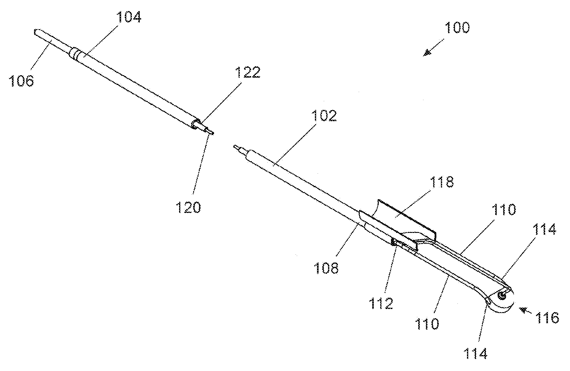
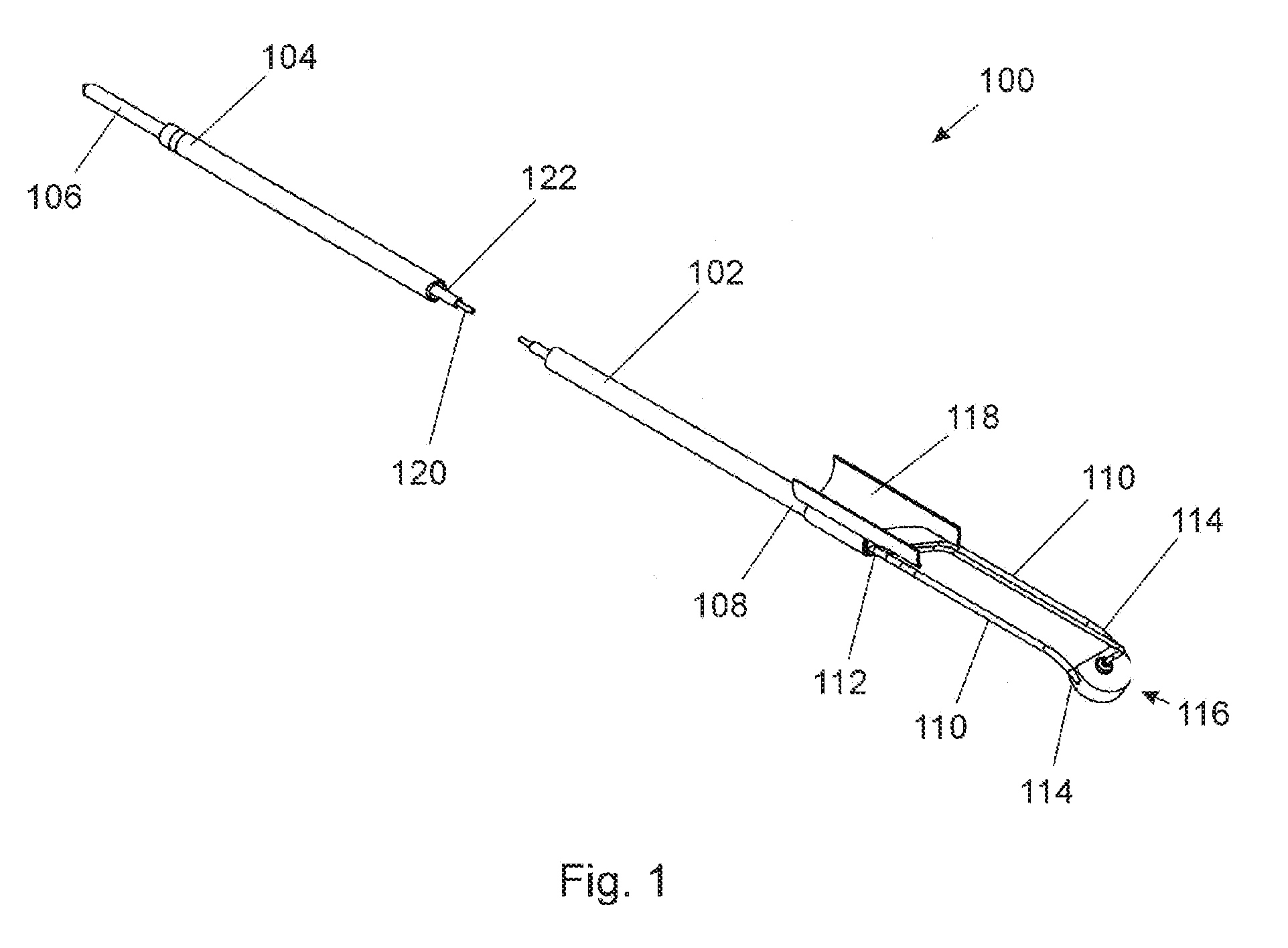
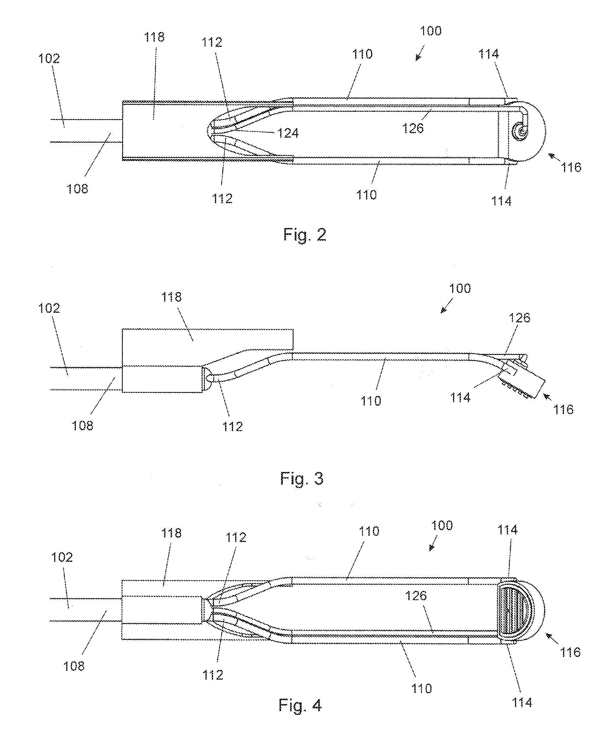
[0132]Referring to FIGS. 1 through 4, which depict an electrosurgical probe specifically configured for use with a resectosope (not shown) and constructed in accordance with the principles of this invention, probe 100 has an elongated tubular member 102 with a proximal end 104 having an electrical connector 106 suitable for connecting via an electrical cable to an electrosurgical generator, and a distal end 108. Members 110 have proximal ends 112 mounted to distal end 108 of elongated tubular member 102, and distal ends 114 to which are mounted electrode assembly 116. Optional electrode stabilizer 11...


PUM


Abstract

Disclosed herein are embodiments of an electrosurgical device that include one or more floating electrodes and are specifically adapted to remove, cut, resect, ablate, vaporize, denaturize, drill, coagulate and form lesions in soft tissues, with or without externally supplied liquids, preferably in combination with a resectoscope, particularly in the context of urological, gynecological, laparoscopic, arthroscopic, and ENT procedures. Specific adaptations for urological and gynecological applications, for example kidney stone removal and BPH treatment, are also described.

Description

PRIORITY[0001]This application claims the benefit of U.S. Provisional Application No. 60 / 847,496 filed Sep. 27, 2006, the contents of which are incorporated by reference herein.FIELD OF THE INVENTION[0002]The present invention relates generally to the field of electrosurgery, and more particularly, to high efficiency electrosurgical devices and methods which use radio frequency (RF) energy to cut, resect, ablate, vaporize, denaturize, drill, coagulate and form lesions in soft tissues, with or without externally supplied liquids. The electrosurgical devices of the instant invention find particular utility in combination with a resectoscope, in the context of urological, gynecological, laparoscopic, arthroscopic, and ENT procedures.BACKGROUND OF THE INVENTION[0003]As compared to conventional tissue removal techniques, electrosurgical procedures are advantageous in that they generally reduce patient bleeding and trauma. More recently, electrosurgical devices have gained significant pop...

Claims


Application Information

Patent Timeline
IPC IPC(8): A61B18/14
CPCA61B18/149A61B2018/00053A61B2018/0016A61B2018/00505A61B2018/00559A61B18/1482A61B2018/1213A61B2018/1407A61B2018/1427A61B2218/002A61B2018/00625
Inventor VAN WYK, ROBERT A.CARMEL, YUVALSHKVARUNETS, ANATOLY
Owner RF MEDICAL
Why Eureka
Try Eureka