
Method and apparatus to guide laser corneal surgery with optical measurement

Inactive Publication Date: 2007-12-06
UNIV OF SOUTHERN CALIFORNIA
View PDF13 Cites 152 Cited by
  • Summary
  • Abstract
  • Description
  • Claims
  • Application Information

AI Technical Summary

Benefits of technology

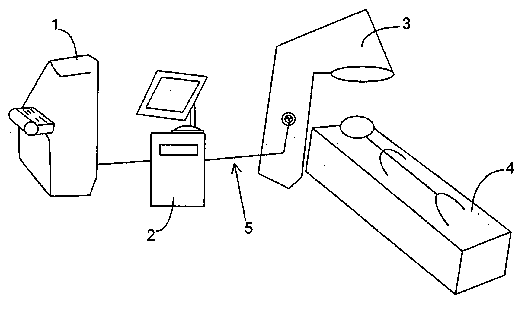
[0031] Major advantages for linking an OCT system to a precise laser surgical system in accordance with the present invention enables the performance of new procedures that are safer, less invasive and produce faster visual recovery than conventional surgical procedures.

Problems solved by technology

Although laser corneal surgery are now routine, current methods still have certain limitations that remain unaddressed, particularly in situations where a subject's eyes have highly irregular cornea geometry or have become opaque.
But these techniques still leave medium to large scale irregularities of the corneal surface untreated.
However, Placido-ring topography can capture data only when there is a relatively smooth surface and good tear film stability.
Most eyes with visually significant corneal scars cannot get a valid topography reading due to excessive surface irregularity or unstable tear film.
Thus topography-guided PTK has only limited applicability and cannot help the patients with more severe corneal problems.
Although the medium term success rate of corneal transplantation for adult patients is good (>90%), it is poor to fair (48-74%) in infants and children.
Graft survival in the very long term may be poor.
A rejection reaction can occur to any layer of the cornea, but most graft failures result from rejection of the corneal endothelium because it has no regenerative capability and its function is critical to corneal transparency and clarity.
Loss of endothelial density beyond a critical value causes swelling and opacification of the cornea.
With graft rejection, there are often changes in the host eye such as abnormal growth of blood vessels into the cornea, adhesion between the iris and the cornea and elevated intraocular pressure.
These inflammation-related changes increase the risk of repeat graft rejection and accelerated graft degeneration.
However, surgeons usually do not choose to perform LK today because the procedure is technically difficult, carrying the risk of penetrating into the eye in uncontrolled fashion or leaving an irregular match between the corneal layers resulting in poor quality of vision.
The primary limitation of LK is the uneven dissection and matching of the donor tissue (graft) and the recipient bed of the host cornea.
Manual dissection of the host stromal bed was the first to be tried and its main draw back is the unevenness of the dissection.
The chief limitations of this approach are the difficulty of the dissection and the increased risk of uncontrolled penetration into the anterior chamber.
However, the technique used so far does not remove the uneven contour in the host cornea.
Without a method to precisely measure corneal depth, applications of femtosecond laser are not feasible.
However, slit-scanning systems tend to underestimate corneal thickness when there is subepithelial haze or stromal opacity.
This is due to the limited axial resolution of slit-scanning technology.
However, it requires immersing the eye in a fluid bath because ultrasound cannot pass through air.
The inconvenience and discomfort associated with the fluid bath makes it unsuitable for clinical applications.
However, the quality of topography data depends on the specular reflection from a smooth tear film and it cannot capture the surface of corneas with severe irregularity or unstable tear film.
Therefore Placido-ring topography-guided laser ablation cannot be applied to highly irregular corneas that need the treatment most.
However, the wavefront sensor cannot obtain a valid measurement on highly aberrated eyes, eyes with extreme refractive error, eyes with corneal opacity or cataract, eyes with unstable tear film and many eyes with intraocular lens implants.
In our experience, the wavefront sensor cannot obtain a valid measurement in the great majority of patients with visually significant corneal scar or irregularity.
Therefore wavefront-guided laser ablation cannot help most eyes with significant corneal irregularity.
Despite the variety of techniques currently available for measuring cornea thickness, none of the currently available prior art methods can meet the requirement of precision, flexibility, and ease-of-use needed to realize routine customized corneal surgeries.

Method used


Image

  • Method and apparatus to guide laser corneal surgery with optical measurement
  • Method and apparatus to guide laser corneal surgery with optical measurement
  • Method and apparatus to guide laser corneal surgery with optical measurement

Examples


examples

1. High-Speed Optical Coherence Tomography Mapping of the Cornea

OCT Image Capture

1. High-Speed OCT Scanning.

[0078] An OCT system with a speed of at least 2 kHz axial scan repetition rate is needed to accurately map corneal thickness. A speed of at least 20 kHz is needed to accurately map the anterior corneal surface elevation. An even higher speed is preferred for the mapping of highly irregular corneas because even a small movement within the scan acquisition period can lead to misregistration of a fine surface irregularity.

2. Meridional Scanning Pattern.

[0079] The strong specular reflection at the corneal vertex (the point on the corneal surface that is perpendicular to the visual fixation axis) is easily visible on the OCT image and serves as a reliable landmark. Radial lines centered on the vertex forms meridians. A meridional OCT scan has the special property that the OCT beam remains perpendicular to the corneal azimuth, and its incidence angle in the meridional plane ...


PUM


Abstract

Optical coherence tomography (OCT) is used to map the surface elevation and thickness of the cornea. The OCT maps are used to plan laser procedures for the treatment of an irregular, opacified or weakened cornea, and in the treatment of refractive errors. In the excimer laser phototherapeutic keratectomy (PTK) procedure, the OCT data is used to plan a map of ablation depth needed to restore a smooth optical surface. In the excimer laser photorefractive keratectomy procedure, OCT mapping of epithelial thickness is used to achieve clean laser epithelial removal. In femtosecond laser anterior keratoplasty procedure, OCT data is used to plan the depth of femtosecond laser dissection to remove an anterior layer of the cornea, leaving a smooth recipient bed of uniform thickness to receive a disk of donated corneal tissue. The linkage of an OCT system to a precise laser surgical system enables the performance of new procedures that are safer, less invasive and produce faster visual recovery than conventional surgical procedures.

Description

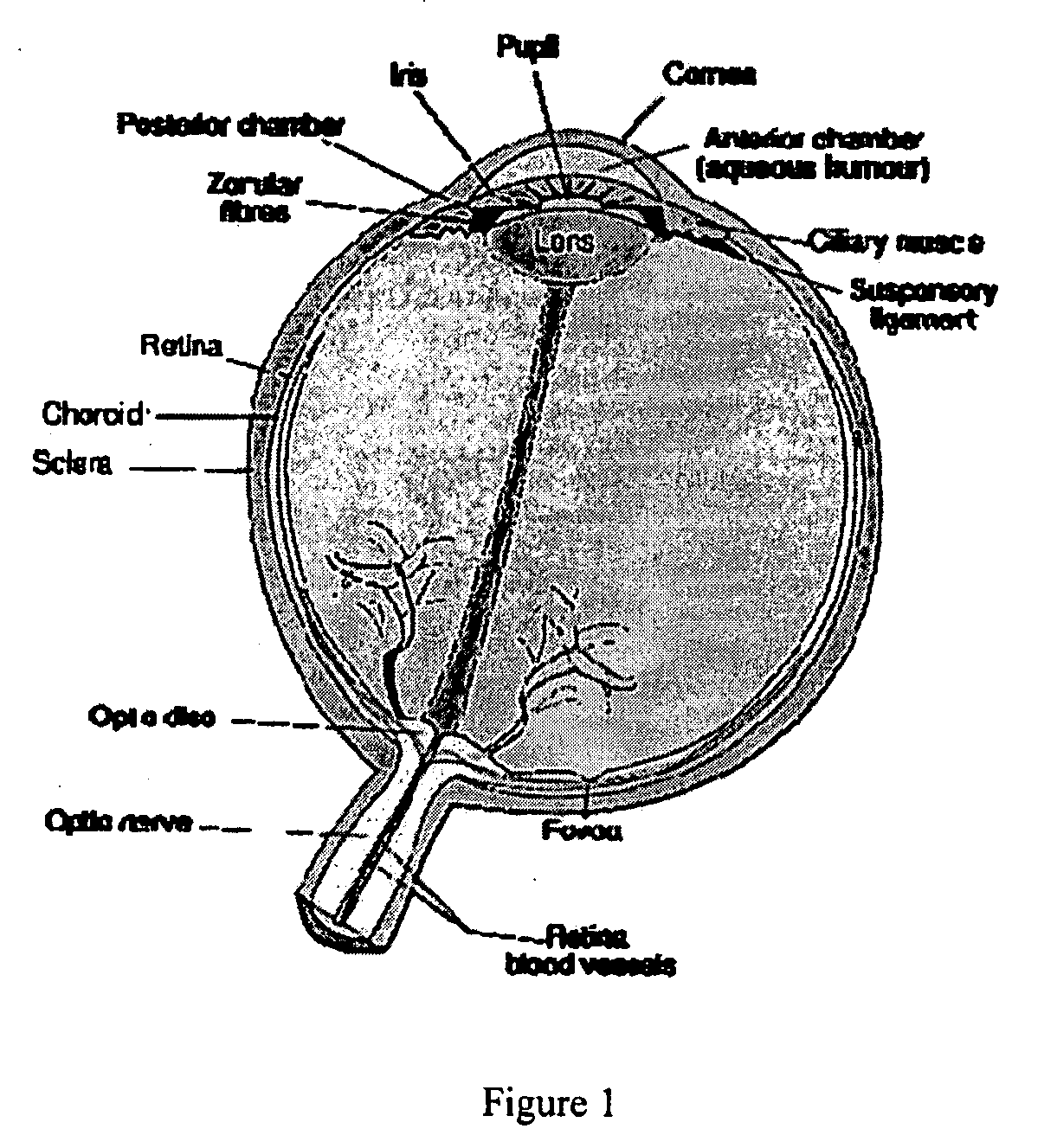
CROSS-REFERENCE TO RELATED APPLICATIONS [0001] This application claims an invention which was disclosed in Provisional Application No. 60 / 810,542 filed Jun. 1, 2006 entitled “METHOD AND APPARATUS TO GUIDE LASER CORNEAL SURGERY WITH OPTICAL MEASUREMENT”. The benefit under 35 USC §119(e) of the U.S. provisional application 5 is hereby claimed. The above priority applications are hereby incorporated herein by reference.STATEMENT REGARDING FEDERALLY SPONSORED RESEARCH AND DEVELOPMENT [0002] Not applicable. FIELD OF THE INVENTION [0003] The invention pertains to the field of opthalmology. More particularly, the invention pertains to methods for guided corneal surgery and apparatuses for performing thereof. BACKGROUND OF THE INVENTION [0004] The cornea is the transparent front part of the eye that covers the iris, pupil, and anterior chamber, providing most of an eye's optical power. Together with the lens, the cornea refracts light, and as a result helps the eye to focus, accounting for ...

Claims


Application Information

Patent Timeline
IPC IPC(8): A61B18/18
CPCA61B3/1005A61B3/102A61F9/00804A61F2009/00882A61F2009/00851A61F2009/00872A61F2009/0088A61F9/00831
Inventor HUANG, DAVIDSONG, JONATHAN C.LI, YANTANG, MAOLONG
Owner UNIV OF SOUTHERN CALIFORNIA
Learn More