High-resolution retinal imaging using adaptive optics and fourier-domain optical coherence tomography

a technology of adaptive optics and optical coherence tomography, applied in the field of retinal imaging, can solve the problems of poor resolution, reduced depth dependent sensitivity, and unsuitable morphological tissue imaging with medical ultrasonography, magnetic resonance imaging (mri) and confocal microscopy, so as to improve lateral resolution and precise positioning of the subject's eye. , the effect of minimizing head and eye motion

Active Publication Date: 2007-11-08
RGT UNIV OF CALIFORNIA
View PDF6 Cites 74 Cited by
  • Summary
  • Abstract
  • Description
  • Claims
  • Application Information

AI Technical Summary

Benefits of technology

[0036] Another object is to provide high lateral resolution of retinal images by using adaptive optics to correct optical aberrations of the human eye in real time.
[0037] Still another object is to provide high speed acquisition of high axial resolution retinal images by the use of Fourier-domain optical coherence tomography.
[0041] The instrument uses adaptive optics (AO) and optical coherence tomography (OCT). An exemplary AO-OCT system according to the present invention has been reduced to practice within a 1 m2 area on a standard laboratory optical table. The sample arm utilizes a Hartmann-Shack wavefront sensor and a 35-electrode bimorph deformable mirror for aberration correction in its present configuration. The lateral resolution is further improved by introducing a second deformable mirror using a commercial microelectromechanical system (MEMs). A key advantage of the present system is that the same light source is used for both OCT and Hartmann-Shack detection channels, allowing simultaneous operation of AO and OCT. Two independent PCs are used, one for AO control (AO-PC) and one for the OCT scanning and detection unit (OCT-PC). Separate sample and reference arms are constructed on the same table to test the OCT system with and without AO. A bite-bar, forehead-rest assembly is mounted on an X-Y-Z translation stage to permit precise positioning of the subject's eye. This may also be accomplished with a chin and forehead rest mounted to a height-adjustable table and slit-lamp-like device. An image from the wavefront sensor is used to monitor the X-Y position of the eye's pupil while fixation is directed to an external target to minimize head and eye motion and to allow precise imaging of different retinal locations. Although not necessary, to ensure the maximum pupil size and minimize fluctuations in accommodation, the subject's eye is dilated and cyclopleged with 2.5% Phenylephrine and 1% Tropicamide.

Problems solved by technology

Medical ultrasonography, magnetic resonance imaging (MRI) and confocal microscopy are not suited to morphological tissue imaging; the former two having poor resolution; the latter lacking millimeter penetration depth2,3.
The drawbacks of this technology are found in a strong fall-off of the SNR, which is proportional to the distance from the zero delay and a sinc-type reduction of the depth dependent sensitivity because of limited detection linewidth.
Therefore the signal has to be resampled before processing, which can not take care of the difference in local (pixelwise) bandwidth, which results in further reduction of the signal quality.
Drawbacks are the nonlinearities in the wavelength, especially at high scanning frequencies.

Method used


Image

  • High-resolution retinal imaging using adaptive optics and fourier-domain optical coherence tomography
  • High-resolution retinal imaging using adaptive optics and fourier-domain optical coherence tomography
  • High-resolution retinal imaging using adaptive optics and fourier-domain optical coherence tomography

Examples


Embodiment Construction

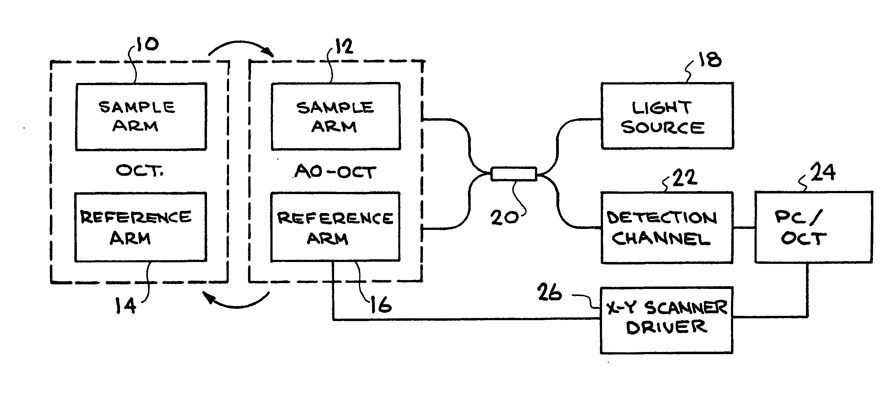
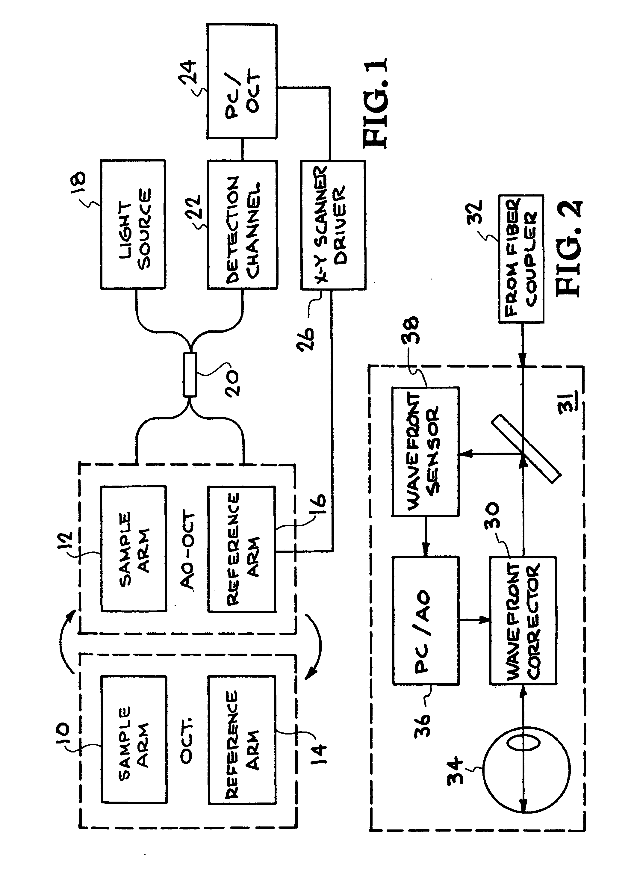
[0048] The Fourier-domain OCT system uses the spectral OCT approach in which a high-efficiency spectrometer measures the spectrum of the light that returns from reference and sample arms. FIG. 1 shows a schematic of the OCT engine according to a test where two sample arms, 10 and 12 and two reference arms 14 and 16 were constructed for AO-OCT and OCT imaging. A standard fiber-based OCT instrument is operated in Michelson interferometer configuration, where FC / APC fiber connectors are used to connect the fiber coupler's (80 / 20 splitting ratio) two outputs to fiber collimators placed at the input of bulk optics of the sample and reference arms. This feature of the OCT engine (light source 18, fiber coupler 20, detection channel 22) allows interchangeable operation for both AO-OCT and standard OCT imaging. Computer 24 controls X-Y Scanner driver 26 and collects and analyzes data from detection channel 22. Simple reconnection of FC / APC fiber connectors allowed rapid switching between th...


PUM


Abstract

This invention permits retinal images to be acquired at high speed and with unprecedented resolution in three dimensions (4×4×6 μm). The instrument achieves high lateral resolution by using adaptive optics to correct optical aberrations of the human eye in real time. High axial resolution and high speed are made possible by the use of Fourier-domain optical coherence tomography. Using this system, we have demonstrated the ability to image microscopic blood vessels and the cone photoreceptor mosaic.

Description

[0001] The United States Government has rights in this invention pursuant to Contract No. W-7405ENG-48 between the United States Department of Energy and the University of California for the operation of Lawrence Livermore National Laboratory.BACKGROUND OF THE INVENTION [0002] 1. Field of the Invention [0003] The present invention relates to retinal imaging techniques, and more specifically, it relates to technology for the acquisition of retinal images at high speed and with unprecedented resolution in three dimensions. [0004] 2. Description of Related Art [0005] Interferometry with broadband light sources has become a widely used technique for imaging in biologic samples using time-domain optical coherence tomography (OCT), optical coherence microscopy (OCM), spectral domain OCT (which encompasses spectrometer based Fourier domain OCT and swept source OCT), color Doppler OCT, and phase-referenced interferometry. In all of these interferometry techniques, light traveling a referenc...

Claims


Application Information

Patent Timeline
IPC IPC(8): G01B9/02G01B11/02
CPCA61B3/1015A61B3/12A61B3/102A61B3/14
Inventor OLIVIER, SCOT S.WERNER, JOHN S.ZAWADZKI, ROBERT J.LAUT, SOPHIE P.JONES, STEVEN M.
Owner RGT UNIV OF CALIFORNIA
Why Eureka
Try Eureka