Eureka AIR delivers breakthrough ideas for toughest innovation challenges, trusted by R&D personnel around the world.

Methods and systems for ultrasound delivery through a cranial aperture

a cranial aperture and ultrasound technology, applied in the field of medical methods and equipment, can solve the problems of affecting the treatment effect of stroke patients, unable to help patients with acute stroke, and almost nothing can be done to help patients, so as to reduce the non-linear acoustic effect of soft tissue, improve ultrasound energy transmission, and minimize or reduce the effect of non-linear acoustic effects

Inactive Publication Date: 2007-02-15
PENUMBRA
View PDF11 Cites 87 Cited by
  • Summary
  • Abstract
  • Description
  • Claims
  • Application Information

AI Technical Summary

Benefits of technology

[0016] In one aspect of the present invention, methods and devices provide intracranial ultrasound delivery while minimizing or reducing non-linear acoustic effects on soft tissue. In one embodiment, for example, non-linear effects are reduced by controlling the temperature of tissue between the ultrasound source and a target area. In some embodiments, targeted areas may include, for example, occluded intracranial blood vessels and / or blood clots in the intracranial space. The present invention provides methods to cool tissue exposed to ultrasound energy. In one embodiment, an acoustical medium used to improve ultrasound energy transmission from the ultrasound source to the tissue may also facilitate temperature modulation or cooling of the tissue. Alternatively, a separate element capable of cooling tissue may be delivered into a burr hole, through the dura, to a targeted location in the brain, penumbra, ventricle and / or along the epidural space to a secondary location on top of the dura mater. In some embodiments, the temperature of brain tissue may be reduced in combination with delivery of ultrasound energy into the intracranial space, which combination may significantly reduce metabolic needs of the affected brain tissue and reduce the severity and / or size of the stroke.

Problems solved by technology

Stroke is characterized by the sudden loss of circulation to an area of the brain, resulting in a corresponding loss of neurological function.
Until very recently, almost nothing could be done to help patients with acute stroke.
Treating patients early enough in the course of stroke, however, is an extremely challenging hurdle to effective treatment of stroke.
Furthermore, t-PA for stroke treatment is much more effective if delivered locally at the site of blood vessel blockage, but such delivery requires a great deal of skill and training, which only a small handful of medical professionals possess.
The primary challenge in using TCD to enhance stroke treatment, however, is that the skull attenuates the ultrasound signal to such a high degree that it is very difficult to deliver high-frequency, low-intensity signals through the skull.
Using higher intensity ultrasound signals, in an attempt to better penetrate the skull, often causes unwanted bleeding of small intracranial blood vessels and / or heating and sometimes burning of the scalp.
There are two main drawbacks to delivering high-frequency TCD through the temporal window.
First, such delivery requires a high level of skill, and only a small handful of highly trained ultrasonographers are currently capable of performing this technique.
Second, not all intracranial blood vessels are reachable with TCD via the temporal window.
For example, although the temporal window approach may work well for addressing the middle cerebral artery, it may not work as well for reaching the anterior cerebral artery or various posterior intracranial arteries.
In any such treatments, however, use of TCD faces the same challenges in that it is very difficult to deliver at safe and effective frequencies to desired locations in the brain and thus can be performed only by a small handful of highly skilled technicians and can be directed only to a few areas in the brain.
Also, the high intensities required to transmit ultrasound through the skull in TCD make its utility for treating any chronic disorder impractical, since any implantable power source used with a chronic, implantable ultrasound delivery device would be depleted too quickly.
Ultrasound energy propagation through soft tissue produces localized heating by ultrasound absorption and thus induces changes in acoustic properties of surrounding tissue, thereby increasing the risk of thermal injury to that surrounding tissue.
This risk of tissue damage is especially important in ultrasound delivery to intracranial tissues, as damage to surrounding soft tissues may compromise the blood-brain barrier.
In addition to the risk of surrounding tissue damage, localized tissue heating during ultrasound treatment typically distorts the acoustic waves intended to treat the target tissue.
Furthermore, nonlinear effects related to acoustic propagation through soft tissue can become significant when higher ultrasound intensities are required for therapeutic action, especially when the therapeutic target is located apart from the energy source and ultrasound energy needs to propagate through soft tissue to reach the target.
Non-linear effects can produce unanticipated effects on soft tissue including unwanted damage to the tissue between the ultrasound source and targeted areas.
In addition, the non-linear effects can limit the effectiveness of treatment, such as tissue lysis, directed at the target area.

Method used


Image

  • Methods and systems for ultrasound delivery through a cranial aperture
  • Methods and systems for ultrasound delivery through a cranial aperture
  • Methods and systems for ultrasound delivery through a cranial aperture

Examples


Embodiment Construction

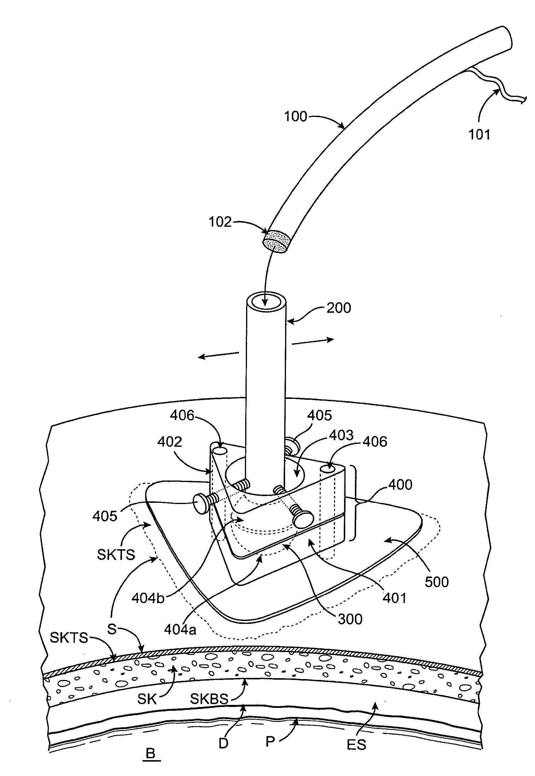
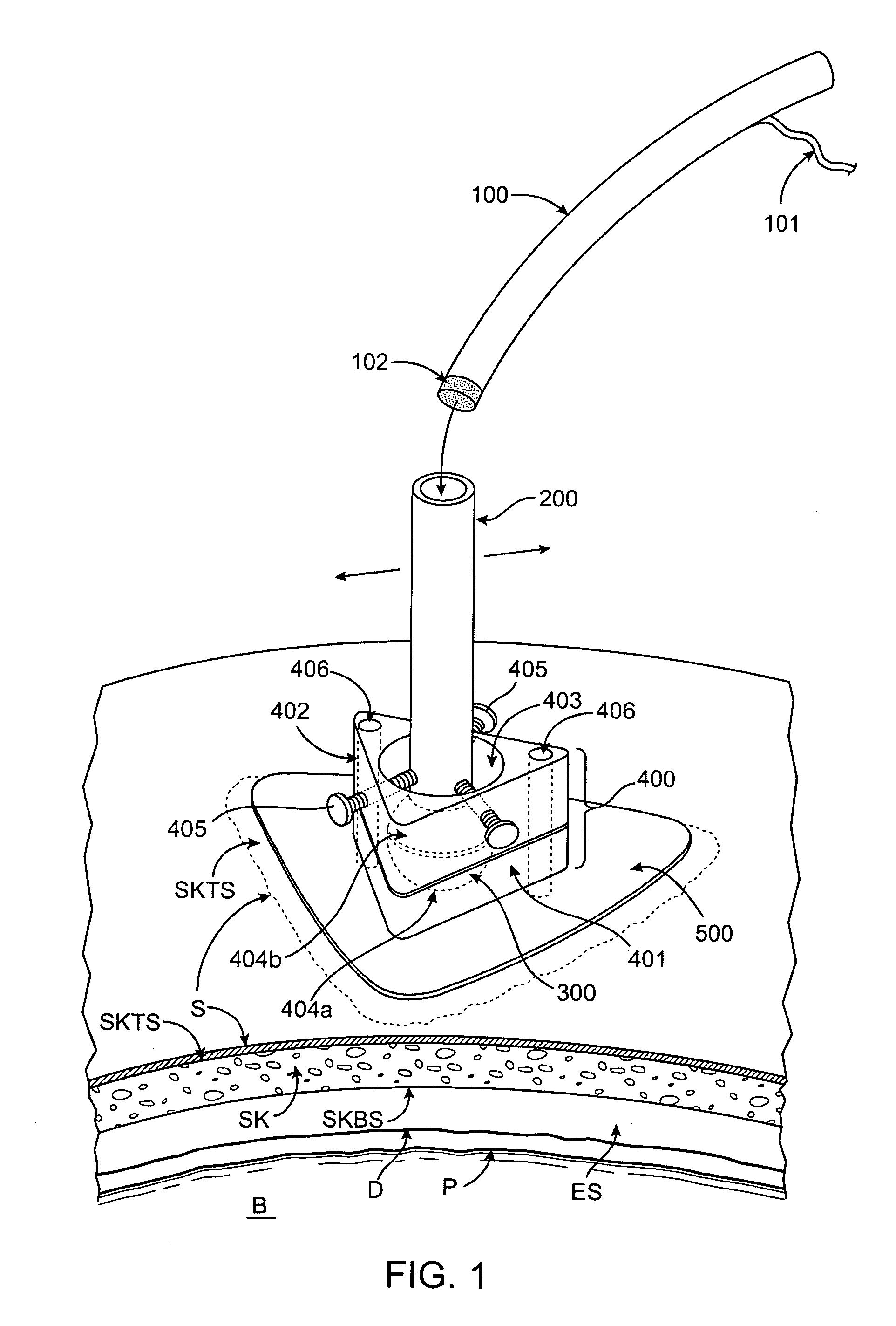
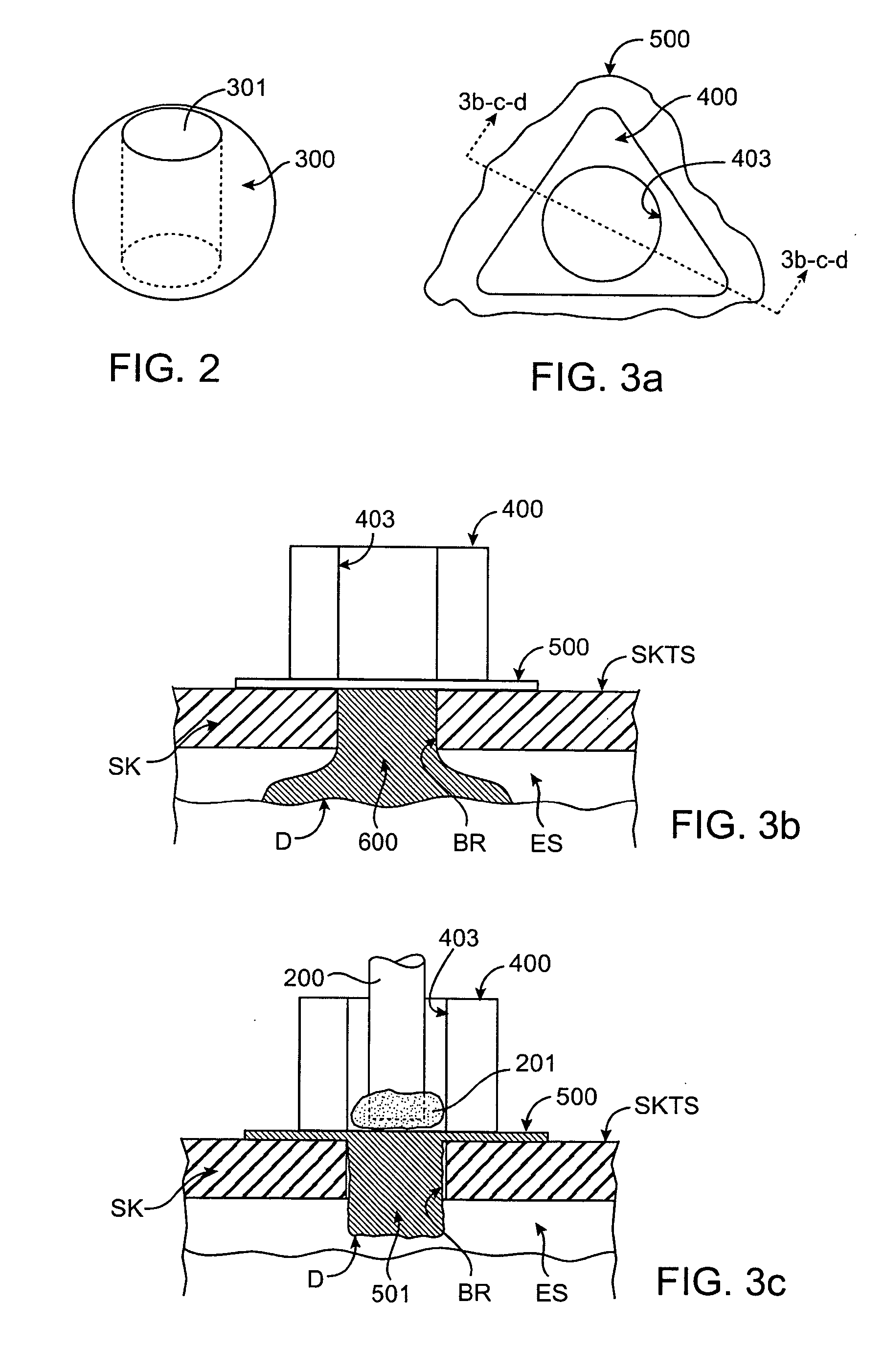
[0037] In one aspect of the present invention, a method for delivering ultrasound energy to a patient's intracranial space involves fixing at least one access device to the patient's skull, advancing at least one ultrasound delivery device at least partway through the access device, and transmitting ultrasound energy from the ultrasound delivery device to the patient's intracranial space. The access device may be fixed in place with screws through the scalp and into the skull, or alternatively the scalp may be retracted so that the base of the access device is located directly on the skull. If the scalp is retracted, it may be advantageous to also drill a hole through the skull near the center location of the access device to provide a route for minimal attenuation of the signals delivered from the ultrasound device to the intracranial space. In some embodiments, one hole is placed in the skull, and one ultrasound delivery device is used. In alternative embodiments, multiple holes a...


PUM


Abstract

A method for delivering ultrasound energy to a patient's intracranial space includes forming at least one aperture in the patient's skull, introducing at least one acoustically conductive medium into the intracranial space to contact brain tissue of the patient, advancing an ultrasound device at least partially through the aperture in the skull, and transmitting ultrasound energy to the intracranial space, using the ultrasound device. In some embodiments, the acoustically conductive medium may be cooled to help regulate the temperature of the patient's brain tissue.

Description

RELATED APPLICATION AND INCORPORATION BY REFERENCE [0001] This is a continuation-in-part of the following co-pending U.S. patent application Ser. No. 11 / 165,872, filed on Jun. 24, 2005; U.S. Ser. No. 11 / 203,738, filed on Aug. 15, 2005; and U.S. Ser. No. 11 / 274,356, filed on Nov. 15, 2005, whose entire disclosures are incorporated by this reference as though set forth fully herein.BACKGROUND OF THE INVENTION [0002] 1. Field of the Invention [0003] The present invention relates generally to medical methods and apparatus. More specifically, the invention relates to methods and apparatus for intracranial ultrasound delivery, which may include diagnostic ultrasound, therapeutic ultrasound, or both, delivered through an aperture or hole in the skull. [0004] 2. Background Art [0005] Stroke is characterized by the sudden loss of circulation to an area of the brain, resulting in a corresponding loss of neurological function. Also called cerebrovascular accident or stroke syndrome, stroke is ...

Claims


Application Information

Patent Timeline
Patent Type & Authority Applications(United States)
IPC IPC(8): A61B8/00
CPCA61B5/6864A61B8/08A61B8/0808A61B8/12A61N7/00A61B8/4209A61B8/4281A61B2018/00011A61M37/0092A61B8/42A61B5/4064A61B5/4076
Inventor NITA, HENRY
Owner PENUMBRA
Why Eureka
Eureka Blog
Learn More