
Methods and nucleic acids for the analyses of cellular proliferative disorders

a proliferative disorder and analysis method technology, applied in the field of gene expression dna sequences, can solve the problems of ineffective implementation of colorectal cancer screening, insensitive to multi-focal small lesions and treatment planning, and high cost of current recommended methods for cancer diagnosis, etc., to achieve the effect of improving sensitivity, specificity and/or predictive value, and improving sensitivity and specificity and likely patient complian

Active Publication Date: 2006-12-21
EPIGENOMICS AG
View PDF14 Cites 32 Cited by
  • Summary
  • Abstract
  • Description
  • Claims
  • Application Information

AI Technical Summary

Benefits of technology

[0030] The present invention provides a method for detecting and / or classifying cell proliferative disorders in a subject comprising determining the expression levels of Septin 9 in a biological sample isolated from said subject wherein underexpression and / or CpG methylation is indicative of the presence or class of said disorder. Various aspects of the present invention provide an efficient and unique genetic marker, whereby expression analysis of said marker enables the detection of cellular proliferative disorders with a particularly high sensitivity, specificity and / or predictive value. Furthermore, said marker enables the differentiation of neoplastic cellular proliferative disorders (including pre-cancerous conditions) from benign cellular proliferative disorders. The marker of the present invention is particularly suited for detection of colorectal and hepatocellular carcinomas. In the context of colorectal carcinoma the inventive testing methods have particular utility for the screening of at-risk populations. The inventive methods have advantages over prior art methods (including the industry standard FOBT), because of improved sensitivity, specificity and likely patient compliance.
[0035] The method is novel and has substantially utility, because, for example, no methods currently exist that enable the detection of cancer by analysis of body fluids, with a sensitivity and specificity high enough for use in a commercially-available and regulatory body-approved assay. For example, current methods used to detect and diagnose colorectal carcinoma include colonoscopy, sigmoidoscopy, and fecal occult blood colon cancer. In comparison to these methods, the disclosed invention is much less invasive than colonoscopy, and as, if not more sensitive than sigmoidoscopy and FOBT. The development of a body fluid assay represents a clear technical improvement and advantage over current methods known in the art in that it is anticipated that, at least for colorectal carcinoma screening, patient compliance for a single body fluid-based test will be higher than the triplicate analysis of stool currently recommended for FOBT.
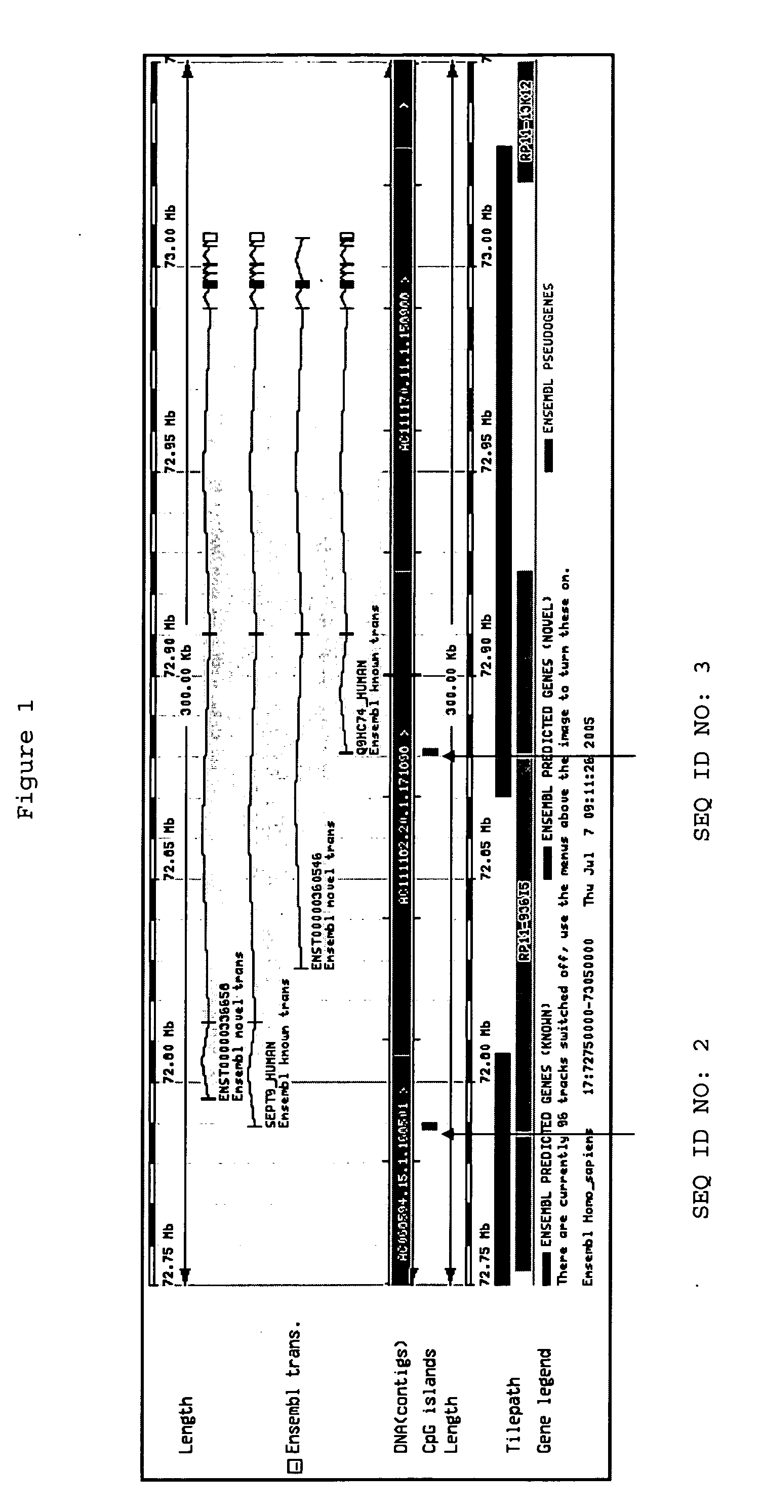
[0037] A particular embodiment the method comprises the use of the gene Septin 9 or its truncated transcript Q9HC74 as a marker for the detection and distinguishing of cellular proliferative disorders. The present invention is particularly suited for the detection of neoplastic cellular proliferative disorders (including at the pre-neoplastic stage). Furthermore, the methods and nucleic acids of the present invention enable the differentiation of malignant from benign cellular proliferative disorders. The methods and nucleic acids of the present invention are particularly effective in the detection of colorectal or liver neoplastic disorders and pre-neoplastic. Furthermore, they have utility in differentiating between neoplastic and benign cellular proliferative colorectal and hepatocellular disorders.
[0040] Aspects of the present invention provide a method for ascertaining epigenetic parameters of genomic DNA associated with the development of neoplastic cellular proliferative disorders (e.g., cancers). In particular aspects, the method has utility for the improved diagnosis, treatment and monitoring of said diseases.

Problems solved by technology

Current recommended methods for diagnosis of cancer are often expensive and are not suitable for application as population wide screening tests.
Ultrasound of the liver, alpha fetoprotein levels and conventional CT scan are regularly obtained in the diagnostic evaluation of HCC (hepatocellular cancer or primary liver cancer), but they are often too insensitive to detect multi-focal small lesions and for treatment planning.
Even though these testing procedures are well accepted by the medical community, the implementation of widespread screening for colorectal cancer has not been realized.
Patient compliance is a major factor for limited use due to the discomfort or inconvenience associated with the procedures.
Sigmoidoscopy has the limitation of only visualizing the left side of the colon leaving lesions in the right colon undetected.
Both scoping procedures are expensive, require cathartic preparation and have increased risk of morbidity and mortality.
A positive FOBT leads to colonoscopic examination of the bowel; an expensive and invasive procedure, with a serious complication rate of one per 5,000 examinations.
A number of studies show that FOBT screening does not improve cancer-related mortality or overall survival.
Compliance with occult blood testing has been poor; less than 20 percent of the population is offered or completes FOBT as recommended.
In reality, physicians frequently fail to instruct patients properly, patients frequently fail to adhere to protocol, and some patients find the task of collecting fecal samples difficult or unpleasant, hence compliance with annual occult blood testing is poor.
Compounding the problem of compliance, the sensitivity and specificity of FOBT to detect colon cancer is poor.
Poor test specificity leads to unnecessary colonoscopy, adding considerable expense to colon cancer screening.
However, no single or combination of marker has been shown to be sufficient for the diagnosis of colon carcinomas.
However its application as a routine diagnostic tool in a clinical environment is impeded by the extreme instability of mRNA, the rapidly occurring expression changes following certain triggers (e.g., sample collection), and, most importantly, the large amount of mRNA needed for analysis (Lipshutz, R. J. et al., Nature Genetics 21:20-24, 1999; Bowtell, D. D. L. Nature genetics suppl.
Recently, further molecular changes and genetic defects have been revealed.
Despite recent progress in the understanding of the pathogenesis of adenomas and carcinomas of the colon and their genetic and molecular changes, the genetic and epigenetic changes underlying the development of metastasis are less well understood.
Unfortunately, cancer is a disease state in which single markers have typically failed to detect or differentiate many forms of the disease.
Thus, assays that recognize only a single marker have been shown to be of limited predictive value.
Historically, many diagnostic tests have been criticized due to poor sensitivity and specificity.
A test having poor sensitivity produces a high rate of false negatives, i.e., individuals who have the disease but are falsely identified as being free of that particular disease.
The potential danger of a false negative is that the diseased individual will remain undiagnosed and untreated for some period of time, during which the disease may progress to a later stage wherein treatments, if any, may be less effective.
This type of test exhibits poor sensitivity because it fails to detect the presence of the virus until the disease is well established and the virus has invaded the bloodstream in substantial numbers.
A test having poor specificity produces a high rate of false positives, i.e., individuals who are falsely identified as having the disease.
A drawback of false positives is that they force patients to undergo unnecessary medical procedures treatments with their attendant risks, emotional and financial stresses, and which could have adverse effects on the patient's health.
A feature of diseases which makes it difficult to develop diagnostic tests with high specificity is that disease mechanisms, particularly in cancer, often involve a plurality of genes and proteins.

Method used


Image

  • Methods and nucleic acids for the analyses of cellular proliferative disorders
  • Methods and nucleic acids for the analyses of cellular proliferative disorders
  • Methods and nucleic acids for the analyses of cellular proliferative disorders

Examples


example 1

[0269] In the following example a variety of assays suitable for the methylation analysis of SEQ ID NO:1 were designed. The assays were designed to be run on the LightCycler™ platform (Roche Diagnostics), but other such instruments commonly used in the art are also suitable. The assays were MSP and HeavyMethyl assays. MSP amplificates were designed to be detected by means of Taqman™ style fluorescent labelled detection probes, HeavyMethyl™ amplificates were designed to be detected by means of LightCycler™ style dual probes.

Genomic Region of Interest:

[0270] SEQ ID NO:1

[0271] Assay type: MSP

Primers:aaaatcctctccaacacgtcSEQ ID NO:121cgcgattcgttgtttattagSEQ ID NO:122Taqman probes:cggatttcgcggttaacgcgtagttSEQ ID NO:123

Temperature Cycling Program: [0272] activation: 95° C. 10 min [0273] 55 cycles: 95° C. 15 sec (20 ° C. / s) [0274] 62° C. 45 sec (20° C. / s) [0275] cooling: 40° C. 5 sec

Genomic Region of Interest: [0276] SEQ ID NO:1

[0277] Assay type: MSP

Primers:aaaatcctctccaacacgtcS...

example 2

[0288] The following analysis was performed in order to select preferred panels suitable for colorectal carcinoma screening and / or diagnosis based on analysis of DNA methylation within whole blood, said panel comprising at least the analysis of SEQ ID NO: 1. The best performing assays from Example 1 were selected for the analyses, in addition to methylation assays suitable for analysis of the genes according to SEQ ID NOS:24 to SEQ ID NO:27 of Table 1.

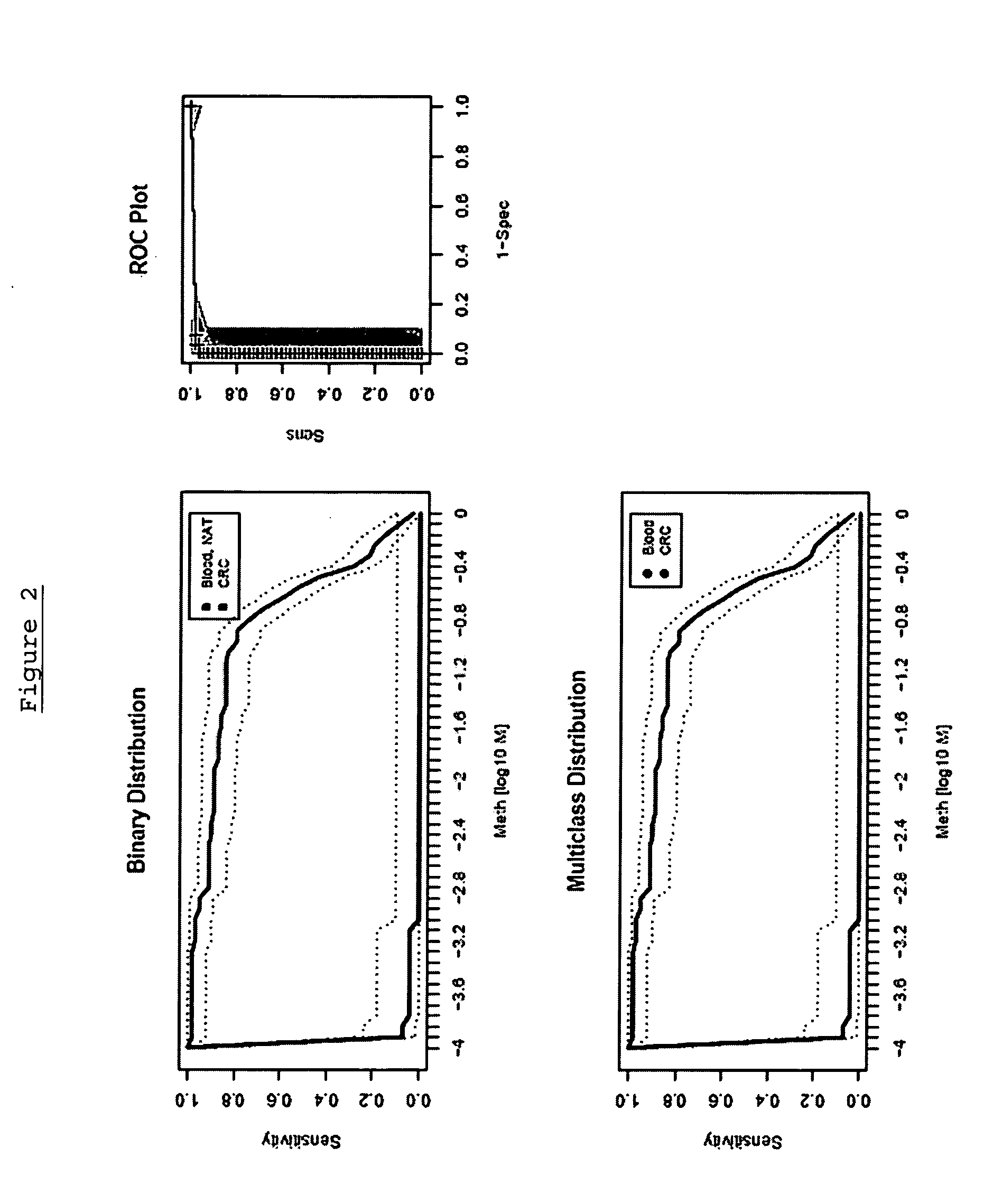
[0289] The performance of each marker was analysed using an assay platform (LightCycler™) and real time assays (MSP and / or HeavyMethyl™) as would be suitable for use in a reference or clinical laboratory setting. The performance of each marker was tested independently in colorectal carcinoma tissue and whole blood, in order to provide an indication of the accuracy of each marker.

[0290] In addition to the analysis of SEQ ID NO:1 the panels comprised further genes selected from the group of markers consisting:

FOX2(SEQ ID NO:24)NGFR(S...

example 3

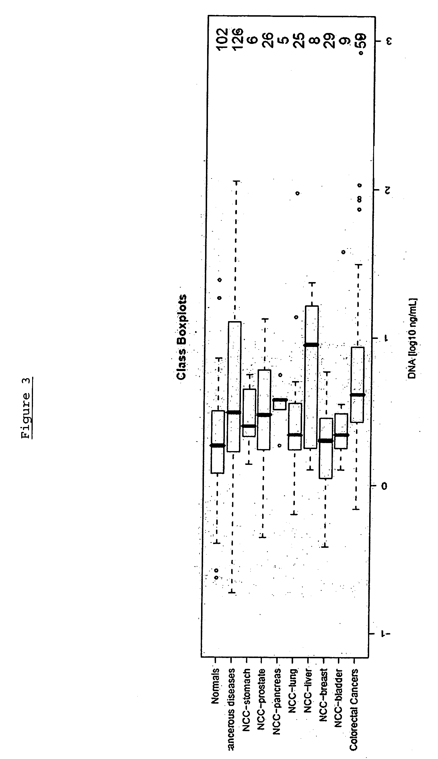
[0309] The following analysis was performed in order to confirm the gene Septin 9, (including its transcript variant Q9HC74) and panels thereof as a suitable marker for colorectal carcinoma screening and / or diagnosis based on analysis of DNA methylation in whole blood by validating the performance of assays in a large sample set.

[0310] The performance of the marker was analysed using an assay platform (Lightcycler) and real time assays (MSP and / or HeavyMethyl) as would be suitable for use in a reference or clinical laboratory setting. The performance of each marker was tested independently in colorectal tissue (normal adjacent tissue), colorectal carcinoma tissue and whole blood, in order to provide an indication of the accuracy of the marker.

[0311] The following primers and probes were used: SEQ ID NO:1 (Assay 7) using the LightCycler™ probes according to Table 2 was performed using the following protocol:

WaterFill up to final volume of 10 μlMgCl23.5Primer forward0.3Primer reve...


PUM

PropertyMeasurementUnit
Temperatureaaaaaaaaaa

Abstract

The invention provides methods, nucleic acids and kits for detecting, or for detecting and distinguishing between or among liver cell proliferative disorders or for detecting, or for detecting and distinguishing between or among colorectal cell proliferative disorders. The invention discloses genomic sequences the methylation patterns of which have utility for the improved detection of and differentiation between said class of disorders, thereby enabling the improved diagnosis and treatment of patients.

Description

CROSS-REFERENCE TO RELATED APPLICATIONS [0001] The present application claims the benefit of priority to U.S. Provisional Patent Applications: 60 / 672,242, filed 15 Apr. 2005; 60 / 676,997, filed 02 May 2005; 60 / 697,521, filed 08 Jul. 2005; 60 / 704,860, filed 01 Aug. 2005; 60 / 709,318, filed 17 Aug. 2005; 60 / 723,602, filed 04 Oct. 2005; and 60 / 787,402, filed 30 Mar. 2006; all of which are incorporated by reference herein in their entireties.FIELD OF THE INVENTION [0002] The present invention relates to genomic DNA sequences that exhibit altered expression patterns in disease states relative to normal. Particular embodiments provide, inter alia, novel methods, nucleic acids, nucleic acid arrays and kits useful for detecting, or for detecting and differentiating between or among cell proliferative disorders. Preferably, the methods, nucleic acids, nucleic acid arrays and kits for the detection and diagnosis of cell proliferative disorders are used for the diagnosis of cancer and in particu...

Claims


Application Information

Patent Timeline
IPC IPC(8): C12Q1/68C07H21/04C12P19/34
CPCC12Q1/6886C12Q2600/112C12Q2600/106C12Q2600/158C12Q2600/154G01N33/57438C12Q1/6813C12Q1/6806
Inventor LOFTON-DAY, CATHYSLEDZIEWSKI, ANDREWLESCHE, RALFSCHUSTER, MATTHIASDISTLER, JUERGENTETZNER, REIMOHILDMANN, THOMASMODEL, FABIANSONG, XIAOLING
Owner EPIGENOMICS AG
Learn More