
Scan parameter setting method for shuttle mode helical scan and X-ray CT apparatus

a technology of x-ray ct and helical scan, which is applied in the field of scan parameter setting method for shuttle mode helical scan and x-ray ct (computed tomography) apparatus, can solve the problem of not taking into account the predetermined range, and achieve the effect of reducing patient exposure and efficient and intelligible setting of imaging parameters

Inactive Publication Date: 2006-11-23
GE MEDICAL SYST GLOBAL TECH CO LLC
View PDF29 Cites 41 Cited by
  • Summary
  • Abstract
  • Description
  • Claims
  • Application Information

AI Technical Summary

Benefits of technology

[0005] Therefore, an object of the present invention is to provide a scan parameter setting method for a shuttle mode helical scan and an X-ray CT apparatus, which are capable of efficiently and intelligibly setting parameters for imaging conditions, such as a helical pitch and a noise index for the shuttle mode helical scan, etc. and reducing exposure to a patient while image quality is being held.

Problems solved by technology

The conventional X-ray CT apparatus is accompanied by a problem that the setting of scan parameters for such a shuttle mode helical scan that one-direction scan or a shuttle scan is repeated with respect to a predetermined range has not been taken into consideration.

Method used

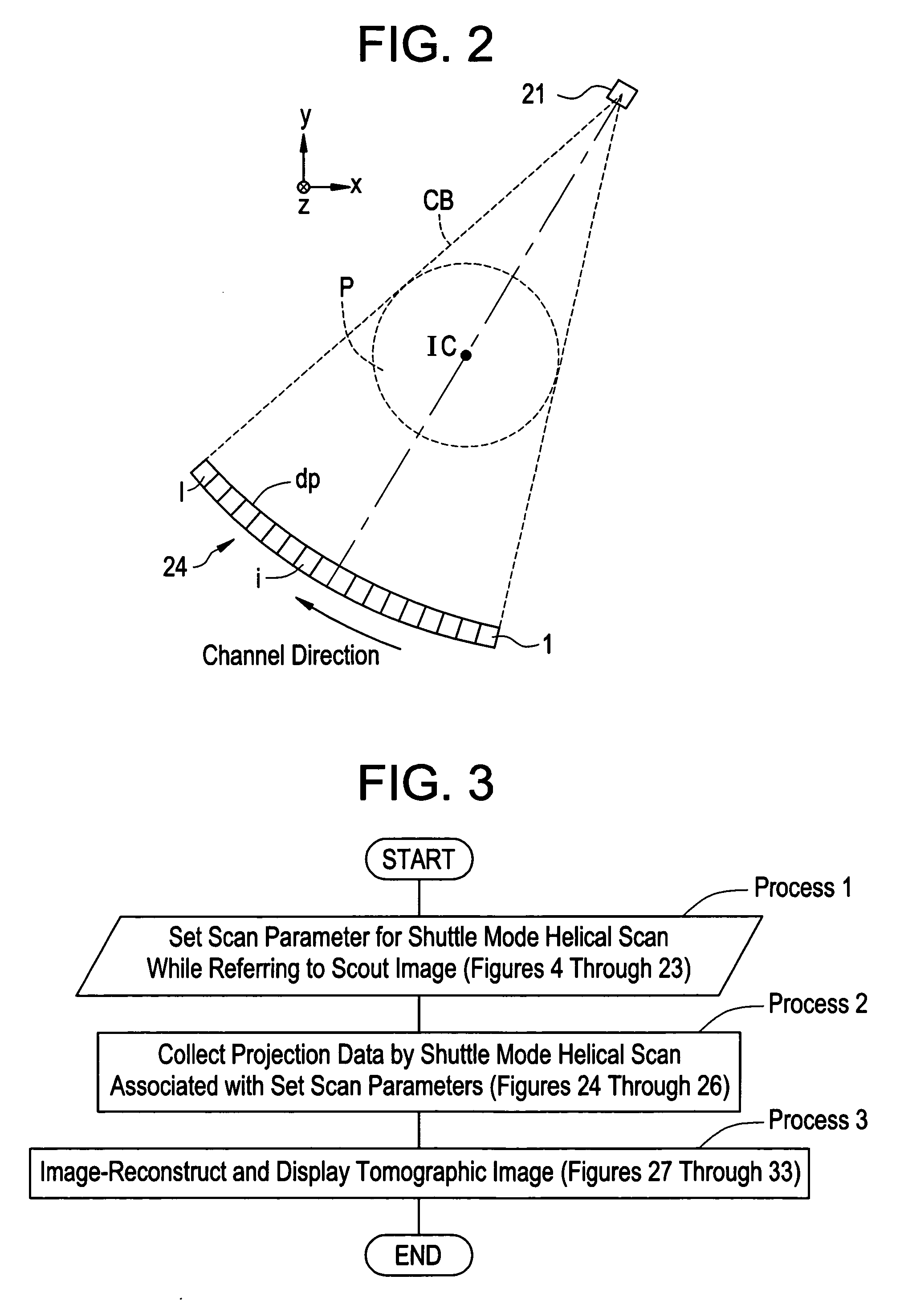

Image

  • Scan parameter setting method for shuttle mode helical scan and X-ray CT apparatus
  • Scan parameter setting method for shuttle mode helical scan and X-ray CT apparatus
  • Scan parameter setting method for shuttle mode helical scan and X-ray CT apparatus

Examples


embodiment 1

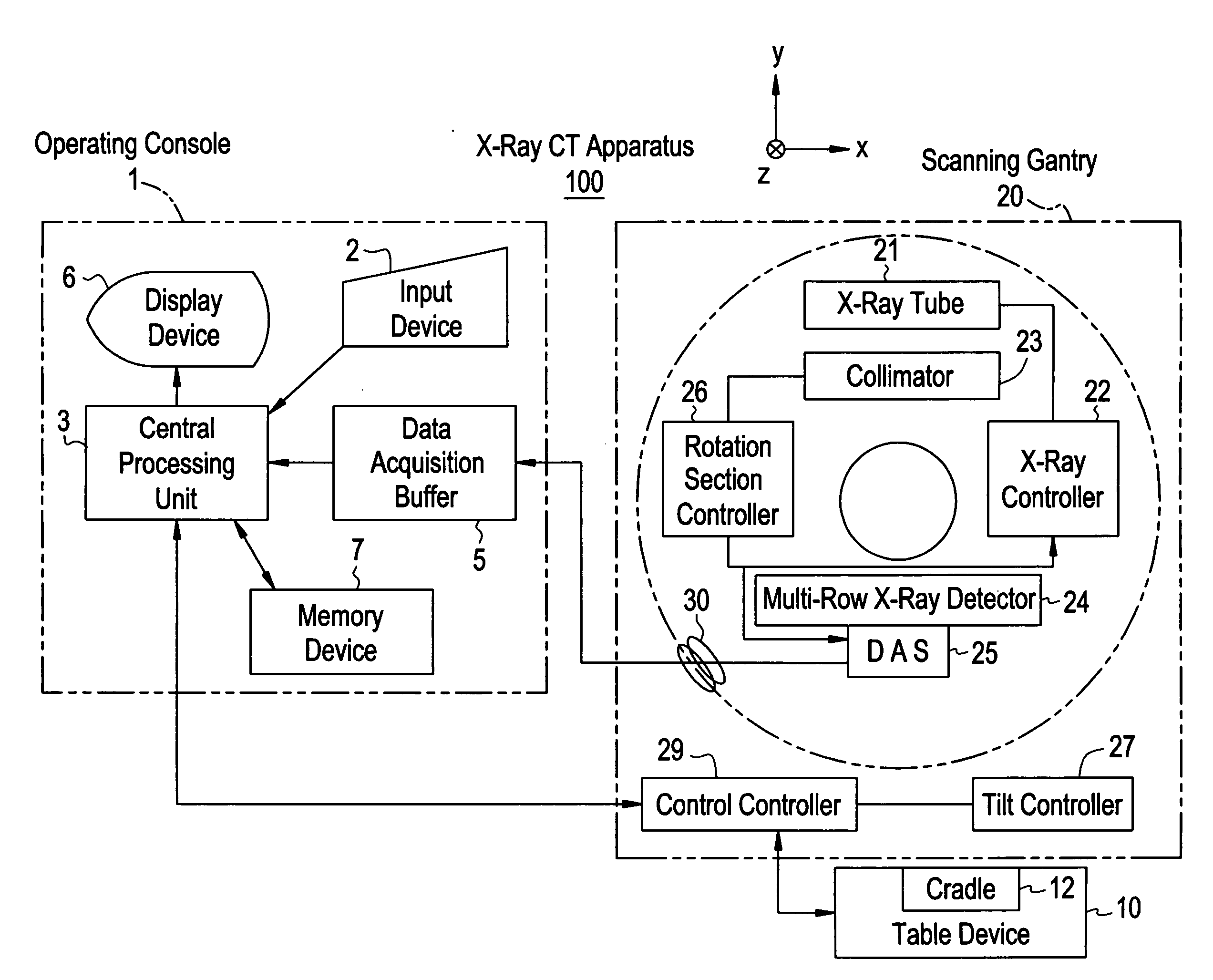
[0099]FIG. 1 is a configurational block diagram showing an X-ray C apparatus 100 according to an embodiment 1.

[0100] The present X-ray CT apparatus 100 is equipped with an operating console 1, a table device 10 and a scanning gantry 20.

[0101] The operating console 1 includes an input device 2 which receives an operator's input, a central processing unit 3 which executes an image reconstructing process, etc., a data acquisition buffer 5 which collects projection data obtained by the scanning gantry 20, a display device 6 which displays a tomographic image reconstructed from the projection data, and a memory device 7 which stores programs, data and an X-ray tomographic image therein. Incidentally, the display device 6 is configured as a multiscreen display having two screens of a right screen and a left screen.

[0102] The table device 10 includes a cradle 12 which places a subject thereon and inserts and draws it into and from a bore (cavity section). The cradle 12 is elevated (in a...


PUM


Abstract

A scout image of a subject is displayed. An operator designates at least one range in a body-axis direction, of the scout image. Further, the operator graphically inputs or key-inputs and sets imaging condition parameters such as a helical pitch, a noise index and the like for a shuttle mode helical scan with being made independent every ranges. Thus, conditions such as helical pitch and noise index for the shuttle mode helical scan can efficiently and intelligibly be set independently for each region or organ, and hence the control / optimization of imaging conditions is enabled.

Description

BACKGROUND OF THE INVENTION [0001] The present invention relates to a scan parameter setting method for a shuttle mode helical scan and an X-ray CT (Computed Tomography) apparatus, and more specifically to a scan parameter setting method for a shuttle mode helical scan and an X-ray CT apparatus, which are capable of efficiently and intelligibly setting parameters for imaging conditions, such as a helical pitch for the shuttle mode helical scan, a noise index corresponding to an index of image noise of each tomographic image, etc. and reducing exposure to a patient while image quality is being held. [0002] There has heretofore been known an X-ray CT apparatus wherein when one reconstruction area is designated based on a scout image (scanogram image) after the completion of input of scan parameters such as a slice thickness, a helical pitch, a tube voltage, a tube current, etc., a scan area or field is calculated from the reconstruction area, and the calculated scan field is helically...

Claims


Application Information

Patent Timeline
IPC IPC(8): A61B6/00G01N23/00G21K1/12H05G1/60
CPCA61B6/032A61B6/463A61B6/466A61B6/027A61B6/488A61B6/542A61B6/469
Inventor NISHIDE, AKIHIKOKATSUKI, AKIESHINYA, MASAE
Owner GE MEDICAL SYST GLOBAL TECH CO LLC
Learn More