
Methods for preparing and analyzing cells having chromosomal abnormalities

a chromosomal abnormality and cell technology, applied in the field of preparing and analyzing cells with chromosomal abnormalities, can solve the problems of limited field of view and image quality, laborious, slow and laborious study of germ line aneuploidy,

Inactive Publication Date: 2006-11-16
AMNIS CORP
View PDF99 Cites 30 Cited by
  • Summary
  • Abstract
  • Description
  • Claims
  • Application Information

AI Technical Summary

Benefits of technology

[0013] In a specific embodiment of this method for determining the presence of a chromosomal abnormality in a cell, the cell is contacted with (a) a first nucleic acid probe and a second nucleic acid probe; (b) a first nucleic acid probe, a second nucleic acid probe, and a third nucleic acid probe; or (c) a first nucleic acid probe, a second nucleic acid probe, a third nucleic acid probe, and a fourth nucleic acid probe, wherein the first nucleic acid probe is capable of hybridizing to a first target chromosomal DNA sequence, wherein the second nucleic acid probe is capable of hybridizing to a second target chromosomal DNA sequence, wherein the third nucleic acid probe is capable of hybridizing to a third target chromosomal DNA sequence, and wherein the fourth nucleic acid probe is capable of hybridizing to a fourth target chromosomal DNA sequence. In a further embodiment, the first probe is detectably labeled with a first reporter molecule, the second probe is detectably labeled with a second reporter molecule, the third probe is detectably labeled with a third reporter molecule, and the fourth probe is detectably labeled with a fourth reporter molecule. In one embodiment, the first reporter molecule is a first fluorochrome, the second reporter molecule is a second fluorochrome, the third reporter molecule is a third fluorochrome, and the fourth reporter molecule is a fourth fluorochrome. In another embodiment, one or more of the first reporter molecule, the second reporter molecule, the third reporter molecule, and the fourth reporter molecule is biotin. In certain embodiments of the method, wherein a reporter molecule is biotin, the method further comprises contacting the cell with streptavidin conjugated to a fluorochrome. In a specific embodiment, the cell is a somatic cell that remains morphologically intact in suspension, and in another specific embodiment, the somatic cell is a tumor cell. In another specific embodiment, the cell is a germ cell. In a certain embodiment, the germ cell is a sperm cell, wherein the sperm cell is a human sperm cell or a sperm cell from a non-human animal (a non-human sperm cell). In a certain embodiment, the chromosomal abnormality detected is sperm aneuploidy, wherein the aneuploidy detected is (a) the absence of a non-sex chromosome; (b) the presence of at least one extra copy of a non-sex chromosome; (c) the presence of more than one sex chromosome; or (d) the absence of sex chromosomes. In a particular embodiment, the cell is obtained from a biological sampl

Problems solved by technology

Despite the development of animal models, the study of germ line aneuploidy remains a slow, labor-intensive, and data-sparse process due to the limitations of the manual scoring of sperm-FISH assays (Schmid et al., Mutagenesis 16:189, 2001).
This technique, as described, is limited for the following reasons: (i) the assessment of aneuploidy is based upon intensity integration of the fluorescent probes rather than the more precise discrete FISH spot detection and enumeration employed in manual scoring; and (ii) the LSC technique uses an air coupled optical objective that limits both its field of view and image quality.

Method used


Image

  • Methods for preparing and analyzing cells having chromosomal abnormalities
  • Methods for preparing and analyzing cells having chromosomal abnormalities
  • Methods for preparing and analyzing cells having chromosomal abnormalities

Examples


example 1

Evaluation of Fixatives for Sperm Cells

[0082] Preservation of cell integrity when analyzing cells by FISH-IS provides optimal results. This Example describes evaluation of various fixatives, including cross-linking fixatives and denaturing fixatives. The cross-linking fixative tested was the zero-length cross-linker formaldehyde, which may be obtained as paraformaldehyde powder or as a liquid product from various commercial vendors (e.g., Fix and Perm® Solution A, Caltag Laboratories, Burlingame, Calif.).

[0083] Several experiments were performed with variations of the procedure for preparing sperm cells for hybridization with a probe. Briefly, the basic procedure included treating sperm cells with dithiothreitol (DTT), followed by centrifugation; treating the cells with lithium diiodosalicylate; removing the cells from the lithium diiodosalicylate by centrifugation; fixing the cells; centrifuging and washing the cells, and then performing a hybridization reaction. Despite varying ...

example 2

Preparation of Sperm Cells in Suspension Without Clumping

[0087] The yield and clumping problems associated with the hybridization step were subsequently examined. Whether losses were due to the high temperatures used during hybridization or to the use of formamide were investigated. Mock hybridizations were performed in which the hybridization mix was incubated at 37° C. overnight. The results showed that the losses were not dependent on temperature, but were likely caused by exposing cells to formamide.

[0088] Because formamide may denature surface proteins, the addition of a detergent during hybridization was examined to determine if the presence of a detergent would minimize cell loss due to clumping. In theory, cells should be relatively stable to detergent once they are fixed. Non-ionic detergents (Triton® and Tween®) and glycosides (e.g. saponin) most effectively minimized loss of sperm cells due to clumping of the cells. In certain embodiments, 1% Saponin was used.

[0089] In...

example 3

Efficient FISH-IS Procedure for Sperm Cells

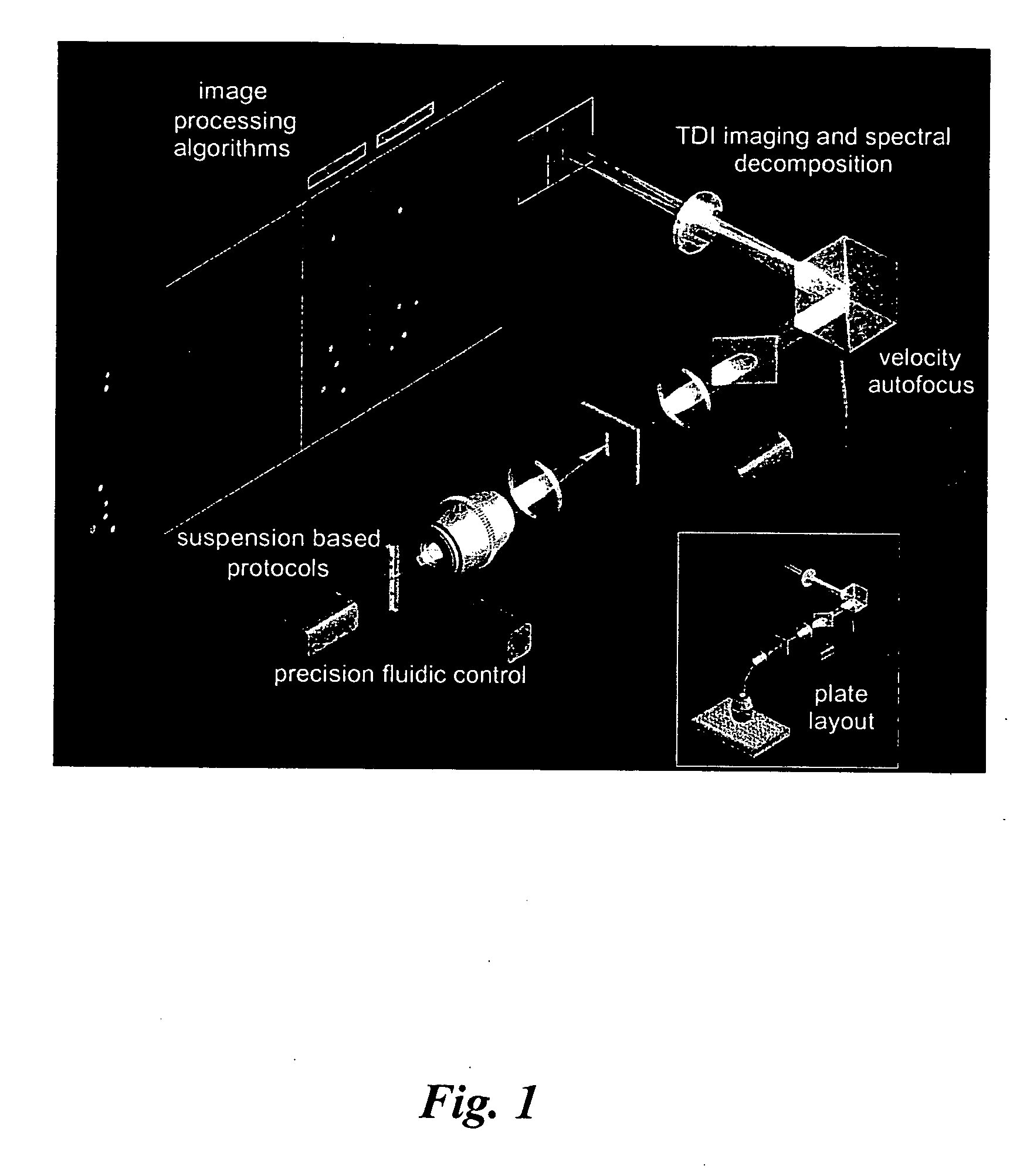
[0090] The standard ImageStream® instrument uses six multi-spectral imaging channels as follows: Channel 1, 470-500 nm, darkfield laser side scatter channel (488 SSC); Channel 2, 400-470 nm, used for brightfield; Channel 3, 500-560 nm, used for brightfield, FITC, Alexa 488, GFP, Syto, or Spec.Green; Channel 4, 560-595 nm, used for brightfield, PE, or Cy-3; Channel 5, 595-660 nm, used for brightfield, 7-AAD, or Alexa610 / PE; and Channel 6, 660-730 nm, used for brightfield, PE-Cy5, Alexa680 / PE, Alexa647 / PE, PerCP, or Draq-5. The dyes listed for each channel represents a partial list of available 488 nm-excitable fluorescent dyes that can be detected in each channel. Because one channel is assigned to brightfield and a second channel is assigned to dark field (laser scatter), four channels remain available for distinct probes. See Table 1.

TABLE 1ImageStream ® Imagine Channels1Channel 1Channel 2Channel 3Channel 4Channel 5Channel 62470-500 nm4...


PUM

PropertyMeasurementUnit
Ratioaaaaaaaaaa
Heterogeneous phase systemaaaaaaaaaa

Abstract

The present invention provides methods for preparing cells with highly condensed chromosomes, such as sperm, and methods for detecting and quantifying specific cellular target molecules in intact cells. Specifically, methods are provided for detecting chromosomes and chromosomal abnormalities, including aneuploidy, in intact cells using fluorescence in situ hybridization of cells in suspension, such as sperm cells.

Description

CROSS-REFERENCE TO RELATED APPLICATION [0001] This application claims the benefit of U.S. Provisional Patent Application No. 60 / 573,775 filed May 20, 2004. This provisional application is incorporated herein by reference in its entirety.STATEMENT OF GOVERNMENT INTEREST [0002] This invention was made with government support under Contract No. N43-ES-35507 awarded by the National Institute for Environmental Health Sciences (NIEHS). The government may have certain rights in this invention.TECHNICAL FIELD [0003] The present invention relates generally to methods for detecting and quantifying specific cellular target molecules in intact cells by multispectral imaging of the cells in flow, and more specifically, to methods of processing an intact cell to facilitate fluorescence in situ hybridization of cells, such as sperm cells, in suspension. BACKGROUND [0004] Aneuploidy, the condition of having more than or less than the normal number of chromosomes, is the most common class of cytogen...

Claims


Application Information

Patent Timeline
IPC IPC(8): C12Q1/68
CPCC12Q1/6841
Inventor BRAWLEY, JAMESMORRISSEY, PHILIP J.FINCH, ROSALYNDE J.BASIJI, DAVID A.LIANG, LUCHUAN
Owner AMNIS CORP
Learn More