
Removeable stents

a biliary system and metallic stent technology, applied in the field of thin wall intraluminal grafts, can solve the problems of inability to remove, poor long-term performance of metallic stents in the biliary system, limiting their widespread application in the treatment of benign biliary diseases, and inability to be removed, and still represent very real problems

Inactive Publication Date: 2006-11-09
KUO MICHAEL D
View PDF19 Cites 27 Cited by
  • Summary
  • Abstract
  • Description
  • Claims
  • Application Information

AI Technical Summary

Problems solved by technology

In contrast, the poor long term performance of metallic stents in the biliary system and their inability to be removed limits their widespread application in the treatment of benign biliary disease.
But, complications such as infection, inadvertent “jailing” of bile ducts, intra-stent sludge accumulation or foreign body occlusion still represent very real problems.
Plastic stents have been routinely used in such settings but unfortunately, experience high rates of malfunction, occlusion and migration.
However, tissue in-growth, inucosal hypertrophy and stent incorporation are very serious and real problems for potential retrieval, particularly in patients with a long life expectancy and thus are generally not advocated for treatment of benign biliary disease.
However, stent grafts in the biliary system can still be complicated by infection or occlusion; thus, the need for removal in this population still exists.
Further, they also reported several cases of device malfunction including rupture of the retrieval-suture in 2 out of 9 (˜22% device retrieval apparatus failure) of their devices which ultimately required tract upsizing and piecemeal extraction of the stent grafts.
While this study work demonstrates the potential feasibility of intentional percutaneous retrieval of stent grafts from the biliary system there are a number of limitations with this approach.
Clearly, the tendency for device migration inherent in this device construction is a significant primary limitation; if the device can not stay in the original location where it is needed to exert its action and thus cannot even perform its primary function, than the need to retrieve it at a later time once it has migrated is clearly immaterial.
Additionally, the use of a retrieval suture as the primary means of retrieving the stent graft is prone to failure as was seen in these cases where the suture ruptured in 22% of cases.
The prominent thickness of the ePTFE used (8-10 mm diameter), and the construction of this stent graft using Gianturco-Rosch Z stents is also a limitation for retrieval in that the primary structure and design of the Gianturco-Rosch stents, the method in which they are appended together (individual Z stents held together in tandem by an outer coating of manually sutured ePTFE) as well as the composition of the stent material (stainless steel), all do not allow for optimal re-compression of the stent grafts once captured, as tract upsizing and piecemeal extraction of the stent grafts was not uncommon in their series.
Further, the porosity of the ePTFE used in these devices does not limit or impede intra-stent sludge accumulation, which can represent a significant obstacle to retrieval as this may effectively increase the total stent graft retrieval volume, thereby making it even harder to re-compress it into the retrieval sheath or may even facilitate adherence of the stent graft to the native duct wall thereby increasing removal friction and the potential for significant iatrogenic injury.
Additionally, the use of retrieval sutures that are exposed with in the native bile duct lumen increases the potential risk for proximal sludge or debris accumulation which may get caught in the retrieval sutures.
This would then impede retrieval by denying access to the device due to the proximal obstruction created by this focus.
This means of capturing, which again inherently relies on the device's tendency to migrate, can cause iatrogenic injury to the bile duct and surrounding structures as the stent graft is manually forced along the bile duct and into the bowel.
Further, the long term primary patency of covered wallstents compared to ePTFE covered stents and the Viabil Biliary Endoprosthesis in particular, is inferior due to the graft material composition which may make it more prone to sludge accumulation.
Indeed, the covered Wallstent has been shown to incite greater neointimal hyperplasia and thrombus induction along the graft surface compared to uncovered wallstents and ePTFE covered stents, supporting the concept that the polyester graft material itself may not be an ideal material for treating stenoses or strictures due to its inherent nature to incite intra-stent graft tissue formation and deposition and induce thrombus formation, all of which as described above would significantly impair attempted stent graft retrieval.
While these modifications may in theory minimize device migration, they significantly deter retrieval due to tissue ingrowth into the uncovered components which can serve to anchor the device to the body.
The difficulty with the use of either the GORE-TEX Vascular Graft® or the Impra graft® as the sleeve component is that the relatively thick, bulky wall of the extruded, longitudinally expanded PTFE tubes limits the ability of the tube to be contracted into a small cross-sectional area for insertion into a blood vessel.
The thinness of the wall is limited by the difficulty of manufacturing an extruded, longitudinally expanded tube having a thin wall of uniform thickness.
This stent is complicated and numerous things could go wrong like leaking of the fluid or a kink being in the stent that would prevent the stent to shrink properly.
The ability to easily and safely remove the above mentioned stents have limited their widespread application to the treatment and management of a large number of diseases.

Method used


Image

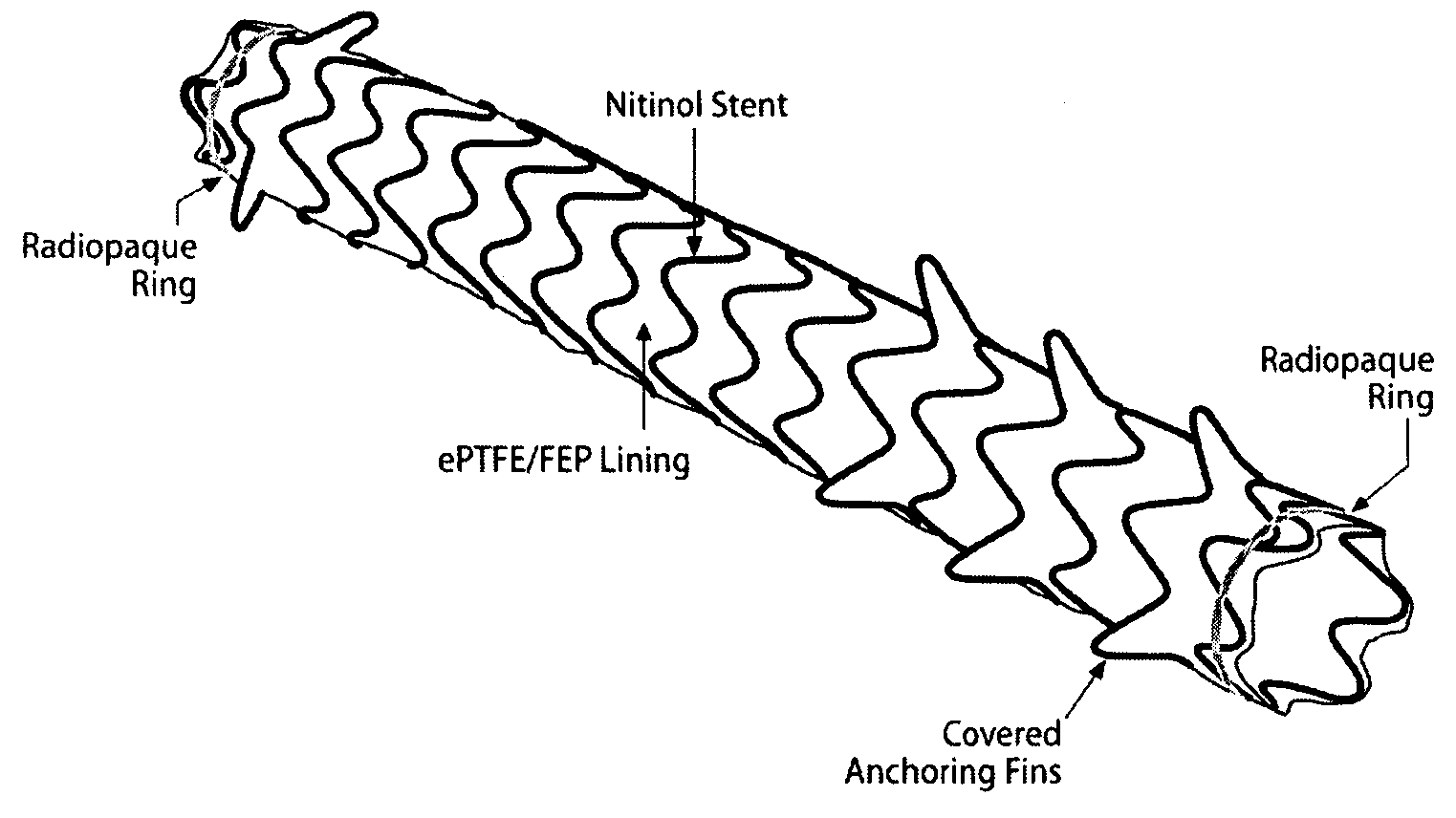
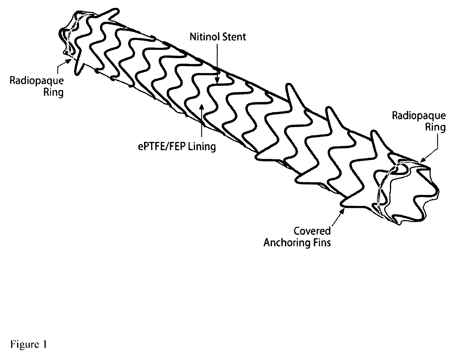
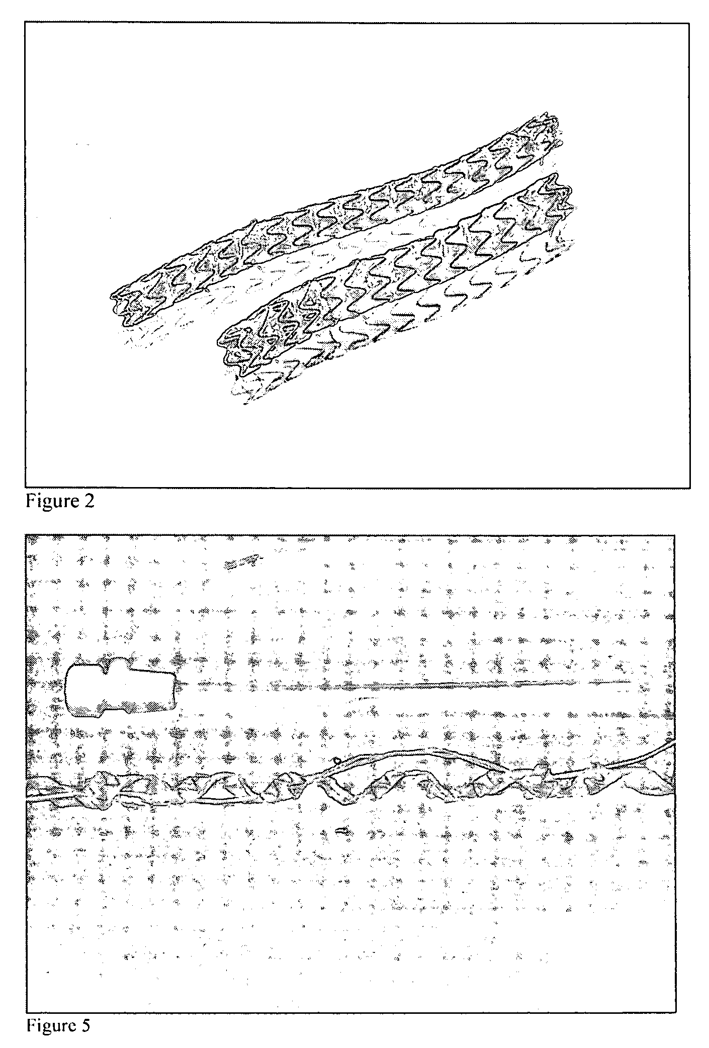
  • Removeable stents
  • Removeable stents
  • Removeable stents

Examples


example 1

[0092] Patients:

[0093] Between December 2004 and October 2005 a 6 patients had Viabil Biliary Endoprostheses placed and retrieved (W. L. Gore Associates, Flagstaff, Ariz.). With respect to the patients described here, three patients had focal anastamotic strictures associated with previous orthotopic liver transplantation. One patient had a chronic recalcitrant focal stenosis of the distal left hepatic duct of uncertain etiology. One patient had a mucinous tumor of her biliary system. The final patient had unresectable metastatic adenocarcinoma involving both the right and left hepatic ducts extending into the proximal common hepatic duct. All patients initially presented with clinical findings consistent with biliary obstruction and infection and were subsequently decompressed with percutaneous biliary drainage and treated with antibiotics.

[0094] At the time of stent graft placement, all patients had internal / external biliary drains in place. Of the 3 patients with transplant-rel...

example ii

[0110] Removal of Stent Grafts From the Vasculature

[0111] Two patients with end-stage renal disease with an arterial-venous graft had covered stents placed because of a recurrent, recalcitrant stenoses that demonstrated repeated restenosis and high elastic recoil despite repeated balloon angioplasty near the venous outflow. In both patients, the stent grafts used was a Viabahn stent graft from W L Gore which was placed in order to serve as a vascular conduit across the recalcitrant stenoses and to also serve as a scaffold for the vessel to remodel around. The first patient subsequently returned for extraction of the implanted stent graft. Using a similar technique as described and detailed above the stent graft was removed approximately 3 weeks later after initial stent graft placement. Namely, in this patient bidirectional vascular access was obtained surrounding the stent graft (a 10 French sheath was placed distal to the proximal margin of the stent graft directed towards the in...


PUM


Abstract

There is disclosed novel removable stent grafts and methods for removing stent grafts. There is also disclosed new diseases and disorders that can now be treated with removable stents.

Description

RELATED APPLICATION(S) [0001] This Application claims priority of U.S. provisional application Ser. No. 60 / 678,843, filed May 6, 2005 and U.S. provisional application Ser. No. 60 / 693,332, filed Jun. 22, 2005.FIELD OF THE INVENTION [0002] This invention relates to the field of intraluminal grafts and particularly to thin-wall intraluminal grafts useful as an inner lining for blood vessels or other body conduits and methods to remove them. BACKGROUND OF THE INVENTION [0003] The percutaneous management of biliary strictures remains poorly defined. Treatment has classically been guided by whether the underlying disease process is benign or malignant. In cases of inoperable malignant disease, bare metal stents are routinely used as the desire for biliary conduit restoration outweighs their relatively poor overall primary patency due to tissue in-growth. In contrast, the poor long term performance of metallic stents in the biliary system and their inability to be removed limits their wide...

Claims


Application Information

Patent Timeline
IPC IPC(8): A61F2/06
CPCA61F2/07A61F2002/075A61F2/89A61F2250/0098A61F2002/9528
Inventor KUO, MICHAEL D.
Owner KUO MICHAEL D
Learn More