
Infectious, chimeric hepatitis C virus, methods of producing the same and methods of use thereof

a technology of chimeric hepatitis c virus, which is applied in the field of infectious recombinant hepatitis c virus (hcv), can solve the problems of limited primary hepatocytes, low and variable levels of replication and virus-induced cytotoxicity, and serious hcv drug search and development difficulties, etc., to achieve the effect of improving cell culture growth

Inactive Publication Date: 2006-09-21
THE ROCKEFELLER UNIV
View PDF4 Cites 25 Cited by
  • Summary
  • Abstract
  • Description
  • Claims
  • Application Information

AI Technical Summary

Benefits of technology

[0016] In one embodiment, the invention provides a method of identifying HCV variants with improved growth in cell culture, the method comprising contacting cells with an isolated nucleic acid molecule encoding an infectious recombinant HCV genome, comprising a chimeric HCV genome contacting cells with an isolated nucleic acid molecule comprising at least one mutation of the chimeric HCV genome, independently culturing the cells and determining HCV infection, replication, or cell-to-cell spread, in cells contacted with the chimeric HCV or the mutated virus, whereby enhanced HCV infection, replication, or cell-to-cell spread in cells contacted with the mutated virus indicates that the HCV variant has improved growth in cell culture.

Problems solved by technology

The search for HCV drugs as well as for the development of an HCV vaccine is severely hampered by the lack of an efficient tissue culture, or robust cellular system that would support virus replication, or a simple animal system for the study of replication and HCV pathogenicity.
Some vitro culture systems attempted for the study of HCV used human cells of hepatocytic and lymphocytic origin, but low and variable levels of replication and virus-induced cytotoxicity posed important problems.
Primary hepatocytes (derived from a human donor) can be infected with HCV isolated from serum of infected patients, and the virus can be detected in the supernatant for several weeks after infection, however, the availability of primary hepatocytes is limited and, their isolation is time-consuming and labor-intensive.
Consequently, such tissue culture systems are generally considered unsuitable for intensive large-scale antiviral studies.
The proposed system was limited, however, by the fact that only non-structural viral proteins were expressed.

Method used


Image

  • Infectious, chimeric hepatitis C virus, methods of producing the same and methods of use thereof
  • Infectious, chimeric hepatitis C virus, methods of producing the same and methods of use thereof
  • Infectious, chimeric hepatitis C virus, methods of producing the same and methods of use thereof

Examples


example 1

Detection of HCV Replication and Spread Using the Culture System

[0096] The FL-J6 / JFH construct comprises the cloned cDNA of a chimeric HCV genome with the sequence as set forth in SEQ ID No: 1.

[0097] Replication of FL-J6 / JFH or FL-J6 / JFH++ can be monitored by various methods. A standard immunohistochemical staining procedure was adapted to detect NS5A expression in cells by using the 9E10 monoclonal antibody (Dr. Tim Tellinghuisen and Dr. Charles M. Rice). Antibody staining of HCV-positive cells was revealed through the use of a horseradish peroxidase-coupled secondary antibody and diaminobenzidine. As can be seen in FIG. 2A, HCV replication in Huh-7.5 cells transfected with the FL-J6 / JFH or the SGR-JFH subgenomic replicon, was detected, however FL-J6 / JFH (GND), which contained a lethal mutation in the HCV RNA polymerase active site was not detected.

[0098] Cells harboring FL-J6 / JFH secreted infectious virus particles (HCVcc) that were capable of transferring NS5A expression to na...

example 2

Quantitation of HCV Infectivity

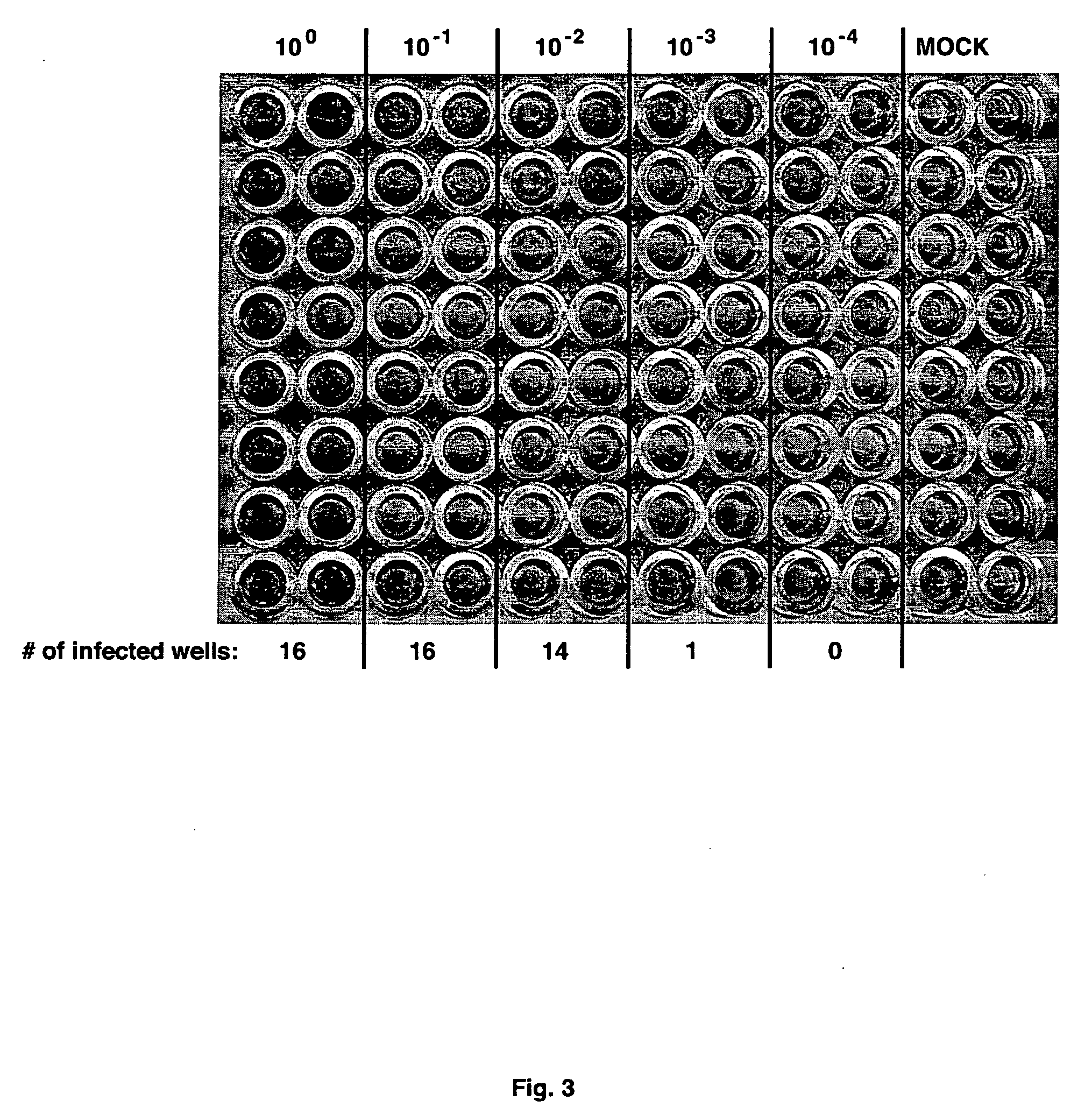
[0101] The procedure for immunohistochemical staining for NS5A has been adapted to determine the infectious virus concentration within samples. In one system, virus-containing samples were subjected to serial 10-fold dilutions (typically, 10−1 to 10−6) in complete growth medium. Each dilution was then used to infect multiple wells of Huh-7.5 cells seeded in 96-well plates. Following 2 or 3 day incubation, the cells are fixed and subjected to immunohistochemical staining for NS5A expression, as above. The assay plate was then scored for the number of infected wells (i.e. at least one infected cell per well) for each dilution (FIG. 3) and the virus titer was then calculated as the 50% end-point tissue-culture infectious dose per ml (TCID50 / ml) according to the statistical method of Reed and Muench (Am. J. Hyg. 27:493-497 (1938)). The sensitivity of this assay was determined by volume of the lowest dilution and the number of replicates tested, and was ty...

example 3

Growth of Virus

[0103]FIG. 4 shows a growth curve of FL-J6 / JFH and FL-J6 / JFH++ following electroporation of Huh-7.5 cells. Both viruses were efficiently released from cells following a 9-12 hour lag phase. Although the kinetics of FL-J6 / JFH release were greater than that of FL-J6 / JFH++, both viruses reached a peak of ≈5×10⁴ TCID50 / ml between 48 and 72 hours post-electroporation.

[0104] Following this initial virus growth phase, virus was continuously produced by transfected cells. Therefore, cells can be passaged as normal and continued to produce virus. Since virus was detected in cells after 5 passages, virus cultures may be conveniently scaled up in this way.

[0105] Additionally, virus can be produced following infection of naïve cells. Typically, cells are infected at a multiplicity of infection (MOI) of less than one to minimize co-infection, washed, and fed with complete growth medium. Virus-containing supernatants are then collected over subsequent days, and infected cells ma...


PUM

PropertyMeasurementUnit
Fractionaaaaaaaaaa
Molar densityaaaaaaaaaa
Concentrationaaaaaaaaaa

Abstract

The present invention provides infectious recombinant Hepatitis C Viruses (HCV), and vectors, cells and animals comprising the same. The present invention provides methods of producing infectious recombinant HCV, and their use in identifying anti-HCV therapeutic agents, as well as sequences of HCV associated with HCV pathogenesis.

Description

CROSS REFERENCE TO RELATED APPLICATIONS [0001] This application claims the priority of U.S. Provisional Application Ser. No. 60 / 658,187 filed Mar. 4, 2005, which is incorporated herein by reference in its entirety.GOVERNMENT INTEREST STATEMENT [0002] This invention was made in whole or in part with government support under grant number CA 579373, awarded by the National Institute of Health. The government may have certain rights in the invention.FIELD OF THE INVENTION [0003] This invention provides infectious recombinant hepatitis C viruses (HCV), and vectors, cells and animals comprising the same. The present invention provides methods of producing the infectious recombinant HCV, and their use in identifying anti-HCV therapeutic and including for use in vaccines and diagnostics and, as well as sequences of HCV associated Shouldn't with HCV pathogenesis. BACKGROUND OF THE INVENTION [0004] Hepatitis C virus (HCV) is a member of the Flaviviridae family of enveloped, positive-strand RN...

Claims


Application Information

Patent Timeline
IPC IPC(8): C12Q1/70C07H21/04C12N7/00C12N15/86C12N5/06
CPCA61K2039/525C07K2319/61C12N7/00C12N15/86C12N2770/24243C12N2770/24251C12N2840/203C12Q1/18G01N33/5767G01N2333/18G01N2500/00
Inventor RICE, CHARLESLINDENBACH, BRETTEVANS, MATTHEWJONES, CHRISTOPHER
Owner THE ROCKEFELLER UNIV
Learn More