Method and apparatus for accelerated spiral-coded imaging in magnetic resonance tomography

a magnetic resonance tomography and spiral-coded technology, applied in the field of magnetic resonance tomography (mrt), can solve the problems of reducing image quality, affecting and limiting the acquisition time of mrt images in many medical diagnostic applications. achieve the effect of reducing the readout duration of spiral-shaped acquisition methods

Inactive Publication Date: 2006-07-20
SIEMENS HEALTHCARE GMBH
View PDF7 Cites 2 Cited by
  • Summary
  • Abstract
  • Description
  • Claims
  • Application Information

AI Technical Summary

Benefits of technology

[0013] An object of the present invention is to significantly reduce the readout duration of a spiral-shaped acquisition method without otherwise having to accept disadvantages.

Problems solved by technology

In spite of the technical progress in the construction of MRT apparatuses, acquisition time of an MRT image remains the limiting factor for many applications of MRT in medical diagnostics.
An unsolved problem in fast MRT imaging (fast single shot spiral scanning and fast multi shot spiral scanning and EPI) is that generally image quality reductions occur due to frequency and phase errors during the readout times of the RF response signal.
This has the severe disadvantage, however, that missing passes result in a lower resolution of the reconstructed image.
Such methods are extremely expensive in terms of calculation time and therefore are not practically applicable at the present time.

Method used


Image

  • Method and apparatus for accelerated spiral-coded imaging in magnetic resonance tomography
  • Method and apparatus for accelerated spiral-coded imaging in magnetic resonance tomography
  • Method and apparatus for accelerated spiral-coded imaging in magnetic resonance tomography

Examples


Embodiment Construction

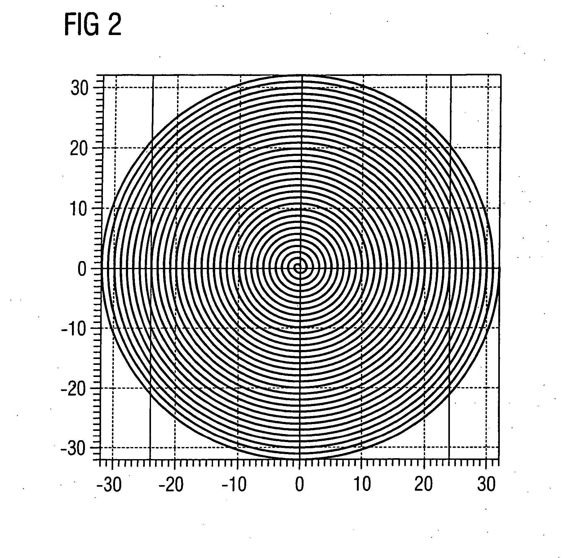
[0024]FIG. 1 schematically illustrates a magnetic resonance imaging or tomography apparatus for generation of a magnetic resonance image of a subject according to the present invention. The design of the magnetic resonance tomography apparatus thereby corresponds to the design of a conventional magnetic tomography apparatus except for the differences described below. A basic field magnet 1 generates a temporally-constant strong magnetic field for polarization or alignment of the nuclear spins in the examination region of a subject such as, for example, a part of a human body to be examined. The high homogeneity of the basic magnetic field necessary for the magnetic resonance measurement is defined in a spherical measurement volume M in which the parts of the human body to be examined are introduced. To support the homogeneity requirements, and in particular for elimination of temporally invariable influences, shim plates made of ferromagnetic material are mounted at a suitable locat...


PUM


Abstract

In a method and apparatus for accelerated spiral-coded imaging (scanning k-space with a spiral trajectory) in magnetic resonance tomography, the user-defined sequence forming the basis of the spiral scanning is modified such that the k-matrix forming the basis of the sequence is scanned only in a sub-region, the sub-region being defined by a symmetrical shortening on both sides of the k-space matrix in a first direction as well as by a one-sided shortening of the k-space matrix in a second direction orthogonal to the first direction.

Description

BACKGROUND OF THE INVENTION [0001] 1. Field of the Invention [0002] The present invention in general concerns magnetic resonance tomography (MRT) as used in medicine for the examination of patients. The present invention in particular concerns a spiral-coded method for accelerated MRT imaging as well as a magnetic resonance tomography apparatus that is suitable for implementation of the method. [0003] 2. Description of the Prior Art [0004] MRT is based on the physical phenomenon of magnetic resonance and has been successfully used as an imaging modality for over 15 years in medicine and biophysics. In this examination modality, the subject is exposed to a strong, constant magnetic field. The nuclear spins of the atoms in the subject, which were previously randomly oriented, thereby align. Radio-frequency energy can now excite these “ordered” nuclear spins to a specific oscillation. In MRT, this oscillation generates the actual measurement signal which is acquired by means of appropr...

Claims


Application Information

Patent Timeline
Patent Type & Authority Applications(United States)
IPC IPC(8): G01V3/00
CPCG01R33/4824G01R33/561
Inventor KRUGER, GUNNARMOHR, CECILETHESEN, STEFAN
Owner SIEMENS HEALTHCARE GMBH
Why Eureka
Try Eureka