
Ultrasound cardiac stimulator

Inactive Publication Date: 2006-03-09
TECHNION RES & DEV FOUND LTD
View PDF39 Cites 54 Cited by
  • Summary
  • Abstract
  • Description
  • Claims
  • Application Information

AI Technical Summary

Benefits of technology

[0005] An aspect of some embodiments of the invention relates to stimulating cardiac tissue, or any other excitable tissue such as muscles and nerves, using ultrasound, for diagnostic and / or therapeutic purposes, using transducers located outside the body, or in the esophagus or other non-invasive body channels, such as the nasal cavities. Optionally, ultrasound contrast agents, for example microbubbles or liposomes, are used to enhance the procedure, for example by increasing the sensitivity of the cardiac tissue to stimulation.
[0006] In some embodiments of the invention, an imaging system is used to track and observe the heart before, during, and / or after the cardiac tissue is stimulated. This is done, for example, in order to observe the local or global mechanical response of the heart to the stimulation, or to focus the ultrasound energy on the correct spot. Optionally, the imaging system is an ultrasound imaging system, using the same transducers as are used for stimulation. Alternatively or additionally, other ultrasound transducers may be used for imaging. Alternatively or additionally, a Computer Aided Tomography system using x-rays, a Magnetic Resonance Imaging system, or any imaging system known to the art may be used. Optionally, contrast agents are used to improve image quality, and / or to distinguish between perfused and non-perfused tissue.
[0007] In some embodiments of the invention, the location and orientation of the heart, or a particular point on the heart, are tracked in real time while ultrasound is used to stimulate the heart. Such tracking makes it possible to repeatedly focus ultrasound on the same spot in the heart, or to successively focus ultrasound on two or more spots with known relative positions.
[0011] In some embodiments of the invention, ultrasound is used to increase membrane permeability at particular locations in the heart and / or particular times in the cardiac cycle, in order to selectively increase the absorption or effect of drugs at those locations and / or times. Optionally, the drugs are delivered to specific locations by a cardiac catheter.
[0025] Optionally, the controller is operative to reduce the power level of ultrasound when the contrast agents are injected.
[0077] Optionally, the at least one ultrasound transducers comprise at least two ultrasound transducers, sized and spaced so that they can be placed between the ribs, in such a way as to avoid blocking of ultrasound by the ribs.

Problems solved by technology

Separately, high energy ultrasound pulses have been reported to shatter kidney stones, while also affecting inner cellular structures and membrane properties.
All of these invasive procedures involve obvious risks and expenses.

Method used


Image

  • Ultrasound cardiac stimulator
  • Ultrasound cardiac stimulator
  • Ultrasound cardiac stimulator

Examples


Embodiment Construction

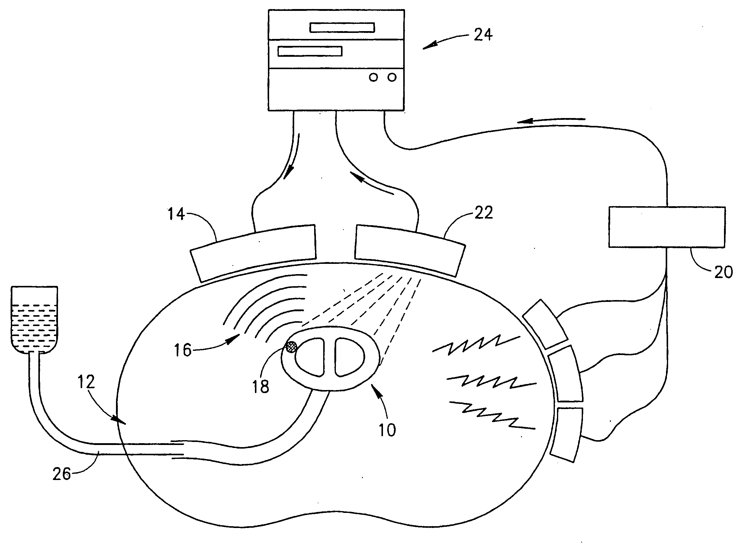
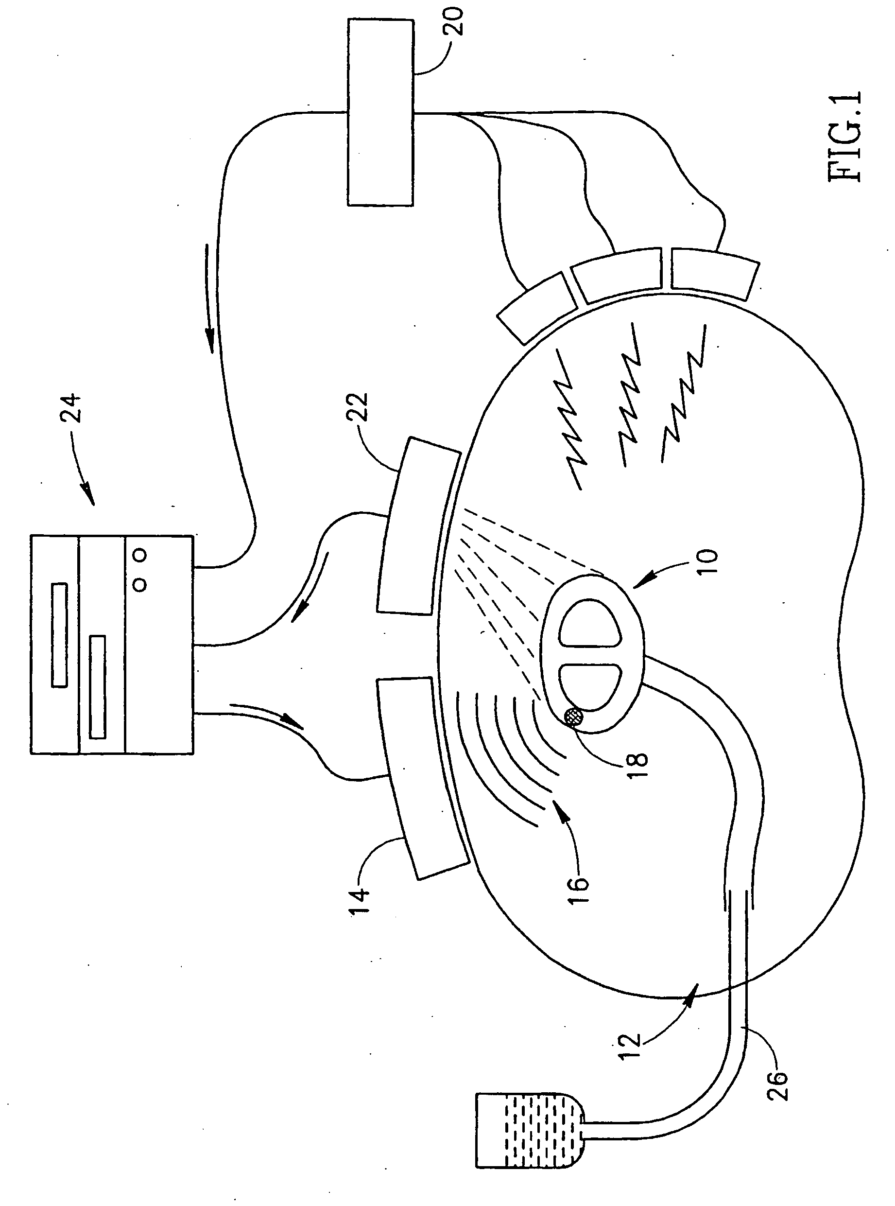
[0115]FIG. 1 schematically shows an ultrasound system according to an exemplary embodiment of the invention. A heart 10 is shown in a cross-section of a patient's chest 12. A phased array 14 of ultrasound transducers focuses ultrasound waves 16 on a spot 18 in the wall of the heart, stimulating cardiac tissue at that spot and possibly inducing action potentials, detected by an electrocardiograph 20. By adjusting the relative phases and amplitudes of the different transducers in the array, the ultrasound can generally be focused on any desired spot 18. Alternatively, any other method of focusing ultrasound known to the art is used to focus the ultrasound waves on spot 18. The diameter of the spot cannot be much smaller than one wavelength, assuming that it is in the far field of the transducers, i.e. at least several wavelengths away from the transducers. If it is desired to focus the ultrasound on a region much smaller than the thickness of the myocardium, the frequency of the ultra...


PUM


Abstract

An ultrasound cardiac stimulation system comprising: a spatially selective ultrasound source comprising at least one ultrasound transducer located outside the circulatory system; and a controller; where the controller generates an electrical response in the heart by directing the ultrasound source to transmit a high enough power level of ultrasound to one or more locations in the heart.

Description

FIELD OF THE INVENTION [0001] The present invention is related to the field of cardiac diagnosis and therapy. BACKGROUND OF THE INVENTION [0002] The myocardium is susceptible to mechanical stimulation: case reports have detailed incidents of cardiac arrest due to a ball or a fist hitting the person's thorax. Physiological studies have reported initiation of an action potential due to a mechanical stimulation such as tapping of the epicardium [Avitall, B., Levine, H. J., Naimi, S., Donahue, R. P., Pauker, S. G., and Adam, D. R., “Local effects of electrical and mechanical stimulation on the recovery properties of canine ventricle,”Am. J. Cardiology 50, 263-270 (1982)], or due to stretch. Separately, high energy ultrasound pulses have been reported to shatter kidney stones, while also affecting inner cellular structures and membrane properties. In particular, it has been reported that ultrasound pulses increase membrane permeability and specific ion flow. Ultrasound transducers attach...

Claims


Application Information

Patent Timeline
IPC IPC(8): A61B8/00A61B17/00A61N1/362A61N7/00
CPCA61B2017/00243A61B2017/00703A61N2007/0078A61N7/00A61N1/362A61N1/3629
Inventor ADAM, DAN
Owner TECHNION RES & DEV FOUND LTD
Learn More