Methods for in vitro expansion of hematopoietic stem cells
a technology of hematopoietic stem cells and in vitro expansion, which is applied in the field of human hematopoietic stem cell expansion, can solve the problem of primitive phenotype expansion of cells
- Summary
- Abstract
- Description
- Claims
- Application Information
AI Technical Summary
Benefits of technology
Problems solved by technology
Method used
Image
Examples
example 1
Materials and Methods
[0092] The present example provides some exemplary materials and methods that may be used in conjunction with the present invention to determine expansion of a given population of HSC cells. Certain of these methods were used to obtain the data presented herein below. However, it should be understood that these methods may be adapted and still produce data useful in interpreting the present invention.
Isolation of Human CD34+ Cells
[0093] Those of skill in the art are aware of numerous techniques by which to isolate cells that have particular markers that identify the cell as being of a certain lineage. HSC cells are often isolated initially as being that population of cells that have a CD34+ phenotype. The following description provides one exemplary methods for such an isolation that was used by the inventors.
[0094] Cryopreserved cadaver adult human bone marrow (BM) mononuclear cells (MNC) were rapidly thawed at 37° C. and slowly diluted dropwise in RMPI (B...
example 2
Initial Studies Showing Role of HSC Methylation and Acetylation in Reprogramming HSC Behavior In Vitro
[0112] Epigenetically mediated loss of gene function has been shown to be accompanied by DNA methylation of a gene's promoter and by histone deacetylation. Thus, conditions previously utilized for in vitro stem cell expansion result in silencing of genes required for hematopoetic stem cells (HSC) to undergo symmetrical cell division.
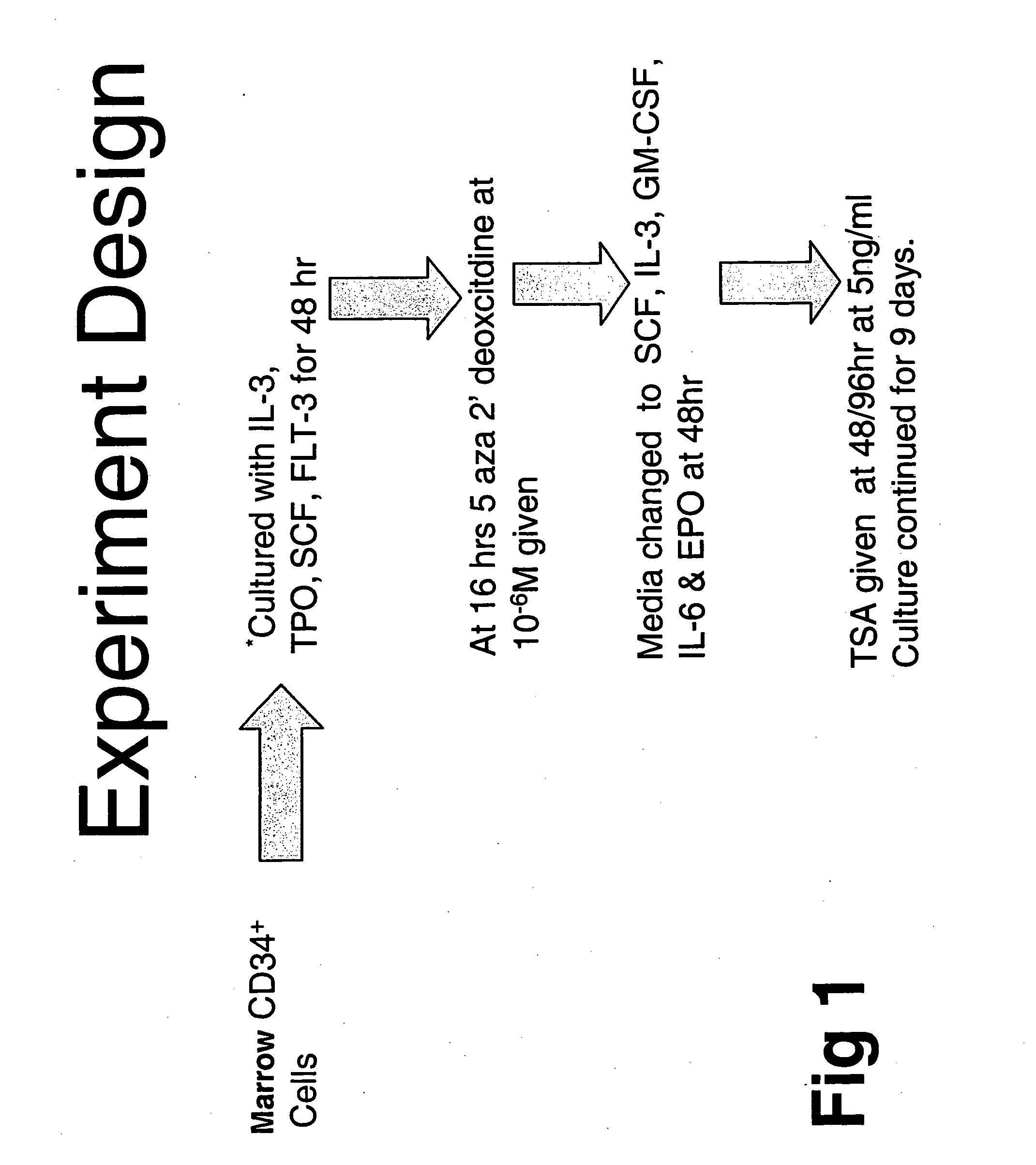
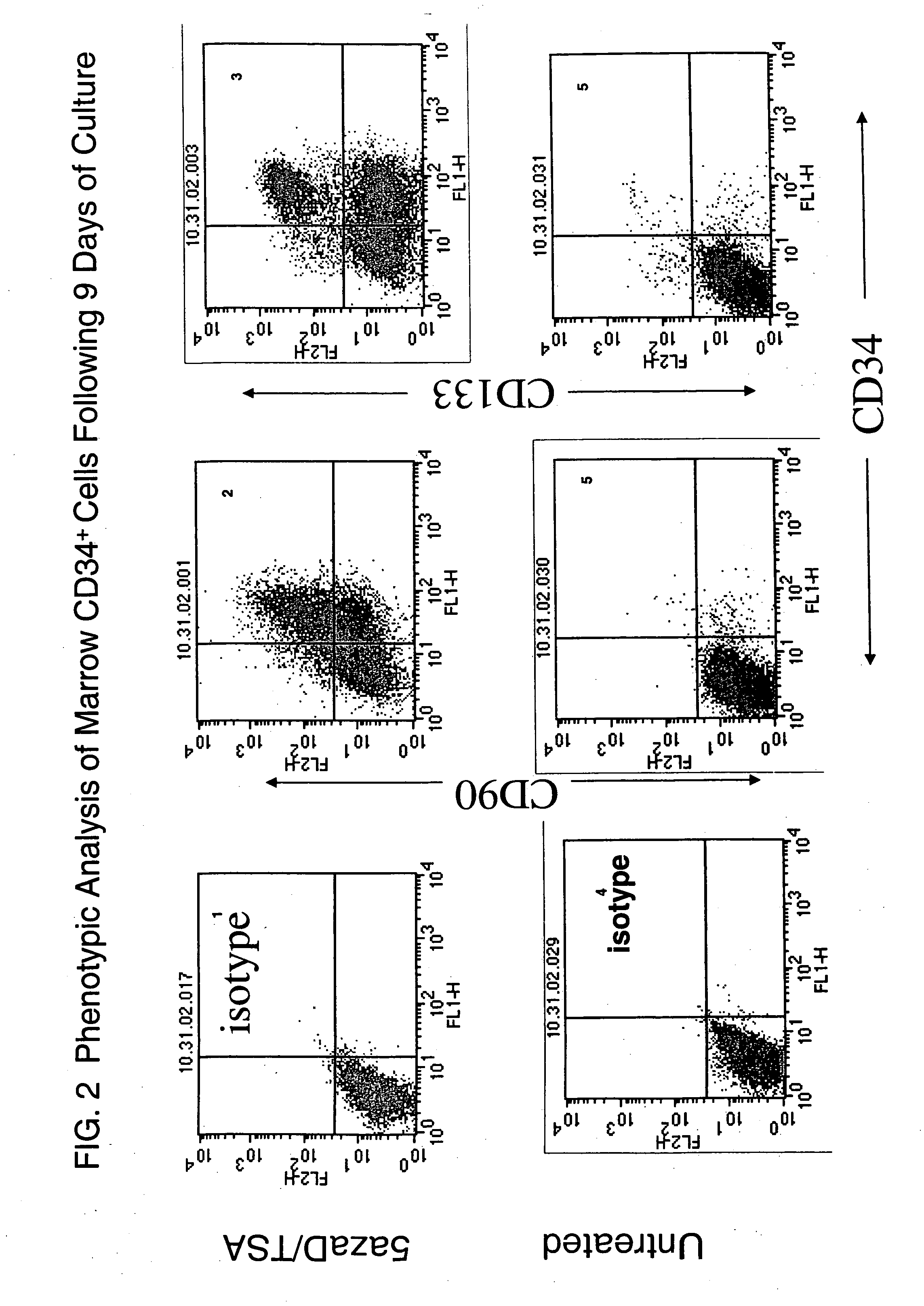
[0113] In order to study this phenomenon further, adult human bone marrow CD34+ cells were cultured in media containing IL3, TPO, SCF and FLT-3 for 48 hours in order to promote cell division. The cells also were exposed to conditions and different time points. The cells were exposed to an inhibitor of DNA methylation (IDM), 5 aza 2′dexoxycitidine (5azaD) or the cytotoxic agent 5-flurouracil (5-FU). The cells were then exposed to conditions (SCF, IL-3, IL-6, GM-CSF and EPO) known to promote cell differentiation. Trichostatin A(TSA), a histone deaceytlas...
example 3
Methylation Status of Marrow CD34+ Cells with and without 5azaD and TSA
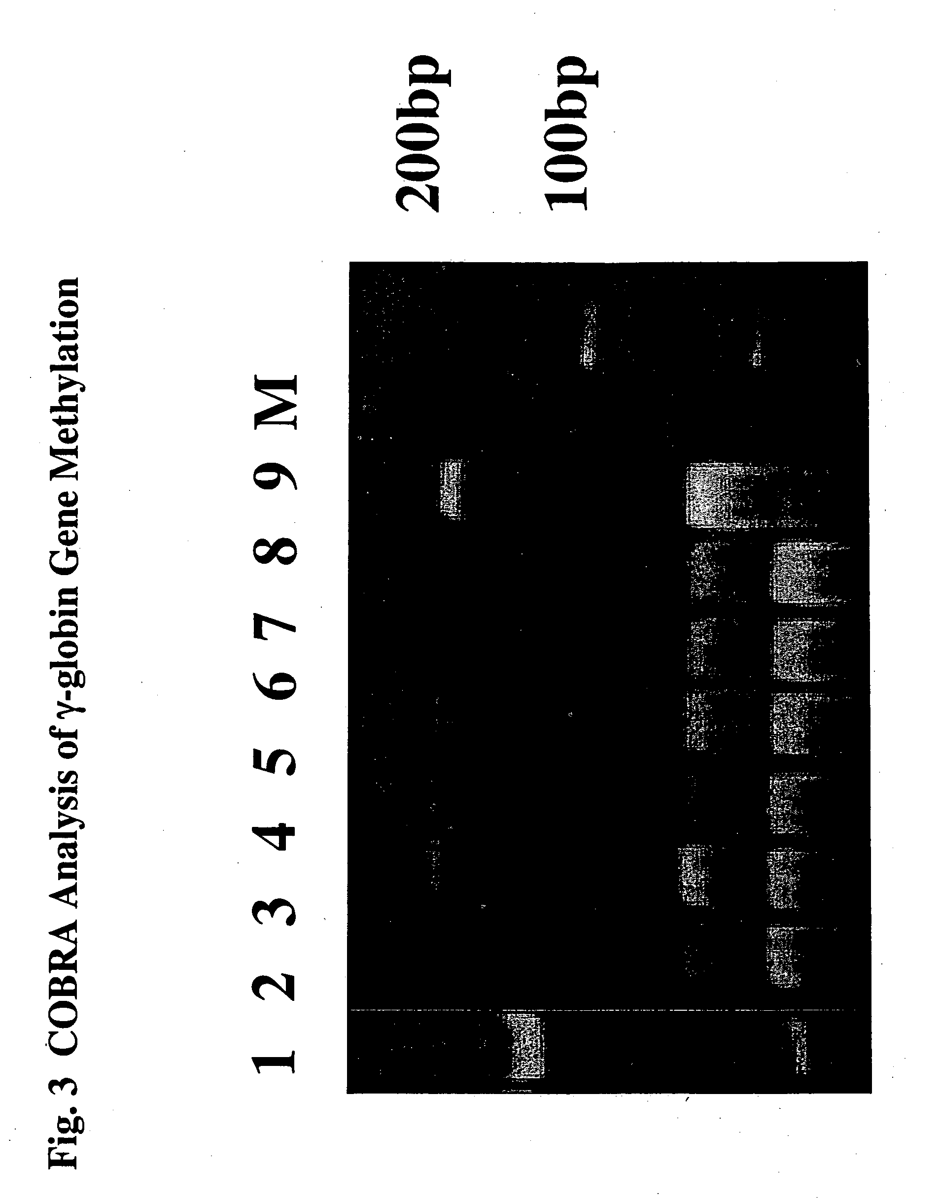
[0118] To determine the methylation status of treated and untreated CD34+ marrow cells COBRA (Combined Bisulfite Restriction Analysis) was used to quantitate the DNA methylation of a CpG residue located at −256th position within the γ-globin promoter region. Primary CD34+ cells (day 0) initially were methylated at this promoter site (90%). These cells were then cultured for 5 days under various conditions outlined (Table 7). 87% of cells cultured for five days with cytokines alone were methylated suggesting that the status of this promoter was unchanged throughout the culture period. By contrast 58% of the cells treated with cytokines and sequential addition of 5aza2′deoxycitidine (5azaD) and Trichostatin (TSA) were methylated after culture (Table 7) indicating that these drugs might be capable of altering their methylation pattern in vitro.
[0119] Bisulfite sequence analysis of 5 CpG sites within the γ-globin g...
PUM
| Property | Measurement | Unit |
|---|---|---|
| concentration | aaaaa | aaaaa |
| concentration | aaaaa | aaaaa |
| concentration | aaaaa | aaaaa |
Abstract
Description
Claims
Application Information
 - R&D
- Intellectual Property
- Life Sciences
- Materials
- Tech Scout
- Unparalleled Data Quality
- Higher Quality Content
- 60% Fewer Hallucinations
Browse by: Latest US Patents, China's latest patents, Technical Efficacy Thesaurus, Application Domain, Technology Topic, Popular Technical Reports.
© 2025 PatSnap. All rights reserved.Legal|Privacy policy|Modern Slavery Act Transparency Statement|Sitemap|About US| Contact US: help@patsnap.com


