
Apparatus and method for creating an arterio-venous connection in hemodialysis maintenance

a technology of arteriovenous connection and hemodialysis, which is applied in the field of apparatus and method for creating an arteriovenous connection in hemodialysis maintenance, can solve the problems of reducing affecting the efficiency of hemodialysis, and affecting the quality of hemodialysis, so as to achieve the effect of reducing the risk of hematocrit, and reducing the risk of hemocri

Inactive Publication Date: 2005-09-15
PERMAGRAFT +1
View PDF9 Cites 55 Cited by
  • Summary
  • Abstract
  • Description
  • Claims
  • Application Information

AI Technical Summary

Benefits of technology

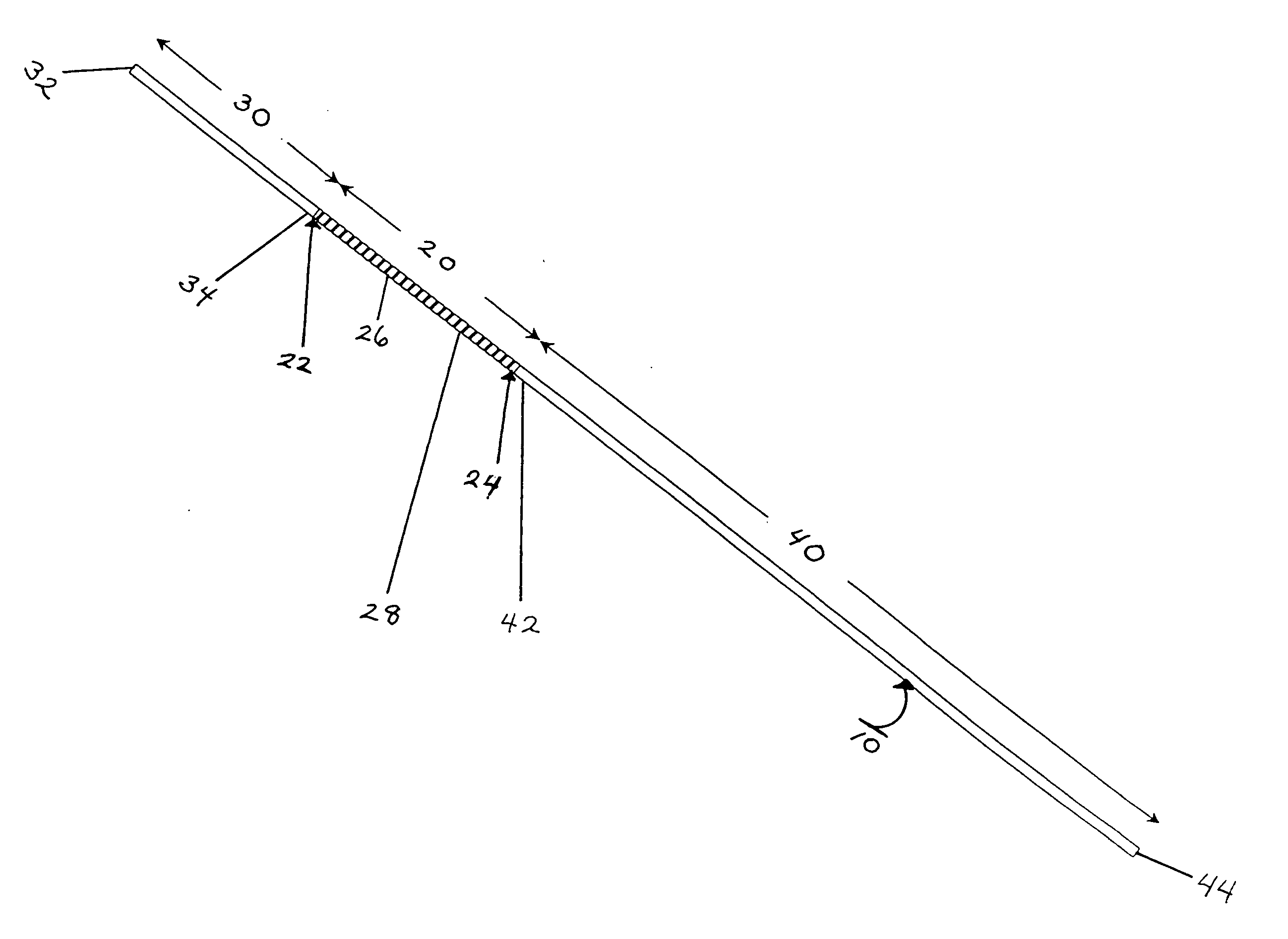
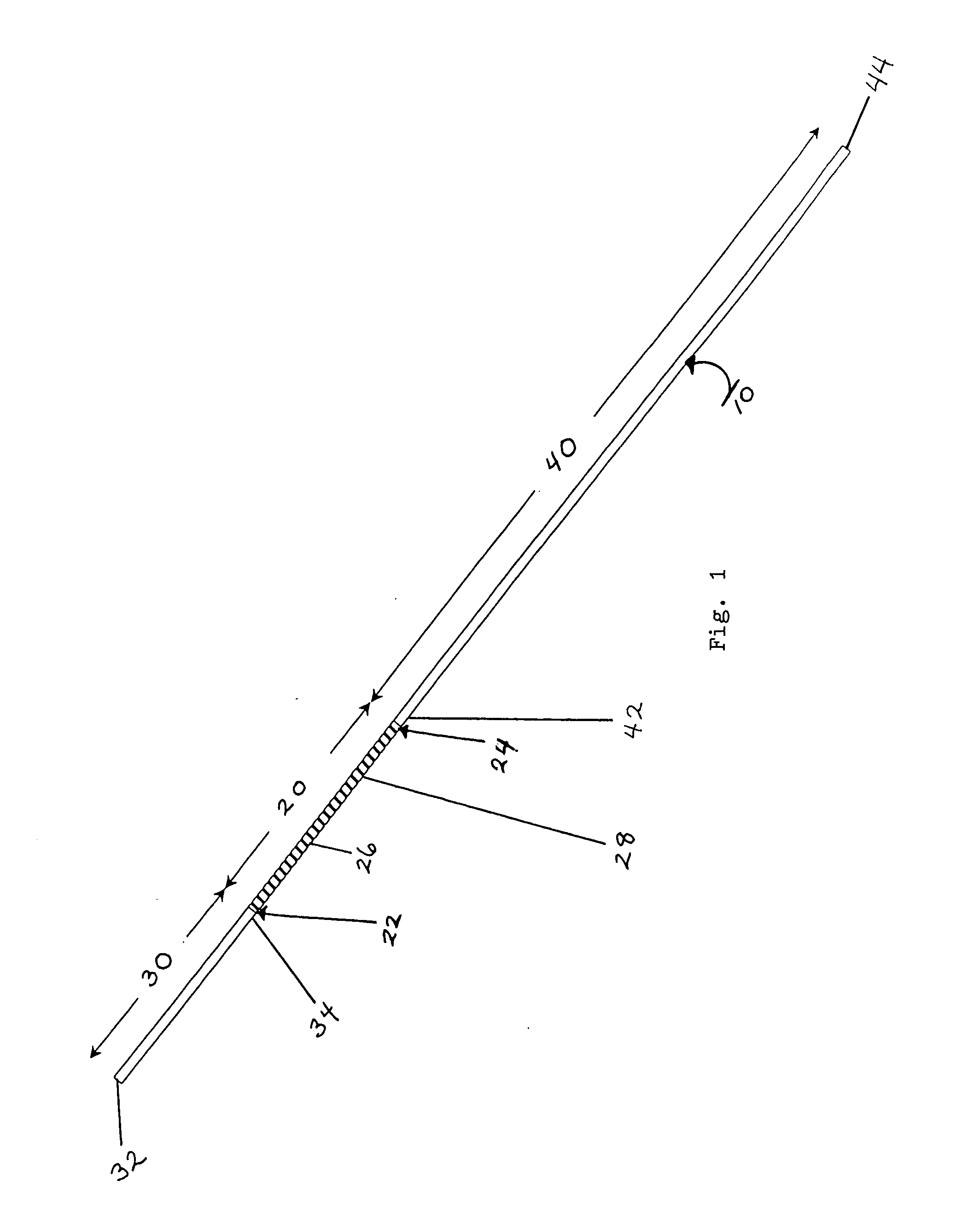
[0052] (ii) a hollow distal conduit arm having two open ends, one open end terminating as a discrete distal conduit end and the other open end being integrally joined to and in fluid flow communication with said ribbed medial section, said distal conduit arm being of predetermined external diameter size, tubular wall thickness, and internal lumen diameter, and having a subject-customized linear length which is to be custom-sized by a surgeon such that after in-vivo insertion of said sized distal conduit arm into a pre-chosen vein, said distal conduit end will float freely within the vein and anatomically lie adjacent to the cavo-atrial junction of the heart in the particular subject; and
[0057] (ii) a hollow distal conduit arm having two open ends, one end terminating as a discrete distal conduit end and the other end being integrally joined to and in fluid flow communication with said ribbed medial section, said distal conduit arm being of predetermined external diameter size, tubular wall thickness, and internal lumen diameter, and having a subject-customized linear length which is to be custom-sized by a surgeon such that after in-vivo insertion of said sized distal conduit arm into a pre-chosen vein in the particular subject, said distal conduit end will float freely within the vein and anatomically lie adjacent to the cavo-atrial junction of the heart in the particular subject,
[0071] (ii) a hollow distal conduit arm having two open ends, one end terminating as a discrete distal conduit end and the other end being integrally joined to and in fluid flow communication with said ribbed medial section, said distal conduit arm being of predetermined external diameter size, tubular wall thickness, and internal lumen diameter, and having a subject-customized linear length which is custom-sized by the surgeon such that after in-vivo insertion of said sized distal conduit arm into a pre-chosen vein in the particular subject, said distal conduit end will float freely within the vein and anatomically lie adjacent to the cavo-atrial junction of the heart in the particular subject,

Problems solved by technology

Renal disease continues to be an important cause of mortality and morbidity in the United States and throughout the world.
Acute renal failure is a worsening of renal function over hours to days, resulting in the retention of nitrogenous wastes (such as urea nitrogen) and creatinine in the blood.
In comparison, chronic renal failure results from a loss of renal function over months to years.
However, with End Stage Renal Disease (“ESRD”), there is chronic kidney failure; and the kidneys progressively fail and stop performing their essential functions over an extended period of time.
Unfortunately, because relatively few kidneys are presently available for transplantation purposes, the overwhelming majority of patients suffering from ESRD must receive regular blood dialysis treatments for the remainder of their lives.
Lastly, the price for medically treating ESRD continues to rise; for example, the cost to the Federal government for the medical management of ESRD is currently 17.9 billion dollars annually.
Hemodialysis is most often performed as an out patient procedure in approximately 3,600 approved centers in the U.S. In comparison, home dialysis is an option that is becoming ever less popular because of the need for a trained helper, large-sized dialysis equipment, and the very high costs.
However, the vascular access is widely called the “Achilles heel of dialysis” because of the markedly high morbidity and mortality among dialysis patients associated with complications of vascular access.
This combination of natural history failures, comorbidity and complications of therapy results in approximately 67,000 deaths attributed to ESRD in the U.S. alone.
The Scribner shunt suffered from major infection and clotting problems; and required extensive post-operative and long-term care of the shunt.
Percutaneous catheter assemblies have been used in hemodialysis since the early 1960's but for many years have been considered to be only a “temporary” form of vascular access because of their concomitant major infection and stenosis problems.
Nevertheless, for many years, the risk of potentially life-threatening infection complications was considered to be so great that the percutaneous catheter assemblies were withdrawn after each dialysis session and re-inserted when necessary to minimize the risk of infection.
Note that subclavian vein stenosis not only blocks blood flow, making it impossible to conduct hemodialysis; but also, catastrophically, can destroy all potential vascular access sites in one or both arms.
In addition, such tissue in-growth is believed by many medical practitioners to retard bacterial travel along the outer surface of the percutaneous catheter assembly, although it does not prevent it entirely.
Yet, while numerous published reports suggest that the cuff has reduced the infection rate, clinical infections remain a major problem even with the use of cuffed percutaneous catheter assemblies.
Furthermore, although it is not medically feasible to repeatedly puncture an artery, formation of the fistula “arterializes” the vein.
Surgeons have found that successful A-V fistula maturation is not possible in most hemodialysis patients because of the greatly increasing number of diabetic and older patients who have cardiovascular disease, which prevents the maturation process.
Another reason for the low rate of usage is that since surgeons have failed so often to achieve fistula maturation after performing the costly A-V fistula surgery, the surgeon often will no longer even try this technique for creating a vascular access.
Since about half of all prospective patients have an immediate and urgent need to start hemodialysis as quickly as possible, the patient often cannot wait for A-V fistula maturation to occur.
Thus critical patients must undergo costly temporary procedures and use percutaneous catheter assemblies to enable dialysis to take place, while waiting for maturation to occur.
In addition, it is one of the unfortunate drawbacks of A-V fistula, even with careful physical examination and / or the use of doppler ultrasound or venography to identify suitable veins, that approximately 40-50% of patients do not have the vascular anatomy sufficient to create a primary A-V fistula.
In addition, many dialysis veterans, for whom the use of an A-V fistula has previously failed, can no longer be considered as candidates for a primary A-V fistula.
Given the fact that A-V fistulas are largely not possible, a subcutaneously implanted PTFE prosthetic graft is today the most common form of permanent vascular access for the overwhelming majority of hemodialysis patients—because, in spite of the some severe limitations and risks for the conventionally known PTFE prosthetic graft, there simply is no better alternative available for them to date.
Despite these recent improvements and advances in prosthetic graft technology, the frequency of PTFE graft failure in-vivo remains very high.
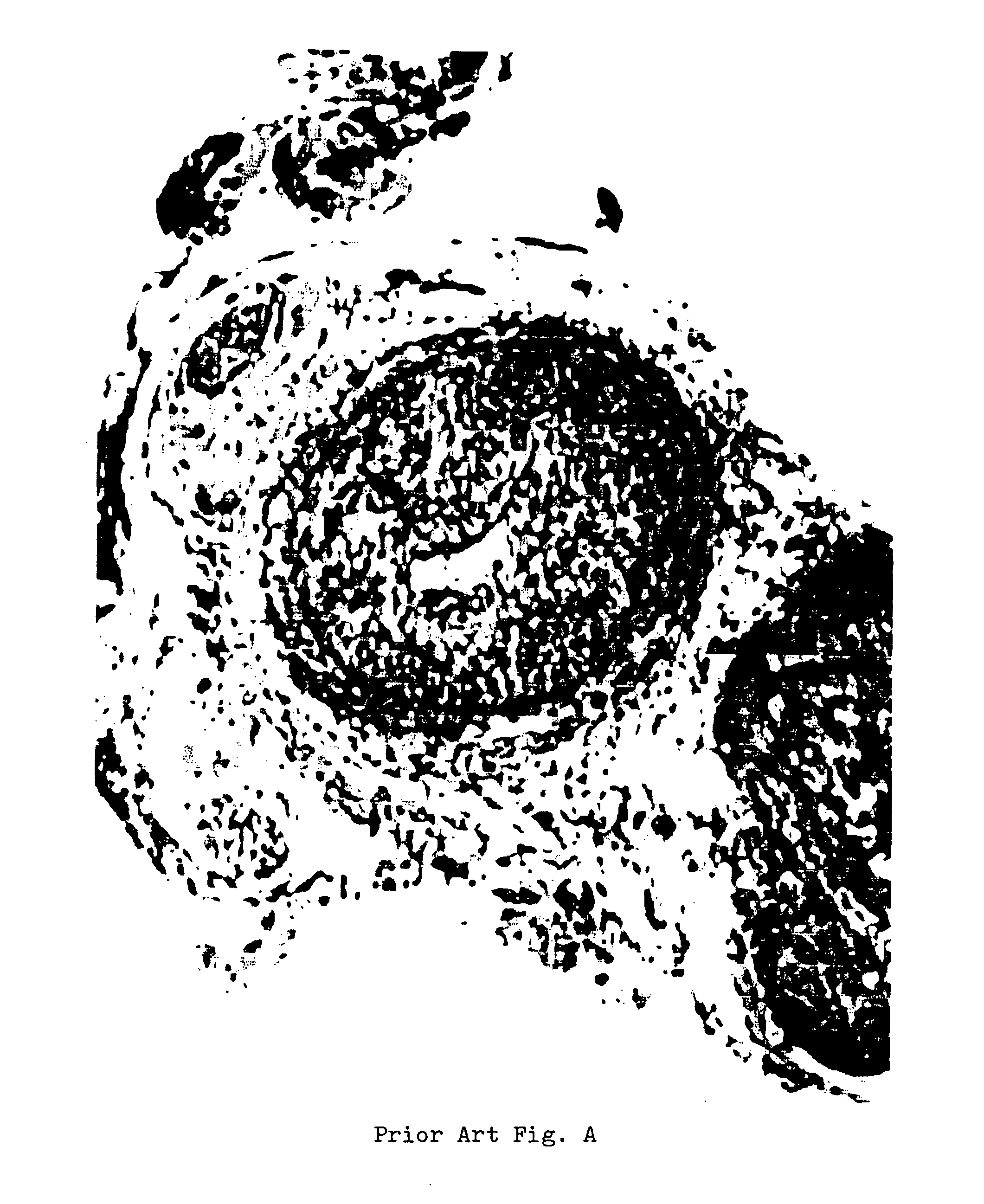
However, the most common cause of failure by far is neointimal hyperplasia—as exemplified by the hyperplasia occurring at the venous side of the access graft anastomosis in an implanted prosthetic graft.
A herein, neointimal hyperplasis results in the narrowing or “stenosis” of the distal outflow portion of the prosthetic graft device, and ultimately causes thrombosis of the entire length of the prosthetic graft, thereby rendering it unusable for dialysis.
Although the thrombus can theoretically be removed, the underlying cause cannot; and thus the patient enters a spiral phase of recurrent failure, hospitalization and surgery.
Despite innumerable attempts of various kinds over the years to prevent this particular cause of graft thrombosis and secondary failure, there have been few substantive advances to date.
Clearly therefore, the major disadvantages of the implanted PTFE prosthetic grafts are stenosis (i.e., closing of the lumen) and thrombosis (i.e., clotting), both of which block the flow of blood.
This dysfunction occurs in almost all graft patients several times during their lives; and, because it interferes with life-sustaining dialysis, must be corrected quickly.
Medical interventions to maintain PTFE prosthetic grafts and to treat patient complications (infection, thrombosis and aneurysm formation) are also expensive.
Although these additional procedures add cost and inconvenience, they have yet to improve significantly the mean time interval between interventional repairs, although they may in fact improve the prosthetic graft survival life as such.

Method used


Image

  • Apparatus and method for creating an arterio-venous connection in hemodialysis maintenance
  • Apparatus and method for creating an arterio-venous connection in hemodialysis maintenance
  • Apparatus and method for creating an arterio-venous connection in hemodialysis maintenance

Examples


Embodiment Construction

[0102] The subject matter as a whole which is the present invention provides a prosthetic endograft article, a surgical insertion kit, and an surgical insertion methodology for creating a vascular access in-vivo. In addition, the present invention is able to prevent a primary cause of arterio-venous graft thrombosis; and provides a novel vascular access construction for successful long term use in maintenance hemodialysis.

[0103] The present invention employs a prosthetic endograft which is patient-customized by the surgeon as an endovascular component; and utilizes a unique surgical method for endovascular insertion of the prosthetic endograft in a manner which does not require a distal anastomosis of the endograft, thereby allowing the distal outflow end of the implanted article to remain unattached and freely floating within the internal lumen of a pre-chosen vein.

[0104] The present invention is therefore able to provide a range of unforeseen advantages and unexpected medical be...


PUM


Abstract

The present invention provides a kit apparatus and a methodology to prevent the primary causes of arterio-venous graft thrombosis; and provides a durable vascular access for successful long term use in hemodialysis. The invention employs a patient-customized prosthetic endograft as an subcutaneously implanted vascular access; and utilizes a surgical method for endovascular insertion of the prosthetic endograft into a pre-chosen vein, which does not require a distal anastomosis, and thus allows the distal outflow end of the implanted vascular access to remain unattached and freely floating at a precisely located anatomic position within the internal lumen the pre-chosen vein.

Description

PRIORITY CLAIM [0001] This invention was first filed as U.S. Provisional Patent Application No. 60 / 553,007 on Mar. 15, 2004. The filing date and priority of this first filing is expressly claimed pursuant to 35 U.S.C. 119(e).FIELD OF THE INVENTION [0002] This invention relates generally to the making of a permanent anatomic connection to access the vascular blood system in-vivo; and is directed specifically to apparatus and methods for creating a vascular access suitable for blood dialysis in humans afflicted with end stage renal disease (or “ESRD”). BACKGROUND OF THE INVENTION [0003] Renal disease continues to be an important cause of mortality and morbidity in the United States and throughout the world. Renal disease may be acute or chronic. Acute renal failure is a worsening of renal function over hours to days, resulting in the retention of nitrogenous wastes (such as urea nitrogen) and creatinine in the blood. In comparison, chronic renal failure results from a loss of renal fu...

Claims


Application Information

Patent Timeline
Patent Type & Authority Applications(United States)
IPC IPC(8): A61M5/00A61M39/10
CPCA61B17/3415A61F2/06A61M25/0668A61M25/0194A61M25/06A61M1/3655
Inventor SMEGO, DOUGLAS R.
Owner PERMAGRAFT
Learn More